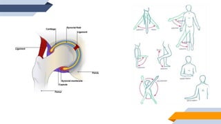
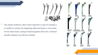
TOTAL HIP
IMPLANT
HIP ANATOMY
 The hip joint connects the lower extremities with the axial skeleton.
 The hip is one of the body’s largest joints. It is a ball and socket joint.
 The socket is formed by the part of large pelvis bone (acetabulum of the pelvis) and the
ball is head of the femur (thigh bone).
 The bone surfaces of the ball and socket are covered with articular cartilage, a smooth tissue that cushions the
ends of the bones and enables them to move easily.
 A thin tissue called synovial membrane surrounds the hip joint.
 Band of tissue called ligaments (the hip capsule) connect the ball to the socket
and provide stability.
NECESSITY OF IMPLANT
 Hip prosthesis or Hip replacement surgery becomes necessary when the
hip joint has been damaged due to arthritis, malformation of the hip
since birth or due to abnormal development or damaged due to injury/trauma.
 Many older people are subjected to degenerative bone and joint diseases
such as osteoarthritis and rheumatoid arthritis. These diseases necessitate joint replacement.
 In an arthritic hip joint, the cartilage has been damaged, narrowed or even lost due to degenerative process or
by inflammation.
 This can make movement gut wrenching for the patient.
Healthy
Hip Joint
Osteoarthritis
 If a part of joint has been damaged, then the surgeon may repair
or replace only the damaged parts. There are basically three types
of hip replacement: total hip replacement(most common), partial
hip replacement and hip resurfacing.
 If the entire joint is damaged, Total Hip Replacement (THR) or
Total Hip Arthroplasty is suggested, in which artificial prosthesis
of the joint is replaced, aimed for pain relief and restoration of
mobility.
Hip Arthroplasty
Acetabular cup
Liner
Femoral
Head
x- ray of THR showing the ball, socket and
stem implants.
HISTORY
 The early methods of correcting hip joint malfunctions involved
only the acetabular cup or femoral head. One technique of
restoring hip joint function is to place a cup over the femoral head
while the surface of acetabulum is also resected to fit the cup.
 During the past 40 years many combinations of materials and
designs have been considered and developed to achieve an ideal
painless, stable and freely mobile joint implant with a longer lifetime.
First total hip
replacement was
performed by Gluck
1890
1938
Philips Wiles used a
stainless steel ball secured
to the femur with a bolt
and a stainless acetabular
liner secured with screws.
1939
Smith Peterson described an mold
arthroplasty, initially made by glass,
modified over a period of time to
celluloid, pyrex, bakelite and finally
vitallium.
1967
Charnley formed a metal
femoral component with a
low friction HMWPE
acetabular cup.
Over the years, superiority
of Co-Cr alloys over any
other steel components
was established.
1968
Ring pointed out disadvantage of
using PMMA cements. He developed
cement-less cup components with
long pelvic anchoring screws.
Meanwhile, charnley introduced cemented
femoral and acetabular components, with a
plastic bearing surface and PMMA for fixation,
and it is his prosthesis that remains the gold
standard in hip replacement even today.
MATERIALS USED
 In any joint replacement surgery, there are three key variables: geometry, choice of material and operative
procedure.
 Ideally, the new joint must exactly match the geometry and performance of natural hip joint or any other joint.
 Choice of material plays a major role as the implant comes in contact with body fluids and tissues, so it should be
biocompatible and also should not harm surrounding tissues.
 Operative procedure is also a vital variable. There are two major operative approach for performing THR: the
posterior approach(more common) and the anterior approach. Both approaches offer pain relief and improvement
in walking and movement within weeks of surgery.
 THR includes the two major components of femoral and acetabular. The
femoral component itself is including femoral head, femoral neck, femoral
collar and femoral stem.
 Each one of these components has its own role in handling load and providing a
smooth movement.
 In order to handle the applied load, the femoral component need to be connected to the adjacent bone using
various fixations. Femoral fixations falls into two general groups of fixation using PMMA bone cement (cemented)
or non-cemented categories.
 The SOCKETS for use with cement are made of special type of polyethylene, which is very tough, and slippery
particularly when wet. It has ridges on the outside and a wire marker so that its position can be seen on the x-ray.
The ridges are designed to improve fixation of the cup by the cement.
 The artificial socket is implanted into the natural socket by first filling the cavity with the
cement and then pushing in the artificial cup, which is held still wile the cement sets.
 The socket for use without cement is made of metal. It is specially designed porous
surface on its outer side to encourage bone growth into it and to join up with the socket
much like broken bone heals.
 This process can take 6-12 weeks to heal, however the cemented socket is held firmly in
place till it sets.
 Machining the natural socket accurately and using an artificial socket that is precisely
1-2mm bigger provides the initial fit.
 The metal socket is then forcibly “jammed” into place and further fixation can be obtained if necessary by
additional screws.
 The next stage is to place PLASTIC INSERT within the metal shell
against which the artificial ball will form the joint.
 The BALL portion of artificial hip consists of metal stem or rod on
top of which the metal ball is attached at an angle to mimic the
shape of the femur bone.
 This is inserted into the thigh bone after first filling the marrow cavity with bone cement and holding it still while
the cement sets.
 The implant for use without cement has a specially fabricated outer surface as in the case of the un-cemented
socket..
 Femoral stems are of materials such as titanium alloys and CoCr alloys.
 Excellent long term results have been obtained with cemented THR.
 In contrast, un-cemented components offer advantages of reduced operating time, reduced initial trauma to
endosteal bone surfaces, long term interface stability, less foreign material.
 Only four broad material combinations have so far found favor in practical use: a metallic femoral head in polymeric
acetabular cup, a ceramic head in a polymeric cup, a ceramic head on ceramic cup and a metallic head on metallic cup.
 The metals used are generally Co-Cr-Mo alloys or in some cases titanium alloys, the polymer used Ultra High
Molecular Weight Polyethylene (UHMWPE) and the ceramic used high purity alumina or in some cases partially
stabilized zirconia.
 Coatings or ion implantations are usually used to improve the surface properties and biocompatibility of implants as
well as decreasing metallic wear and corrosion.
 One simple method to allow tissue ingrowth (a type of coating) is
to modify its surface by implanting spherical beads or wire mesh.
On the other hand, coating of hydroxyapatite (HA) onto a bioinert
metallic implant is an effective method.
POSSIBLE COMPLICATIONS AFTER THR
 The complication rate following hip replacement surgery is low. Sometimes it can occur in less than 2% patients.
1. BLOOD CLOTS: it can occur in the leg veins or pelvis. These clots can be life threatening if they break free and
travel to lungs or rarely brain.
2. INFECTION: it can occur superficially near your incision or deep around the prosthesis. It may even occur after
days, weeks or years later too.
3. FRACTURE: during surgery, healthy portions of hip may damage or fracture. Sometimes there are small fractures
or large fractures can occur too.
4. DISLOCATION: this occurs when the ball comes out of the socket. The risk is greatest in first few months.
5. LEG-LENGTH INEQUALITY: sometimes after surgery, one leg may feel longer or shorter than the other. This may
occur due to surrounding muscles, strengthening and stretching those muscles may help.
6. LOOSENING: over the years the him implant may wear out or loosen. This is most often due to everyday activity. It
can also occur due to osteolysis. Revision surgery is required to cure this.
7.NERVE DAMAGE: rarely, nerves in the area where the implant is placed can be injured. This can cause numbness,
pain and weakness.
BLOOD CLOT
DISLOCATION
THANK YOU!

Total Hip Implant

  • 1.
  • 2.
    HIP ANATOMY  Thehip joint connects the lower extremities with the axial skeleton.  The hip is one of the body’s largest joints. It is a ball and socket joint.  The socket is formed by the part of large pelvis bone (acetabulum of the pelvis) and the ball is head of the femur (thigh bone).  The bone surfaces of the ball and socket are covered with articular cartilage, a smooth tissue that cushions the ends of the bones and enables them to move easily.  A thin tissue called synovial membrane surrounds the hip joint.  Band of tissue called ligaments (the hip capsule) connect the ball to the socket and provide stability.
  • 4.
    NECESSITY OF IMPLANT Hip prosthesis or Hip replacement surgery becomes necessary when the hip joint has been damaged due to arthritis, malformation of the hip since birth or due to abnormal development or damaged due to injury/trauma.  Many older people are subjected to degenerative bone and joint diseases such as osteoarthritis and rheumatoid arthritis. These diseases necessitate joint replacement.  In an arthritic hip joint, the cartilage has been damaged, narrowed or even lost due to degenerative process or by inflammation.  This can make movement gut wrenching for the patient. Healthy Hip Joint Osteoarthritis
  • 5.
     If apart of joint has been damaged, then the surgeon may repair or replace only the damaged parts. There are basically three types of hip replacement: total hip replacement(most common), partial hip replacement and hip resurfacing.  If the entire joint is damaged, Total Hip Replacement (THR) or Total Hip Arthroplasty is suggested, in which artificial prosthesis of the joint is replaced, aimed for pain relief and restoration of mobility. Hip Arthroplasty Acetabular cup Liner Femoral Head
  • 6.
    x- ray ofTHR showing the ball, socket and stem implants.
  • 7.
    HISTORY  The earlymethods of correcting hip joint malfunctions involved only the acetabular cup or femoral head. One technique of restoring hip joint function is to place a cup over the femoral head while the surface of acetabulum is also resected to fit the cup.  During the past 40 years many combinations of materials and designs have been considered and developed to achieve an ideal painless, stable and freely mobile joint implant with a longer lifetime.
  • 8.
    First total hip replacementwas performed by Gluck 1890 1938 Philips Wiles used a stainless steel ball secured to the femur with a bolt and a stainless acetabular liner secured with screws. 1939 Smith Peterson described an mold arthroplasty, initially made by glass, modified over a period of time to celluloid, pyrex, bakelite and finally vitallium. 1967 Charnley formed a metal femoral component with a low friction HMWPE acetabular cup. Over the years, superiority of Co-Cr alloys over any other steel components was established. 1968 Ring pointed out disadvantage of using PMMA cements. He developed cement-less cup components with long pelvic anchoring screws. Meanwhile, charnley introduced cemented femoral and acetabular components, with a plastic bearing surface and PMMA for fixation, and it is his prosthesis that remains the gold standard in hip replacement even today.
  • 9.
    MATERIALS USED  Inany joint replacement surgery, there are three key variables: geometry, choice of material and operative procedure.  Ideally, the new joint must exactly match the geometry and performance of natural hip joint or any other joint.  Choice of material plays a major role as the implant comes in contact with body fluids and tissues, so it should be biocompatible and also should not harm surrounding tissues.  Operative procedure is also a vital variable. There are two major operative approach for performing THR: the posterior approach(more common) and the anterior approach. Both approaches offer pain relief and improvement in walking and movement within weeks of surgery.
  • 10.
     THR includesthe two major components of femoral and acetabular. The femoral component itself is including femoral head, femoral neck, femoral collar and femoral stem.  Each one of these components has its own role in handling load and providing a smooth movement.  In order to handle the applied load, the femoral component need to be connected to the adjacent bone using various fixations. Femoral fixations falls into two general groups of fixation using PMMA bone cement (cemented) or non-cemented categories.  The SOCKETS for use with cement are made of special type of polyethylene, which is very tough, and slippery particularly when wet. It has ridges on the outside and a wire marker so that its position can be seen on the x-ray. The ridges are designed to improve fixation of the cup by the cement.
  • 11.
     The artificialsocket is implanted into the natural socket by first filling the cavity with the cement and then pushing in the artificial cup, which is held still wile the cement sets.  The socket for use without cement is made of metal. It is specially designed porous surface on its outer side to encourage bone growth into it and to join up with the socket much like broken bone heals.  This process can take 6-12 weeks to heal, however the cemented socket is held firmly in place till it sets.  Machining the natural socket accurately and using an artificial socket that is precisely 1-2mm bigger provides the initial fit.  The metal socket is then forcibly “jammed” into place and further fixation can be obtained if necessary by additional screws.
  • 12.
     The nextstage is to place PLASTIC INSERT within the metal shell against which the artificial ball will form the joint.  The BALL portion of artificial hip consists of metal stem or rod on top of which the metal ball is attached at an angle to mimic the shape of the femur bone.  This is inserted into the thigh bone after first filling the marrow cavity with bone cement and holding it still while the cement sets.  The implant for use without cement has a specially fabricated outer surface as in the case of the un-cemented socket..  Femoral stems are of materials such as titanium alloys and CoCr alloys.
  • 13.
     Excellent longterm results have been obtained with cemented THR.  In contrast, un-cemented components offer advantages of reduced operating time, reduced initial trauma to endosteal bone surfaces, long term interface stability, less foreign material.  Only four broad material combinations have so far found favor in practical use: a metallic femoral head in polymeric acetabular cup, a ceramic head in a polymeric cup, a ceramic head on ceramic cup and a metallic head on metallic cup.  The metals used are generally Co-Cr-Mo alloys or in some cases titanium alloys, the polymer used Ultra High Molecular Weight Polyethylene (UHMWPE) and the ceramic used high purity alumina or in some cases partially stabilized zirconia.  Coatings or ion implantations are usually used to improve the surface properties and biocompatibility of implants as well as decreasing metallic wear and corrosion.
  • 14.
     One simplemethod to allow tissue ingrowth (a type of coating) is to modify its surface by implanting spherical beads or wire mesh. On the other hand, coating of hydroxyapatite (HA) onto a bioinert metallic implant is an effective method.
  • 15.
    POSSIBLE COMPLICATIONS AFTERTHR  The complication rate following hip replacement surgery is low. Sometimes it can occur in less than 2% patients. 1. BLOOD CLOTS: it can occur in the leg veins or pelvis. These clots can be life threatening if they break free and travel to lungs or rarely brain. 2. INFECTION: it can occur superficially near your incision or deep around the prosthesis. It may even occur after days, weeks or years later too. 3. FRACTURE: during surgery, healthy portions of hip may damage or fracture. Sometimes there are small fractures or large fractures can occur too. 4. DISLOCATION: this occurs when the ball comes out of the socket. The risk is greatest in first few months. 5. LEG-LENGTH INEQUALITY: sometimes after surgery, one leg may feel longer or shorter than the other. This may occur due to surrounding muscles, strengthening and stretching those muscles may help.
  • 16.
    6. LOOSENING: overthe years the him implant may wear out or loosen. This is most often due to everyday activity. It can also occur due to osteolysis. Revision surgery is required to cure this. 7.NERVE DAMAGE: rarely, nerves in the area where the implant is placed can be injured. This can cause numbness, pain and weakness. BLOOD CLOT DISLOCATION
  • 17.