
Recommended
More Related Content
What's hot
What's hot (20)
Viewers also liked
Viewers also liked (16)
Similar to Wavelength April 2015 Volume 19 No.1
Similar to Wavelength April 2015 Volume 19 No.1 (20)
More from Jerry Duncan
More from Jerry Duncan (8)
Wavelength April 2015 Volume 19 No.1
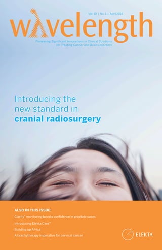
- 1. Introducing the new standard in cranial radiosurgery Vol. 19 | No. 1 | April 2015 Pioneering Significant Innovations in Clinical Solutions for Treating Cancer and Brain Disorders ALSO IN THIS ISSUE: Clarity® monitoring boosts confidence in prostate cases Introducing Elekta Care™ Building up Africa A brachytherapy imperative for cervical cancer
- 2. 2 Volume 19 | Number 1 | April 2015 An Elekta Publication | www.elekta.com All letters, comments or suggestions for future articles, requests for reprints and permissions are welcome. Contact Wavelength: Jerry Duncan, Global Communications Writer Tel: +1- 770 670 2341 | (time zone: Eastern Standard) Email: jerry.duncan@elekta.com The products and product clinical indications for use described within this magazine may not have clearance or registration in certain countries. Please consult Elekta in your country for questions concerning a product or clinical use subject. Art. No. 1518099 ©Elekta AB (publ). All mentioned trademarks and registered trademarks are the property of the Elekta Group. All rights reserved. No part of this document may be reproduced in any form without written permission from the copyright holder. Contents The new standard in intracranial radiosurgery Practical stereotaxy with Versa HD™ at Danish medical center Leksell Gamma Knife® Registry turns big data into clinical intelligence Introducing Elekta Care™ Intra-fraction Clarity® monitoring boosts confidence in prostate cases Building up Africa Gentler skin cancer therapy with Esteya® in Las Vegas Elekta around the world A brachytherapy imperative for cervical cancer “Leading Edge©” Gamma Knife radiosurgery for glioblastoma multiforme Q&A with brachytherapy pioneer Eric van ‘t Hooft Sweden’s MEG center, NatMEG, marks first year 4 19 6 22 10 25 13 28 14 31 16 34
- 3. 3 Approaching my first full year as President and CEO of Elekta has me reflecting on what I’ve learned about Elekta and about you, our customers. During my four-month learning phase, before I officially took the reins of this company, I traveled around the world asking customers why they chose Elekta as their partner. A consistent response was that our employees are thinking about how our solutions impacted patients. Indeed, this is something that I found to be ingrained in Elekta culture; the Elekta people always ask themselves the question: ‘Which alternative is better for the patient?’ In that sense, we share a common mission when it comes to your patients. Not surprisingly, Wavelength once again provides many examples of this patient-centric mindset. We start out with the introduction of Leksell Gamma Knife® Icon™ , a novel Gamma Knife platform with an advanced imaging workflow to make radiosurgery even more precise and safe for the patient. Then, there are articles on how brachytherapy should be used for the patient’s benefit in cervical cancer cases, one on patient-friendly Clarity® , a story on the role of Leksell Gamma Knife to help patients with a particularly virulent tumor type, our efforts to improve access to radiation therapy in Africa, and more. I hope you enjoy reading them! Niklas Savander President and CEO of Elekta Greetings,
- 4. Leksell Gamma Knife Icon* – Elekta’s latest generation stereotactic radiosurgery system for the brain – not only maintains the unparalleled precision for which Gamma Knife is renowned, but it also presents new possibilities for treating virtually any brain target, regardless of size or location. This capability is accomplished through new functionality, fully integrated in Leksell Gamma Knife. Elekta unveiled Leksell Gamma Knife Icon at the 3rd ESTRO Forum, April 25, in Barcelona, Spain. “Leksell Gamma Knife Icon starts a new chapter in cranial radiosurgery, in which a broad range of targets in the brain, large or small, can be treated with extreme accuracy and absolute control of dose delivery,” says Peter Fröberg, Senior Product Manager Radiosurgery. “Equally important, Icon represents the most clinically flexible brain radiosurgery platform ever developed, giving clinicians the option to use frame- based or frameless fixation, single or multiple fractions, traditional radiosurgery or ultra-precise microradiosurgery, all meeting the degree of precision needed for each patient’s case.” The technological evolution of cranial radiosurgery in Leksell Gamma Knife Icon introduces the concept of precision radiosurgery with adaptive DoseControl. 4 The new standard in cranial radiosurgery Elekta introduces Leksell Gamma Knife® Icon™ , featuring precision radiosurgery with adaptive DoseControl™ “Tools used by the surgeon must be adapted to the task and where the human brain is concerned, no tool can be too refined.” Prof. Lars Leksell
- 5. 5 DoseControl™ DoseControl is a comprehensive solution entailing both absolute motion management – whether for frame-based or frameless treatments – and real dose delivery, all seamlessly integrated in the Icon™ control system and ensuring the highest possible treatment precision. Whether using the frame-based or the frameless option, motion management in Icon ensures that patient motion is effectively achieved to facilitate a successful treatment. For frameless treatments, the high-definition motion management solution monitors the patient in real time during treatment with 0.1 mm resolution, enabling the same precision for frameless treatments as for frame-based treatments. The system includes gating functionality; the collimation system sectors instantly block the radiation beams from encountering healthy tissue if the patient moves outside defined limits. To ensure accurate delivery of the actual dose to the patient, the real dose delivery solution was developed. A core component of this solution is the stereotactic cone beam CT (CBCT) workflow integrated in Icon® that allows physicians to adapt the treatment plan online, at the time of treatment. “A unique stereotactic CBCT imaging solution, calibrated to the delivery system, permits the stereotactic reference to be set and verified prior to treatment,” Fröberg explains. “The system automatically adapts the dose delivery to match present patient position.” Also part of the DoseControl solution, a unique feature in Icon enables the clinician to compare the dose that is about to be delivered with the planned dose at the console, based on the actual patient position, before or even during treatment, and to adapt the treatment plan if needed. “In the design of Leksell Gamma Knife Icon, we aimed to develop a comprehensive solution that would be the preferred modality for treating almost any intracranial target,” Fröberg adds. “That meant we needed to provide an effective option for treating large lesions over multiple fractions – which we addressed with superior real- time high-definition motion management technology. The integration of the CBCT workflow ensures the most accurate treatment possible. The complete solution is intended to give clinicians the ability and confidence to treat virtually any pathology found in the brain, and the efficient workflows will allow them to do this every day in the clinical setting.” • * Leksell Gamma Knife Icon is not for sale or distribution in the US and is not CE marked.
- 6. 6 As a major Danish medical center with multiple linear accelerators and other RT technology, Odense University Hospital (OUH) has had experience in SBRT/SRS for nearly a decade. However, it wasn’t until it acquired its Versa HD systems in 2013 that it became practical for OUH clinicians to implement the techniques for several patients per week, mostly for lung SBRT and brain SRS. “For the first stereotactic treatments in 2005, we scheduled one-and-a-half hours for everything, and managed over time to drop down to 45 minutes,” says Olfred Hansen, MD, Head of the OUH Radiation Therapy Department. “Today, with Versa HD and beam-on times as much as 60 percent shorter – which we confirmed with our own internal research1 – the total time slot is down to 20 minutes.” OUH began using the first of its three Versa HD systems in April 2013, with the first lung SBRT patient treated using the system’s High Dose Rate mode on June 3, 2013. “We delivered a single fraction of 22 Gy in about same time it would take to deliver a 2 Gy dose on another of our linacs,” Dr. Hansen recalls. The second and third Versa HD systems went clinical in June and October of 2013 and today all SBRT (mostly lung) and SRS (brain) cases are treated with these systems, in addition to non-stereotactic treatments for head-and-neck and lung cancer cases. “The acquisition of the Versa HD systems was justified as replacements for older linacs and to advance the department’s aim to efficiently perform stereotaxy,” says Knud Aage Werenberg, Head Physicist at OUH. “Since then, SBRT and SRS have undergone a huge expansion at OUH. In 2012, we treated 145 patients stereotactically, 206 patients in 2013 and 267 patients in 2014.” Stereotactic radiotherapy at OUH in 2014 SBRT/SRS Number of patients Number of fractions Lung 174 705 Liver 2 12 Bone 2 6 Brain 89 156 Total 267 879 The suitability of Versa HD for stereotactic cases lies mainly in the Agility™ MLC, according to Dr. Hansen. “Agility has 160 0.5 cm wide leaves for more conformal beam shaping for smaller targets and a leaf design for low radiation leakage,” he says. “The Agility leaf speed of 6.5 centimeter per second means that the Versa HD system’s High Dose Rate [FFF] mode can be delivered in a VMAT treatment even when high modulation is required. The combination of rapid leaf speed and high Versa HD™ provides the speed for routine SBRT and SRS treatments at Odense University Hospital Practical stereotaxy
- 7. 7 dose rate delivery provides the speed we need to reduce beam-on time so that it’s practical to use SBRT and SRS for several patients each week.” Lung SBRT The high rate of smoking in Denmark contributes to among the world’s highest rates of lung cancer for men and women, with Danish women having the highest rate in the world in 2012.2 “Many people with lung cancer have poor lung function and other co-morbidities that make them ill-suited for surgery,” Dr. Hansen notes. “The dilemma has been that – despite the fact that stereotactic radiotherapy has been shown to be a good alternative to surgery for these patients – only three centers in Denmark have been allowed to perform it. Conversely, fractionated radiotherapy is available at more locations, but most patients are reluctant to travel sometimes long distances for many therapy sessions. It’s a fantastic opportunity for lung cancer patients – particularly those in poor condition – that OUH is now an additional stereotactic resource in Denmark, and one that is very convenient for patients, involving just three 20-minute fractions.” A typical beneficiary of Versa HD SBRT capabilities for lung cancer patients who are poor surgical candidates was a 71-year-old female patient with a 3.8 cm NSCLC tumor located in the left upper lobe, but also close to the middle lobe. Surgery would have entailed a double lobectomy, an unfeasible procedure for this patient, who had an unusually low FEV1* of only 19 percent. The patient had SBRT (22 Gy X 3) and 10 months post-therapy – while her FEV1 has not improved – the patient is doing well, Dr. Hansen notes. “In the past, a patient like her would not even have had radiotherapy,” he says. “We have a limit of an FEV1 of 80 percent for treating lung cancer with fractionated radiation therapy in a curative setting. But “The 0.5 cm MLC leaf width of Agility makes it easy to conform not only to small mets, but even up to a field size of 40 cm X 40 cm for larger malignancies.” Olfred Hansen, MD, OUH Radiation Therapy Department Odense University Hospital
- 8. 8 with SBRT, clearly there is no lower limit for FEV1. Patients with serious co-morbidities also have a curative option. Before SBRT, the surgeons often had to take undesirable risks because surgery was the only option. Five to six percent of those patients died within one month of their treatment.” The protocol for lung SBRT involves an initial 4D CT scan for planning purposes. The mid-ventilation phase is chosen as the planning CT and serves as the reference CT in the image registration process. Before treatment delivery a 4D CBCT (Symmetry™ XVI 4.5) scan is done to localize the tumor and to quantify how the tumor moves with respiration, followed by the first VMAT arc (200°). A second “midway” 4D CBCT is performed before the next VMAT arc (180°) to ensure the patient hasn’t moved. Typical prescriptions are 66 Gy X 3 or 56 Gy X 8. With the High Dose Rate mode of Versa HD, total beam-on time has been reduced from about seven minutes (with FF) to approximately three minutes. This lung SBRT delivery speed could lead to the elimination of the three-minute midway 4D CBCT scan at OUH, Werenberg notes. “When we started with SBRT on lung patients with the flattening filter [FF], and therefore longer beam-on time, we decided to verify the intrafractional motion with a CBCT between the two arcs,” he explains. “Another reason was because the high dose and fewer fractions make SBRT more sensitive to intrafraction motion. With the introduction of Versa HD, the High Dose Rate mode can reduce the beam-on time by 60 percent1 , which could reduce the likelihood of intrafraction motion. This in turn could make it unnecessary to pause the beam to perform the midway 4D CBCT scan.” The 2015 implementation of 4D Intra- Fraction Imaging (XVI 5.0) at OUH, however, could obviate the need to pause the beam for intrafraction CBCT scans. This solution enables clinicians to acquire images during beam delivery, providing a verification of real tumor motion during treatment. “That will definitely speed things up, “The acquisition of the Versa HD systems was justified as replacements for older linacs and to advance the department’s aim to efficiently perform stereotaxy.” Knud Aage Werenberg, Head Physicist Odense University Hospital TOP: SBRT of a patient with NSCLC localized disease treated with 66 Gy/3 fractions with 2 arcs. BOTTOM: SRS of a patient with one brain metastasis treated with 20 Gy/1 fraction with 3 arcs
- 9. 9 because we could omit the midway CBCT and continue treating without a break,” Dr. Hansen says. “When you have finished your first arc, then you already have the answer about whether the patient position is still stable.” At its present pace, OUH is using its Versa HD systems for SBRT treatments of 180 new patients per year or about four patients per week and 730 fractions per year. Brain SRS OUH clinicians use their Versa HD systems for SRS treatments of patients with oligometastatic disease or brain metastases of any histology. They will treat two metastases in a single fraction if each lesion is less than 3 cm in size, or a single met up to 4 cm. All brain SRS patients are treated with a single full coplanar VMAT arc and two non-coplanar half arcs. For head fixation, a thermoplastic mask system (Orfit Industries) for the head, neck and shoulders is used and the HexaPOD™ patient positioning system is used for correcting patient position in six independent axes. “Use of HexaPOD and its 6D functionality is standard for our SRS treatments,” Werenberg notes. “You could argue that, for small conformal lesions, the advantage of the rotational correction is insignificant, but if you are treating two targets with one isocenter – which we often do – then it’s possible to position the patient with respect to both targets.” In a routine 20 Gy single fraction treatment (or 27 Gy X 3), the patient is positioned on the HexaPOD system for an initial CBCT scan. Treatment staff make any needed corrections to patient position on the HexaPOD. The first VMAT arc is delivered (360°, table = 0°) and the customary midway CBCT scan is acquired to determine if the patient’s position has changed, and any needed changes are executed on the HexaPOD. The second VMAT arc is delivered (212°, table = 45°), followed by the third arc (212°, table = - 45°). “The 0.5 cm MLC leaf width of Agility makes it easy to conform not only to small mets, but even up to a field size of 40 cm X 40 cm for larger malignancies,” Dr. Hansen observes. “And with the narrower leaves, it’s also easier to avoid giving a dose to nearby vital organs like the optic chiasm and brain stem, which makes the treatment safer.” As in the lung SBRT cases, using the High Dose Rate (FFF) mode on Versa HD for brain SRS has reduced beam-on time significantly versus when the flattening filter (FF) is used. “The beam-on time is decreased from an average of 10 minutes to a little over six minutes,” Werenberg says. “That translates into a 20-minute treatment slot, from when the patients enter to when they leave.” OUH uses its Versa HD for brain SRS of 90 new patients each year or about two patients every week and 162 fractions annually. A busy stereotactic future for OUH OUH’s acquisition of its Versa HD systems has served as the launch pad for a thriving SBRT and SRS practice at the center, and Werenberg and Dr. Hansen predict that their stereotactic patient volume and indications will continue to grow in the coming years. “Our increasing confidence in safely delivering these high stereotactic doses means we can expand lung SBRT to treat more centrally located lung lesions with a modified fractionation schedule,” Dr. Hansen says. “We will add more spinal lesions, because this has been working very well in the carefully selected cases we’ve done, in addition to more liver SBRT using gold markers. In addition, we plan to grow our palliative indications, particularly bone mets. If we can control these sites in patients with oligometastatic disease, the patients have the potential for longer disease-free survival.” • References 1. Hansen, Christian R, Bertelsen A, Lynggaard, H. et al. Plan quality and delivery accuracy of flattening filter free beam for SBRT lung treat- ments. Acta Oncologica, 2014; Early Online: 1-6. 2. Ferlay J, Soerjomataram I, Ervik M, Dikshit R, Eser S, Mathers C, Rebelo M, Parkin DM, Forman D, Bray, F. GLOBOCAN 2012 v1.0, Cancer Incidence and Mortality Worldwide: IARC CancerBase No. 11 [Internet]. Lyon, France: International Agency for Research on Cancer; 2013. Available from: http://globocan.iarc.fr. * FEV1: Forced Expiratory Volume1, the volume exhaled during the first second of a forced expiratory maneuver started from the level of total lung capacity.
- 10. 10 Turning big data into clinical intelligence Leksell Gamma Knife® Registry designed to bring Gamma Knife users together to improve outcomes, drive the future of Gamma Knife® radiosurgery To provide Gamma Knife practitioners with an unparalleled opportunity to advance clinical research and further optimize patient care, Elekta recently launched Leksell Gamma Knife Registry. This solution enables clinicians to leverage the enormous volume of data created by the more than 70,000 patients each year who receive Gamma Knife radiosurgery. Designed as a centralized, cloud-based solution, Leksell Gamma Knife Registry will allow Gamma Knife practitioners to store, retrieve, process, and analyze Gamma Knife radiosurgery treatments, as well as patient outcomes and operational data, in a common format. “By enabling uniform data entry and indication-specific analysis of Gamma Knife radiosurgery outcomes, clinicians now have a powerful tool to clarify how Gamma Knife should be used to deliver better outcomes for patients and to expand the use of this proven technology for more indications,” says Catherine Gilmore-Lawless, Elekta Vice President, Clinical Intelligence. “While Gamma Knife users have been documenting their cases for decades, the Registry provides a much more systematic, standardized way to perform this activity, which will make the data even more meaningful and accessible. It also will satisfy increasing demands by health authorities and payers for information on clinical efficacy.” Currently deployed at several top academic medical centers, Leksell Gamma Knife Registry aims to: • Identify patterns in global care delivery methods with Leksell Gamma Knife, issues in quality, comparative effectiveness, and connections between care and outcomes • Provide integrated longitudinal data that could help clinicians develop insights on treatment efficacy and cost variations • Advance collaboration in clinical research “As a part of Elekta’s Knowledge Management solution, the Registry is critical to deliver on our vision of Information- guided care™,” says Richard Stark, Senior Vice President, Elekta Software. “The Registry will allow users to better aggregate, analyze and gather insights needed to deliver the most effective and personalized care for each patient. Users can share their experiences in optimizing Gamma Knife usage to make better-informed clinical decisions for their patients’ care and increase quality and efficiency in their practices. The underlying goal is to drive better patient outcomes.” Data aggregation and storage* The Registry encompasses a secure HIPAA- compliant data warehouse and advanced analytics. Aggregated, de-identified data can be used for studies undertaken by the global Gamma Knife community. Interactive self- service dashboards for visual discovery and reports are included, in addition to Leksell Gamma Knife® Society statistical reports.
- 11. 11 Collaborative development Development of Leksell Gamma Knife Registry was a joint effort between Elekta and world-renowned Gamma Knife centers, including the University of Pittsburgh Medical Center, and veteran Gamma Knife radiosurgery practitioners, notably, Douglas Kondziolka, MD, Professor of Neurosurgery and Radiation Oncology at New York University School of Medicine. “Our goal was to create new data sets, specific for Gamma Knife radiosurgery, that are updated continuously and securely over the web,” Dr. Kondziolka says. “This will allow clinicians to obtain data queries in real time to benchmark their own data with those nationally or globally. Importantly, underlying these high level views is a rich database with detailed treatment and follow-up data, which is ideally suited for research.” The national professional societies AANS and ASTRO also are developing a national brain SRS registry for outcomes research Figure 2 Figure 1 Figure 3
- 12. 12 and quality improvement initiatives. Elekta has committed to support this effort, which includes facilitating data upload from Leksell Gamma Knife Registry. The Leksell Gamma Knife Registry was created as essentially a stereotactic radiosurgery medical record with point-of- care data capture, he adds. “That means when you take care of the patient, the data go in,” Dr. Kondziolka explains. “We broke down the data collection fields to demographics, disease features, treatment features and follow-up features, covering 19 disease categories and 47 different disorders. “For many indications, it can be broken down even further,” he adds. “For instance, in the glioma category, there are nine subsets of data that pertain to that specific indication. Another example is vestibular schwannomas, with fields for tinnitus subscores, prior surgeries and other details [Figure 1]. For demographics, users can store referring doctor, patient geographic data, employment status, and other relevant data [Figures 2-3]. Treatment and follow-up features are equally detailed. Notably, all of this rich detail on patients is completely de-identified before it is sent to the secure hosting center.” In Dr. Kondziolka’s experience during Registry development, it takes only minutes to enter the initial patient data and follow-up entries only seconds. Point-of-care data entry helps to maintain data quality. “The potential of the Registry for research applications alone is tremendous,” he says. “Imagine basing a study on not just 50 or 100 patients, but 10,000 or 50,000 patients on these ‘Big Data’ verified data sets. With that kind of powerful information, we may be able to really effect change in medicine.” “This effort will yield the ‘Big Data’ required for meaningful improvements in the quality of care that we deliver to our patients,” adds neurosurgeon Jason Sheehan, MD, PhD, director of the University of Virginia’s Gamma Knife Center, and a supporter of the Registry project. “Leksell Gamma Knife Registry, in conjunction with the AANS and ASTRO national registry, should also afford significant scientific advances. Participation in the Registry by many Gamma Knife users will be critical to its success. The data provided by each additional Registry user further strengthens its clinical and investigational utility.” • *Data, including protected health information, will be stored in the United States. “The potential of the Registry for research applications alone is tremendous. Imagine basing a study on not just 50 or 100 patients, but 10,000 or 50,000 patients on these ‘Big Data’ verified data sets.” Douglas Kondziolka, MD, Professor of Neurosurgery and Radiation Oncology New York University School of Medicine “This effort will yield the ‘Big Data’ required for meaningful improvements in the quality of care that we deliver to our patients.” Jason Sheehan, MD, PhD, Director University of Virginia’s Gamma Knife Center
- 13. 13 Introducing Elekta Care™ Technical Service Remote Service Software Support Application Support Physics Support ONE SERVICE AGREEMENT ONE PHONE NUMBER ONE POINT OF CONTACT By Chris La Fratta, VP of Service, Europe Elekta solutions and faster response and issue resolution times. To address these expressed needs, Elekta is phasing in Elekta Care, the first Elekta-wide program that offers customers an integrated package of service and support for all Elekta products in all regions throughout the world. The essence of Elekta Care is customers will get one service agreement, one phone number to call and a single point of contact for all their service and support needs. Customers should feel they’re dealing with one company, not a linac or software or brachytherapy company. We are confident that implementing this service/support structure will help our customers access the appropriate Elekta people faster and more easily. Most importantly, it will improve the patient experience by shortening care waiting times, transforming care routines and simplifying workflows. Elekta Care is divided into Elekta Care support and Elekta Care contracts. Elekta Care support was introduced to our customers in Western Europe during the fall of 2014, and implementation is ongoing presently for customers in Europe, Africa, Latin America and the Middle East. Elekta’s Region North America, which has been practicing the “single link” has recently renamed and harmonized its activity in line with Elekta Care support. Our next focus for introduction of Elekta Care will be Asia. This spring, we are implementing Elekta Care contracts in Western Europe, Africa, Latin America and the Middle East. Region North America and region Asia will follow later in 2015 and early 2016. • Once again, the insight of Elekta customers has resulted in an innovation that will benefit patients. Customers indicated in recent surveys that they wanted service and support to be integrated within Elekta, including seamless communication between software and hardware teams, a single link for complete site support for all New program delivers integrated service for better patient care
- 14. 14 Clinicians at Bristol Haematology and Oncology Centre (Bristol University Hospitals NHS Trust) are confronting the problem of intra-fraction prostate motion with Elekta’s Clarity® solution for prostate monitoring. This advanced ultrasound- based system not only enables the acquisition of exceptional soft tissue images of the prostate, but also allows continuous tracking of the prostate’s position during beam delivery. The Centre, which began using Clarity monitoring in December, is the first in the United Kingdom to use the solution. Clarity uses an innovative transperineal ultrasound approach to scanning. “It’s a different way to view the prostate. You have to relearn the anatomy,” says Petra Jacobs, Deputy Radiotherapy Services Manager. “But you can see so much with Clarity. In addition to the prostate you can visualize the penile bulb and the bladder, and check how full the bladder is on a daily basis. You also can see whether the patient is compliant with bladder fill protocols. Amazingly, in some patients you can visualize internal ‘fiducial’ markers – the calcifications or biopsy scar tissue unique to the patient – and you can line up to the same marker every day.” Additional imaging capacity The Centre, which operates five linear accelerators, uses Clarity with an Elekta system not equipped with IGRT technology, thereby adding 3D imaging capacity in the department, she says. Before Clarity, Centre staff acquired pre-treatment 3D prostate images exclusively with cone beam CT (CBCT) on the other linacs, in combination with CT simulation. “With a high dose CBCT you can get an improved image of the prostate and visualization of rectal fill,” Jacobs says. “But daily imaging frequently exposes the patient to a high radiation dose. So, for patients imaged on the CBCT linacs, we use a medium dose and verify the prostate position on days one, two and three, and then weekly. However, with a medium dose, sometimes the resolution of the prostate isn’t sufficient to plan off of it. With Clarity ultrasound, the anatomy is brilliant – there’s lots to look at and the radiographers can use anatomical pattern recognition.” [Figure 1] Bristol radiographers began training on Clarity in early 2014. Jacobs set a high standard for the radiographers’ ability to match the CT images with the Clarity images. “We allowed the radiographers to match online – with a patient in the treatment position on the linac – when they could prove to us they could achieve 95 percent confidence of a match within 3 mm offline,” Jacobs explains. The latest version of Clarity has greatly simplified the acquisition of high quality ultrasound images, she adds. “The first version had multiple ultrasound scanning pre-sets, but we’re down to about six presets now,” Jacobs says. “Elekta made it simple for therapy radiographers who have had limited ultrasound training.” Simple workflow After diagnosis, the patient arrives for the planning CT simulation. The transperineal ultrasound (TPUS) probe and knee supports are set up on the CT scanner patient couch Intra-fraction monitoring boosts confidence in prostate cases
- 15. 15 and the patient is positioned on the couch with the probe against the perineum. The ultrasound and CT scans are performed consecutively and the two data sets are fused. “Once we fuse them, the physician contours the prostate and surrounding sensitive structures on CT,” Jacobs says. “We then download the plan back to the Clarity workstation to develop the Clarity IGRT volume that the radiographers use to line up.” Subsequently, the patient enters the treatment room for therapy and is positioned on the linac couch with the TPUS probe in place for a second Clarity scan. The IGRT template is used to match to their current scan and compare it with the reference ultrasound scan acquired at the same time as the planning CT scan. “Once we’ve matched the planning scan, the Clarity cart will send MOSAIQ® the couch shifts we need to perform for the volumes to be matched,” she says. “It’s touch screen and very easy to use.” Just before treatment begins, the radiographer initiates real-time ultrasound scanning by touch screen for the duration of the treatment to detect prostate motion. “The biggest move I’ve seen is 18 millimeters,” Jacobs recalls. “The prostate moved anteriorly about 18 millimeters and returned, all within five seconds.” Physicians at Bristol University Hospital typically define their PTV for prostate cases as CTV +10 mm. If movement of bowel contents causes the prostate to shift outside the PTV, Clarity will prompt the radiographer – who can also see this prostate motion on the monitor – to pause the treatment beam. Once the prostate settles back inside the PTV, the radiographer will restart the beam. Jacobs and her team have created another PTV they call the “Clarity Cloud,” a CTV + 7 mm volume they’re using non-clinically to evaluate the magnitude of prostate motion. “In the future, we would like to hypofractionate,” she says. “We want to see how frequently the prostate moves outside the seven-millimeter Clarity Cloud. If we can reliably predict that these movements are infrequent, we could adopt reduced target volume margins, which would mean fewer long-term side effects and the ability to increase the dose and decrease the number of fractions.” Until then, prostate cancer patients at Bristol University Hospital will receive the standard 74 Gy total dose over 37 fractions. The difference is that many patients now benefit with non-invasive, non-ionizing Clarity monitoring to help increase the accuracy and safety of prostate treatments. • Figure 1. Radiographers looking for pattern recognition should be watching for: symphysis pubis shadowing (A); large black (i.e., echo-free) area on the left hand side of the image (bladder); the “Boot of Italy” appearance, indicating the penile bulb (B) and the urethra going into the prostate (C); circle “ball” shape of the prostate as the patient is scanned left to right. In repeat scans of the same patient, the radiographer can detect a pattern of internal “fiducial” markers some patients have that are unique in each individual. A B C
- 16. 16 Determined to change the outlook for millions of cancer patients in Sub- Saharan Africa, Elekta is focusing intensely on building up the radiation therapy infrastructure in several African countries – with 2015 shaping up to be a particularly busy year in that effort. “In Africa, cancer kills more people than HIV/AIDS, malaria and tuberculosis combined, so Elekta is working more closely than ever with the ministries of health in African nations to develop and improve access to radiation therapy,” says Erik Leksell, Managing Director, Sub-Saharan Africa. “As many as 60 percent of all cancer patients worldwide will need radiation therapy during the course of their disease, either as the sole therapy or in combination with surgery and chemotherapy. In African countries, the situation is critical. Despite being home to 85 percent of the world’s population, less than 35 percent of the world’s radiotherapy facilities are in low- income countries. Africa is a prime example of the shortfall that leaves most cancer patients in low-income countries without access to potentially life-saving radiation therapy treatment.” He adds that Africa will face a nearly 70 percent increase in cancer incidence by 2030 and approximately 40 percent of those cancers can be prevented and 40 percent can be cured. In addition, effective prevention and early detection are either absent or substandard in most sub-Saharan countries, which results in a higher proportion of cancers being detected at an advanced stage, making treatment more complex. “Given these challenges, Elekta feels committed to improving these circumstances and is investing substantially in a training center in Cape Town (South Africa) for linac, OIS and TPS training. Providing affordable solutions is another commitment to make radiation therapy more accessible across Africa,” Leksell says. “It is totally unacceptable that 40 percent of cancer patients in Africa don’t have access to any treatment services.” In 2015, Elekta is engaged in providing cancer management solutions in seven sub-Saharan countries: Mozambique Hospital Central de Maputo will install the country’s first Elekta Synergy® Platform and Flexitron® remote afterloading platform in the first half of 2015. Zimbabwe Harare Onco Care will site an Elekta Synergy system with Agility™, the country’s first private sector linac, in the first half of 2015. The customer has five hospitals and is planning to become the leading provider of private radiotherapy services in the country. Angola In the first half of 2015, Hospital da Casa de Seguranca de Presidente da Republica Luanda will install a Versa HD™ system, an Elekta Synergy system with Agility and a Flexitron remote afterloading platform. The linacs and brachytherapy systems represent the first such units in Angola. Kenya Kenyatta National Hospital has installed Kenya’s first Elekta Synergy Platform system, which also represents the first public sector linac in the country. Elekta boosts radiotherapy capacity in sub-Saharan countries Building up Africa 1 2 3 4
- 17. 17 “In Africa, cancer kills more people than HIV/AIDS, malaria and tuberculosis combined, so Elekta is working more closely than ever with the ministries of health in African nations to develop and improve access to radiation therapy.” Erik Leksell, Managing Director, Sub-Saharan Africa Radiotherapy center currently under construction in Dakar, Senegal. Officials gathered January 24, 2015 for a ceremony on the occasion of the placement of the “first stone.” 1 2 8 3 5 4 6 7
- 18. 18 Namibia Namibian Oncology Center is the site of the nation’s first-ever linac, an Elekta Synergy with Agility, currently being installed and scheduled for clinical start-up in April 2015. Uganda A Flexitron remote afterloading platform, Uganda’s first brachytherapy system, was in the process of installation in March 2015 at Mulago University Hospital. Senegal By mid-2015, an Elekta Synergy with Agility will be installed in a new, as yet unnamed, radiotherapy center currently under construction in Dakar. The facility will be the first in Africa to use VMAT delivery exclusively for all patient cases. Guillaume Faure, MD, co-director of radiation oncology at Clinique Claude Bernard’s Centre Privé de Radiothérapie de Metz (Metz, France), will develop and transmit treatment plans remotely from France during the center’s first year of operation. South Africa In March 2015, the country’s first Versa HD system was installed at The Oncology Centre (Durban), a clinic of Equra Healthcare, a key Elekta customer. Equra also is using MOSAIQ Oncology Information System at 26 sites with remote treatment planning, uniting all sites under a single database and making it the single largest MOSAIQ cluster in the world. • Radiation Therapy in Africa • The yearly incidence of cancer in Africa in 2008 was about 713,000 • Approximately 198 million people (20 percent of continental population) live in one of the 29 African countries that have no teletherapy facilities • Africa has an average of less than one teletherapy machine per one million people. Ethiopia has 0.02 machines (two machines to serve a population of over 80 million). By contrast, North America and Western Europe have 14.89 and 6.12 systems, respectively • The largest gap between the need for radiotherapy machines and availability is Nigeria, where there are seven teletherapy systems, with an estimated need of 145 machines • Most radiation oncology centers in Africa are fairly basic, delivering mostly palliative services and simple, curative treatments based on 2D imaging and treatment planning • About 80 percent of radiotherapy centers in Africa have only one or two radiation treatment systems; 56 percent operate one. Some South African and Egyptian centers have five or more radio- therapy machines, constituting only two percent of African radiotherapy centers Source: Abdel-Wahab M, Bourque J-M, Pynda Y, Iżewska J, Van der Merwe D, Zubizarreta E, Rosenblatt E. Status of radiotherapy resources in Africa: an International Atomic Energy Agency analysis. Lancet Oncol 2013; 14: e168-75. 5 6 7 8
- 19. 19 Continuing a tradition of gentler skin cancer therapy Thomas Dermatology acquires Esteya® electronic brachytherapy In December 2013, seven months before the expiration of a one-year contract with its brachytherapy technology provider, Thomas Dermatology (Las Vegas, Nevada) hired radiation oncologist Anita Pomerantz, MD to guide the dermatology clinic on its options. Although the clinic did not intend to renew its contract, the issue was whether to keep the center’s current electronic brachytherapy system or acquire a different brachytherapy unit. Dr. Pomerantz subjected the current system and Elekta’s Esteya electronic brachytherapy system to a four- month side-by-side evaluation, ultimately selecting Esteya in April 2014. Time for a change Serving Las Vegas for eight years, Thomas Dermatology added electronic brachytherapy to its skin cancer treatment options in July 2013, offering patients a gentler alternative to address their disease. Before that, the clinic had only been providing Mohs micrographic surgery and traditional surgery for its patients with skin cancer. With the Xoft® DermEbx contract on its existing electronic brachytherapy system only months away from conclusion, Thomas Dermatology physicians wanted expert guidance on equipment choice. “The current system was functioning adequately and there were no problems,” says Dr. Pomerantz. “But I had become aware that a new solution called Esteya was available.” Dr. Pomerantz began a four-month evaluation process that included meeting with the company’s current vendor and
- 20. 20 Elekta representatives, inspections of each electronic brachytherapy unit and an assessment of the companies themselves. “As a radiation oncologist, I’m very familiar with Elekta’s reputation and that it recently added Nucletron brachytherapy systems to its portfolio,” she observes. “It’s just the knowledge that came behind that history that made me very comfortable from a clinical standpoint. I made a list of pros and cons of each vendor and their technology, and the pros definitely favored Elekta.” In April 2014, Dr. Pomerantz chose Esteya. She admits there were some concerns about switching brachytherapy providers among the clinic’s dermatologists, but Elekta representatives were quick to put them at ease. “Elekta representatives flew to Las Vegas to meet the medical staff,” she recalls. “To have that support, that Elekta ‘had our back,’ was really a big deal. It showed the people at the clinic that I might not be crazy thinking that you need a good company behind you, and that that matters more than almost anything else.” Esteya enters clinical service Esteya became clinically operational at Thomas Dermatology on July 14, 2014, and in August the clinic officially hired Dr. Pomerantz to manage its Esteya treatments. Indications are typical for electronic brachytherapy: non-melanoma basal cell and squamous cell carcinomas. At the clinic’s two practice sites, treatments are frequently for lesions on the face, hands and distal lower extremities (e.g., below the knee). The dermatologists and Dr. Pomerantz assess each patient’s case and discuss whether surgery or electronic brachytherapy is the best option. “Having electronic brachytherapy as a treatment alternative is important,” she says. “Some patients simply don’t want to have surgery, while others have lesions in difficult operative sites that may require skin grafting, or areas where there are a lot of tendons, blood vessels and nerves. These patients are interested in retaining as much function as possible. For example, we just finished using Esteya to treat a lesion on the corner of a patient’s lip. Surgery might have limited his ability to open his mouth. So, it’s not always about less scarring and better cosmesis with electronic brachytherapy – many times it’s about preserving function.” In addition, some Thomas Dermatology patients are on anti-coagulant medications. The patient’s cardiologist or primary physician will often advise against the temporary discontinuation of these drugs to permit surgery. In these cases as well, the clinic’s dermatologists can recommend electronic brachytherapy. Advantages of Esteya revealed In a short time, the technological advantages of Esteya – those that contributed to Dr. Pomerantz’s decision – became clear to Thomas Dermatology’s staff. “The quality assurance process is so much easier than with the previous system,” she says. “Previously, QA was performed before every fraction, but with Esteya it is done just once per day and it takes only two minutes.” Dr. Pomerantz adds that the durability of the Esteya source represents a major benefit over their former system. The Esteya source “There is a tremendous benefit to be able to offer a non-surgical, non-invasive approach to the treatment of non-melanoma skin cancer.” Anita Pomerantz, MD Thomas Dermatology
- 21. 21 is expected to last for 4,000 fractions (i.e. 12,000 minutes), which is 15 times greater than existing technology. Set-up time with Esteya is slightly more involved than with the clinic’s previous system, she says, but the rapid treatment time makes up for that. “Treatment times are about two to three minutes. The patients really appreciate that they’re in and out fast.” For treatment documentation, Thomas Dermatology uses the eClinicalWorks Comprehensive Electronic Health Records (EHR) Solution, which provides an information management format similar to that of a radiation oncology department. Its laptop-based control streamlines the workflow and increases efficiency. “We didn’t have that with the other unit,” she says. “It was a flash drive system that involved transfer of records in between patients. With eClinicalWorks, we can get all the physics documentation and electronically send it to the EMR.” Another key Esteya feature is automated applicator detection. “There are several different applicator (or cap) sizes, so if the wrong one is on, the system will not let you treat,” she explains. “We did not have any such interlock on the last system. Automated applicator detection brings to life the whole idea that Elekta, as a radiation oncology company, understands that there are situations in which human error can occur.” Continuing a gentle tradition With the acquisition of Esteya, Thomas Dermatology continues its tradition of offering skin cancer patients a gentle option to treat their disease. Esteya has earned a reputation among Thomas Dermatology clinicians and their patients as a worthy unit to begin the clinic’s second year of electronic brachytherapy. “There is a tremendous benefit to be able to offer a non-surgical, non-invasive approach to the treatment of non-melanoma skin cancer, Dr. Pomerantz says. “The patients’ response has been phenomenal, and from the physician’s perspective, since we know the outcomes will be favorable in comparison to Mohs surgery or external beam radiation therapy, then why not go this route?” • “We wanted to provide Dr. Pomerantz with the best equipment to do her job in a timely and efficacious manner. So, when she came to us indicating she wanted to acquire Esteya, we were more than happy to go in that direction. I know she has been happy with the service and the responsiveness of Elekta, and if Dr. Pomerantz is happy and taking good care of her patients with Esteya then we’re happy.” Doug Thomas, MD Dermatologist and founder of Thomas Dermatology
- 23. 23 ATLANTA, GEORGIA, USA MOSAIQ® secures top rank in KLAS report MOSAIQ earned the number one ranking among OISs, according to a survey by KLAS, an independent research firm. KLAS announced in January that MOSAIQ had been selected as KLAS Category Leader for Oncology in its 2014 Best in KLAS Awards: Software and Services report. It was the fifth straight year that MOSAIQ was named either a KLAS Category Leader or Best in KLAS. KLAS presented Elekta with the award for MOSAIQ during HIMSS15 in Chicago. “Recognition of the KLAS award is continued validation that our significant development efforts with MOSAIQ are considered extremely valuable to our customers,” says Todd Powell, Executive Vice President, Elekta Software. ANKARA, TURKEY Turkish Republic, Ministry of Health to acquire Elekta systems Elekta received an order from the Republic of Turkey’s Ministry of Health for nine Elekta Synergy® linear accelerators, each including Agility™ multileaf collimator, as well as Monaco® 5 treatment planning system and MOSAIQ Oncology Information System. “We are delighted to provide the treatment solutions that will allow the Turkish Ministry of Health to offer state-of- the-art cancer care. We’re especially pleased that many of the hospitals are new Elekta customers,” says Ian Alexander, Executive Vice President, Region Europe, Africa, Latin America and Middle East. “This is the first major deal procured by our new entity, Elekta Turkey, which was developed through the acquisition of Mesi Medikal.” AUSTRALIA Australian presence of Versa HD™ radiation therapy system grows Versa HD is making its clinical debut or preparing to treat patients at three Australian centers. In October 2014, Macarthur Cancer Therapy Centre at Campbelltown Hospital became the first clinic in Australia to treat patients on one of its two Versa HD systems. On November 10, Adelaide Radiotherapy Centre followed suit with its first Versa HD patient treatments. Nelune Comprehensive Cancer Centre at Prince of Wales Hospital is making preparations to begin treating patients on its Versa HD system. CRAWLEY, UNITED KINGDOM Elekta wins the Queen’s Award for Enterprise for third year in a row, sixth overall November 3, 2014 was a day of royal celebration at Elekta’s UK headquarters. HRH, The Duke of Gloucester, was on site to officially present Elekta with The Queen’s Award for Enterprise 2014, International Trade. “Last year’s second Queen’s Award in a row was a historic moment for us at Elekta,” says Bill Yaeger, Executive Vice President, Oncology. “But to receive our third Award in as many years – making it the sixth Award overall – is an exceptionally proud achievement for this company and the employees who made it a reality.” BEIJING, CHINA Chinese clinics and cancer patients to benefit from Versa HD In 2014, the China Food and Drug Administration cleared Versa HD for sale and marketing. According to the International Agency for Research on Cancer, well over three million people are diagnosed with cancer every year in China, the most common types including lung, gastric, colorectal, liver and esophageal cancers. “As cancer rates continue to rise in China, the need for advanced and effective treatment becomes even greater,” says Anming Gong, Managing Director for China. “Versa HD will help support the government’s health care reform, which includes upgrading community and rural health care facilities by 2020.”
- 24. 24 STOCKHOLM, SWEDEN Elekta donates $35,000 to prostate cancer research More than 150 Elekta employees from offices in a dozen countries participated in Movember (moustache [mo] + November) to raise awareness and funding in support of men’s health – prostate and testicular cancer in particular. “Elekta’s commitment to improving the lives of people with cancer goes far beyond just selling hardware or software,” says Niklas Savander, President and CEO. “I’m extremely proud of our colleagues who participated and especially those who donated to this cause. Their actions demonstrate that our dedication really is part of the Elekta culture.” TAURANGA, NEW ZEALAND Versa HD™ is centerpiece in New Zealand cancer therapy center New Zealand’s Kathleen Kilgour Centre in Tauranga, began treating patients with the first Versa HD system in the country on September 18, 2014. The clinic acquired two Versa HD systems, in addition to MOSAIQ Oncology Information System, Monaco® treatment planning system and microSelectron® brachytherapy system. “Kathleen Kilgour Centre is now among the most sophisticated and outstanding hospital facilities in New Zealand,” says Centre managing director Mark Fraundorfer, MD. “Radiation therapy is a critical service for Bay of Plenty patients. Having this advanced equipment here will enable patients to get their treatments closer to home without having to travel to other more distant centers.” • HAPPY VALLEY, HONG KONG Hong Kong medical center unites radiotherapy systems with MOSAIQ® In their efforts to better integrate the treatment and planning systems of multiple providers, clinicians at Hong Kong Sanatorium Hospital replaced ARIA® with MOSAIQ. Integrated with the hospital’s Varian and Accuray linacs and planning systems, MOSAIQ went live in August 2014. “Radiotherapy planning and treatment need to be seamless processes, but that can be difficult to achieve with operating systems from multiple vendors,” says George Chiu, Head, Department of Radiotherapy at Hong Kong Sanatorium Hospital. “In 2011, when we began to explore how we could integrate the different systems under one database, we discovered that Elekta had a solution for that in MOSAIQ.” SAN FRANCISCO, CALIFORNIA, USA More than $50,000 raised for women with cancer at ASTRO Elekta activities at ASTRO raised more than $50,000 for the Women’s Cancer Resource Center, a San Francisco-based organization that provides services to help clients cope with the MANAMA, BAHRAIN A new Elekta era of radiotherapy unfolding at Salmaniya Medical Complex Salmaniya Medical Complex is in the process of retiring its Varian radiotherapy equipment and implementing an array of Elekta solutions. “As in many Persian Gulf countries, Bahrain is experiencing rising cancer incidence due to its aging population,” says Andy Scott, operations manager for Elekta’s Middle East business unit. “The dilemma is that these countries are at a disadvantage in terms of general medical care and cancer care technology. We’re gratified that health care professionals in an experienced reference center such as Salmaniya Medical Complex recognize this need and have chosen to work with us to address the cancer management requirements of the citizens in their region.” physical and emotional changes caused by treatment, the disease, and the challenges of caring for someone with cancer. “The Women’s Cancer Resource Center is part of a critical support network for families that are impacted by cancer,” adds Jay Hoey, Executive Vice President, Elekta North America. “We take great pride in annually hosting this charitable event to ensure WCRC and similar charitable organizations can maintain their vital work.”
- 25. 25 Three studies1-3 and counting – and the American Brachytherapy Society (ABS)4 itself – have reminded the US radiotherapy community that brachytherapy should be a critical component of the definitive treatment of locally advanced cervical carcinoma. Yet the same three studies – one1 (Int J Radiation Oncol Biol Phys) co-authored by ABS chairman, Akila Viswanathan, MD, MPH – not only point to the survival benefits of using a brachytherapy boost in treating cervical cancer, but also discover an alarming trend of decreasing use of brachytherapy for these cases over the last two decades. “I don’t think anyone realized how prevalent the use of external beam radiation therapy [EBRT] alone was,” says Dr. Viswanathan, who also serves as Director, Gynecologic Radiation Oncology at Dana-Farber Cancer Institute (Boston, Mass. USA). “This was a shock to us as researchers, to the radiation oncology community, and certainly to the brachytherapy community.” USA use of modality in cervical cancer drops despite clear survival benefits A brachytherapy imperative
- 26. 26 The seminal 2013 retrospective study1 on utilization trends for cervical cancer brachytherapy in the United States from 1988-2009 was based on the Surveillance, Epidemiology, and End Results (SEER) database. The researchers identified 7,359 patients with stages IB2-IVA cervical cancer treated with EBRT; 63 percent had received brachytherapy as well, while 37 percent had EBRT only. “Brachytherapy was associated with higher four-year cause-specific survival (CSS) versus the EBRT cohort; 64.3 percent and 51.5 percent, respectively,” Dr. Viswanathan notes. “Overall survival [OS] for the EBRT plus brachy group was 58.2 percent, while the OS for EBRT alone was 46.2 percent. The results were statistically significant and the study was controlled for many variables, including age, marital status, race, ethnicity and region of country.” Reinforcing the imperative to use brachytherapy for locally advanced cervical cancer, an editorial5 , “Curative Radiation Therapy for Locally Advanced Cervical Cancer: Brachytherapy is NOT Optional,” appeared in a subsequent issue of the International Journal of Radiation Oncology • Biology • Physics. Sharp drop-off in brachy use Despite the survival advantages of receiving brachytherapy, however, the US brachytherapy utilization rate decreased since 1988 (83 percent in 1988 to 58 percent in 2009), with a precipitous decline to 43 percent in 2003. Dr. Viswanathan attributes the reduced utilization to a number of factors. “Brachytherapy requires significant resources and it is a technically demanding technique to perform,” she notes. “The sharp drop in brachytherapy use in 2003 was likely due to the rapid implementation of IMRT when it was introduced. Many clinicians thought they could use IMRT instead of brachytherapy, even though there was a lack of published data; there was no evidence that IMRT was contraindicated, but there also was no evidence showing that it was inferior to brachytherapy if used alone. It took about decade for that to come out.” Ample medical evidence confirms brachytherapy’s clinical value for treating cervical cancer due to its dosimetric benefits, Dr. Viswanathan adds. Brachytherapy can deliver a locally high and conformal dose to the disease site with a surrounding rapid dose fall-off that spares nearby critical structures, such as the small bowel, rectum, bladder and sigmoid. “While IMRT can spare the adjacent organs-at-risk [OARs] better than conventional EBRT, brachytherapy remains the only way to deliver very high radiation dose to the center of the tumor with maximum sparing of OARs,” she notes. Two additional studies2-3 published in 2014 confirm Dr. Viswanathan’s findings, both the declining US use of brachytherapy for cervical cancer and the superiority of EBRT with a brachy boost versus EBRT alone. “Given these data, physicians should use brachytherapy whenever possible,” she says. “Educational programs are available for clinicians who would like to use brachytherapy, but who need more confidence to perform the treatment for patients with cervical cancer. In centers without brachytherapy programs, patients with locally advanced cervical cancer should be referred to a center of excellence.” • References 1.Han K, Milosevic M, Fyles A, et al. Trends in the utilization of brachytherapy in cervical cancer in the United States. Int J Radiat Oncol Biol Phys 2013;87:111-119. 2.Gill BS, Lin JF, Krivak TC, et al. National Cancer Data Base analysis of radiation therapy consolidation modality for cervical cancer: the impact of new technological advancements. Int J Radiat Oncol Biol Phys 2014 Dec 1;90(5): 1083-90. 3.Bagshaw HP, Pappas LM, MStat. Patterns of care with brachytherapy for cervical cancer. Int J Gynecol Cancer 2014 Nov;24(9):1659-64. 4.Viswanathan AN, Thomadsen BC. American Brachytherapy Society cervical cancer recommendations, American Brachytherapy Society consensus guidelines for locally advanced carcinoma of the cervix. Part I: general principles. Brachytherapy. 2012;11:33-46. 5.Tanderup K, Eifel PJ, Yashar CM, et al. Curative radiation therapy for locally advanced cervical cancer: brachytherapy is NOT optional. Int J Radiat Oncol Biol Phys. 2014;88(3):537-9.
- 27. 27 Two new brachytherapy white papers are available and ready for ordering. Send an email to brachytherapy@elekta.com and include “ACE white papers” in the subject field. You will receive the white papers in PDF format. If you prefer a hard copy, indicate “hard copy ACE white papers” in the subject field and add your address information in the message body. ACE within Oncentra® Brachy The Advanced Collapse Cone Engine (ACE) in Oncentra Brachy is a Model Based Dose Calculation Algorithm (MBDCA). ACE discriminates between tissue, air and bone and accounts for tissue heterogeneities, lack of backscatter from areas where there is no tissue, applicator attenuation and shielding. This dose calculation algorithm for brachytherapy is based on the TG-186 recommendations as published by the AAPM workgroup in 2012. The white paper explains the algorithm, its operation and the outcomes. ACE in combination with the new Fletcher CT/MR Shielded Applicator This white paper describes the clinical workflow for the use of Oncentra Brachy ACE with the new Fletcher CT/MR Shielded Applicator, including CT and MR imaging. • NEW Elekta Brachytherapy white papers BRACHYTHERAPY RESOURCES Health care providers can obtain more information on the effective utilization of brachytherapy through the BrachyAcademy (https://www.brachyacademy.com/), supported by Elekta, which comprises a peer-to-peer medical education portfolio and includes programs such as clinical workshops and training visits to leading medical centers and clinics worldwide. The BrachyAcademy also facilitates on-site expertise provided by independent medical consultants at your own hospital. All programs are developed by leaders in the field of brachytherapy, thereby ensuring appropriate expertise and quality of medical and clinical content.
- 28. 28 In 2004, the RT 93-05 protocol1 ostensibly removed all doubt about the role of stereotactic radiosurgery for patients with glioblastoma multiforme (GBM), a tumor that carries with it a dismal prognosis with standard therapies. The 203-patient study showed that stereotactic radiosurgery did not improve outcomes in patients with GBM, nor could it change the patient’s quality of life or cognitive functioning. Veteran Leksell Gamma Knife® user and neurosurgeon Christopher Duma, MD, however, maintains his clinical findings demonstrate that the radiosurgery target in RT 93-05 and many other studies has been wrong all along. “They were targeting the part of the tumor that is enhancing in MRI scans,” asserts Dr. Duma, a neurosurgeon with Brain and Spine Surgeons of Orange County (Newport Beach, California). “The real target should be the leading edge, the area of the lesion that has already wended itself away from the tumor’s epicenter. Where the GBM begins is not an issue with this tumor. You could irradiate or extirpate the enhancing area ad infinitum and still lose the patient to the leading edge.” And doctors have been losing patients by the thousands to GBM. WHO-classified grade IV astrocytomas, GBMs are highly malignant and aggressive and, at about five percent, have among the worst three-year survival rates for human cancers. A GBM grows energetically in brain tissue, sending out tendrils of cancer cells from its central mass to other parts of the brain. Conventional treatment (the so-called Stupp protocol2 ), which cures less than five percent of patients, includes gross total resection, concomitant radio-chemo-therapy with 60 Gy and Temozolomide, followed by six cycles of Temozolomide. The median survival time is about 15 months if this standard protocol is followed. “Everything we do with respect to GBMs is based on that benchmark,” he says. “If you do any better than that then you’re doing OK.” With his Leading Edge Gamma Knife radiosurgery technique, Dr. Duma has been doing more than “OK.” His cumulative results in the treatment of 109 patients since 1998 show major increases in two- and five-year overall survival rates; 43 percent and 12 percent respectively. “These numbers are astounding,” he says. “Compared to EORTC and looking at RPA class, median survival with radiotherapy alone is 13 months, and Temozolomide plus radiation is 16 months, whereas the median survival for our patients was 23 months. Our two-year survival of 43 percent is significantly higher than that of the best standard therapy – radiation and Temozolomide – at 28 percent. For the first time, we’re finally making an impact on GBMs.” GBM behavior In the early 1990s, Dr. Duma conceived of his idea of Leading Edge Gamma Knife radiosurgery for GBMs by contemplating the lesion’s behavior. GBMs are unique among tumors in the way they grow and in their motility. They don’t add mass uniformly from the first few cells, like a snowball rolling downhill picking up snow. In fact, it’s the opposite. “As a GBM grows, it fades out, theoretically to that very last cell,” he says. “In terms of motility, GBM cells change with time – they actually deform and become amoeba-like, enabling them to move in a way that other tumor cells don’t. Their invadopodia allow them to contract and move like an inch-worm through the brain, so a GBM can cross over Dr. Christopher Duma’s “Leading Edge© ” Gamma Knife radiosurgery technique stops glioblastoma multiforme in its tracks Taking new aim at an old target
- 29. 29 the corpus callosum and go between the temporal and frontal lobes using the white matter pathways. “Once I learned how a GBM moves through the brain, it made sense why we always knew that – if a patient had a tumor in the frontal lobe on the right side of the brain – in about six months a GBM will appear in the left side of the brain,” he adds. “Yet no one really thought about it in a therapeutic context. They looked at in a fatalistic way, like: ‘If it crosses the brain, we’re done, it’s over.’” The therapeutic way to think about a GBM is that it moves because it has to, using up the blood supply in one area and then migrating to a region of higher oxygen tension. The GBM genotype is such that if its cells are inhibited from migrating, it will die. “We can use this fact to our advantage when treating this tumor,” he says. “The concept is to apply radiosurgery to establish a ‘firebreak,’ a situation in which the tumor cells can’t penetrate anymore; they can’t get through the white matter tracts. So, it would be a matter of visualizing the cells making up the leading edge of the tumor and treating that area, in effect ‘cutting it off at the pass.’” He first presented his Leading Edge strategy at the American Association of Neurological Surgeons in 1995, having treated a handful of patients successfully during the preceding two years at Hoag Memorial Hospital Presbyterian, Neurosciences Institute (Newport Beach, California). “The AANS talk was the first time I exposed Leading Edge Gamma Knife radiosurgery to the world,” Dr. Duma says. “I’ve exposed it a lot to the world since then, but no else has had the fortitude to carry forth with it.” Targeting for Leading Edge Therapy Rather than observing T1-weighted gadolinium enhancement zone on MRI images, the way to visualize the GBM leading edge is by noting an abnormality on (1a). Post-operative tumor bed of GBM and a Gamma Knife “leading edge” plan ostensibly treating normal brain tissue. (1b). MR FLAIR sequence revealing white matter tracking of GBM and appropriate coverage with a Gamma Knife “leading edge” plan (1c). 5-year follow-up of same patient with GBM showing no evidence of active tumor and only scar tissue at the Gamma Knife “leading edge.” (2a). Gamma Knife leading edge plan, revealing portion of tumor treated with leading edge Gamma Knife plan missed on routine review of gadolinium- enhanced T1-weighted images (2b). This is the cause of failure in the treatment of most GBMs. Christopher Duma, MD, FACS 1a 1c 1b 2a 2b
- 30. 30 FLAIR sequences outside of the enhancing zone. Similarly, Dr. Duma also uses magnetic resonance spectroscopy (MRS) to detect metabolic evidence of tumor activity away from the enhancing zone; the choline- creatinine ratio and NAA levels. “If the volumetric analysis of the FLAIR abnormality outside of the enhancing area is less than 80 cc in volume, the patient is a good candidate for Leading Edge therapy,” he says. All of Dr. Duma’s 109 GBM patients from 1998 to 2013 received involved field radiation therapy and Temozolomide (if clinically available). The median age was 59 (range 25-87) and the leading edge median volume was 33.5 cc (range 2.5-220). The median radiosurgical dose was 8 Gy at the 50 percent isodose line (range 6-10 Gy). He has used both Leksell Gamma Knife B and then Leksell Gamma Knife® Perfexion™ systems during that period. The mechanism by which Leading Edge therapy succeeds in halting the GBM’s spread is unclear. “It’s purely theoretical, because we don’t know exactly what’s happening,” he explains. “In the center of the 50 percent isodose, these patients are getting 13-14 Gy. That might be enough to kill tumors. Then the other effects from the lower isodose, like at the 50 percent isodose line treating at 6 Gy or 7 Gy, that may just be purely a white matter scarring phenomenon – the white matter tracts no longer accommodate the migration of these cells through them.” Regardless of the mechanism, Dr. Duma is convinced that in patients who do well with Leading Edge radiosurgery, the therapy has “caught” the leading edge of the GBM, and that this is a function of the extent of tumor growth and the GBM’s location. “I know how the patient will do based on how far the tumor has spread and where it’s located in the brain,” he says. “If it’s locked in the temporal lobe, it has no other place to go but the temporal lobe and backward down toward the occipital lobe. And, if you block the backward path, you will achieve long survival for that patient.” Treatment failures are correlated with large tumor volume (with a correspondingly large leading edge), GBM location – a GBM in the thalamus, for example, has perhaps 20 different white matter routes with which to spread – and the inability to image the leading edge due to insufficient cell populations. Predicting where a GBM will spread might be improved with the creation of an atlas of tumor cell migration, a project Dr. Duma is eager to initiate. “It would involve examining patterns of failure, and noting where the tumor is at point A in the brain and where it is six months later,” he explains. “So, for example, if the majority of the tumor is in quadrant 31.2, and an atlas indicates the patterns of failure are in quadrants 31, 32 and 34, then that could guide your treatment.” The future of GBM treatment The addition of six cycles of Temozolomide in 1999 as a component of the Stupp protocol gave patients just two additional months of median survival, a feeble improvement over pre-Temozolomide therapy. Leading Edge therapy offers the potential for much longer disease-free survival. “If I were the patient, I’d say don’t bother giving Temozolomide to me – I don’t need the side effects and the extra two months that badly,” Dr. Duma says. “But if you can nearly double my survival time with a single shot of radiosurgery, then I’m all for it. In 10 years I predict Leading Edge radiosurgery will be part of the standard of care. GBM can be stopped if the leading edge is cut off early enough.” In the meantime, Dr. Duma is attempting to organize a multicenter trial on Leading Edge Gamma Knife radiosurgery. “If we can get the funding for it, it has to be done, because there is nothing else on the horizon for GBM – zero,” he says. “We believe that we can gain the power of 90 percent with about 215 patients and a trial length of about 25 months.” • References 1.Souhami L, Seiferheld W, Brachman D, et al. Randomized comparison of stereotactic radiosurgery followed by conventional radiotherapy with carmus- tine to conventional radiotherapy with carmustine for patients with glioblastoma multiforme: report of Radiation Therapy Oncology Group 93-05 protocol. Int J Radiat Oncol Biol Phys. 2004;60:853-860. 2.Stupp R, Mason WP, van den Bent MJ, et al. Radiotherapy plus concomitant and adju- vant temozolomide for glioblastoma. N Engl J Med. 2005;352:987-996.
- 31. 31 With the start of Selectron® development in 1975, engineer Eric van ‘t Hooft revolutionized brachytherapy. Selectron became the world’s first microprocessor controlled radiotherapy device, a remote afterloading machine with programmable miniature spherical sources. The system represented a quantum leap in treatment precision over existing technology. In that year, he founded Nucletron, which, in a couple of years, became the world-leading brachytherapy company. Over time, Nucletron grew to be a worldwide and valued player in the field of brachytherapy, and developed and invented many additional innovative brachytherapy devices, often many years ahead of its competitors. This spectacular growth was the combined result of innovative products and user-focused programs in support of the clinical application of brachytherapy. International scientific educational conferences and workshops, brachytherapy procedure videos, and at one point even an international brachytherapy journal (Activity), were part of the “complete package” that Nucletron offered. At the end of 1994, Nucletron merged with Netherlands-based Delft Instruments NV, where Eric continued as member of the executive board and as chairman of three medical companies and an optical defense company. Eric retired from Delft Instruments in 1999. Five years later, with two former Nucletron colleagues, Eric founded Isodose Control, where the next-generation Flexitron® remote afterloading platform was invented and developed. In 2009, Nucletron and Isodose Control merged. Eric joined Nucletron’s executive team and also continued as an advisor. In 2011, Elekta acquired Nucletron’s stock and merged the two firms. Since then, van ‘t Hooft has served as a valued consultant for Elekta as it continued a tradition of brachytherapy innovation and leadership. As he retires from a career that has included stints as an inventor, patent holder, engineer, designer, marketer, salesman, consultant and brachytherapy educator, van ‘t Hooft’s mark on brachytherapy, Nucletron and Elekta is profound. Wavelength editor Jerry Duncan spoke with him about the milestones of the last four decades. Brachytherapy pioneer Eric van ‘t Hooft bids farewell An enduring legacy 40 years 40 years
- 32. 32 What is the history of remote after- loading technology and how did you get involved? Remote afterloading devices started in the late 1950s with heavy machines that moved cesium- or cobalt-filled tubes into applicators, enabling remote controlled brachytherapy mainly for cervical cancer. When I joined a medical equipment company in 1973, 18 companies were already manufacturing and selling brachytherapy devices. The company I worked for represented a brachytherapy machine, but it was unreliable – it would work for an hour and then break down. The company decided not to start their own development despite my recommendation, which inspired me to start Nucletron. The Netherlands Cancer Institute/Antoni van Leeuwenhoek Hospital [NKI/AvL, Amsterdam] became the first customer. How was this new system different from existing afterloaders at the time? Working for IBM before made working with processors and software to control devices the logical way to go. Ready-made processor boards were becoming available at that time, so instead of developing all electronics of the device from scratch it became logical to use ready-made Intel® boards. When we debuted Selectron at a first radiotherapy conference everyone was flabbergasted. MD’s said things like: ‘I’m too old for this computer technology age, I no longer understand how it’s done!’ But once they looked twice they realized it was actually helping them work faster and safer. This new brachytherapy device had many mechanical refinements, providing clinicians with – at that time – an exceptionally precise dose delivery, and a software controlled treatment to make its use safe and simple for patients and medical staff. How did you market Selectron? Although Selectron was a completely novel device, more advanced than anything preceding it, we realized we needed to market it more innovatively as well. So, we established our own international sales organization and visited radiation therapy clinics and conferences all over the world. We spoke repeatedly with many leading brachytherapy focused radiation oncologists and physicists worldwide, embraced their suggestions, and turned them into significant product innovations all the time. It resulted in increased sales year after year and made the company grow to over 400 employees and achieve a dominant market share in brachytherapy devices. Most other brachytherapy companies discontinued brachytherapy. An important part of marketing Selectron was the creation of training videos for users. Again, we worked with experienced brachytherapists who were the authors of medical training material. In the early 1980s, we started our scientifically- focused user meetings and brought leading clinicians from all over the world to share knowledge and experience about how to better treat patients. These educational conferences helped widely disseminate brachytherapy best practices and expertise. We included many countries that had never done brachytherapy before. Spreading shared knowledge in this way was one of the key elements in helping brachytherapy and Nucletron grow and create leadership for decades. In 1998, after having built Nucletron to a position of global dominance in brachytherapy, you felt the call to pursue other interests. What did you do? After an intense 25-year period in the field of brachytherapy, I really needed some time to breathe. Brachytherapy was certainly my passion, but making time for my family was always difficult due to the never-ending travel. We took our sailboat to the Mediterranean Sea, but I remained involved in other medical companies in a supervisory role. Full of ideas again, two former Nucletron colleagues and I founded a new company named Isodose Control in 2004. It gave us the opportunity to start from scratch and develop a true next generation brachytherapy device called Flexitron®, based on the latest insights and technologies available. It included a user-friendly, metric- based source positioning system and the total treatment procedure became more intuitive and logical, a huge improvement
- 33. 33 for medical staff and patients that can contribute to safer and faster treatments. How did Flexitron fare in a market with such established brands as your competitor? Selectron had always had an excellent reputation, but users noticed that the technology wasn’t progressing as fast. Flexitron showed them what the future of brachytherapy delivery could look like and therefore was a perfect answer to that desire. In 2011, Nucletron merged with Elekta. What are your thoughts about that? An interesting question! In my opinion the merge was a logical step forward for both companies. The product programs are totally complementary and an ideal fit. Nucletron’s and Elekta’s management knew each other for so many years. We often participated in the same scientific meetings where we must have dreamed about merging. We have done plenty of projects together in the past all over the world in which the clinic preferred a combined offer. We knew that we were in the same “blood group” even though we had a different technique to deliver radiation to cancer cells each with its own specific advantages. In the end it’s the clinician’s responsibility, of course, to choose the best possible technique for each patient. Brachytherapy is an effective technique, yet not as widely practiced as other radiation therapy techniques. Why is that? Brachytherapy was the first form of radiation therapy. It has always been successful and improved continuously to an exceptional level of quality. It is recommended in gynecology treatment guidelines for cervical and endometrial indications, and can play an excellent role either as a stand-alone or as boost option in treatment of breast and prostate cancers if the disease is not too advanced, as it provides high cure rates combined with low complication risk compared to many other techniques. On top of that, it is also inexpensive. The one disadvantage that is hard to understand, however, is the therapy’s lower reimbursement compared to other treatment techniques, which sometimes guides clinical choices away from brachytherapy. Education about the clinical value of brachytherapy can play a key role in this. And Elekta has unique access to many of the world’s top specialists in radiation oncology who are extremely interested in supporting such education. Therefore, I am happy to see initiatives such as the BrachyAcademy.com, through which my legacy of educational programs to advance the application of brachytherapy lives on. From a technological standpoint, what are customers asking for in brachytherapy systems? They require best-in-class applicators, and a simple, fast workflow. Physicians want to implant the applicator in the shortest possible time, and minimize preparation and measurement time. They prefer to treat patients in 10 minutes versus 20 minutes. If you keep working on such basic questions, you excite and help the clinic and the patient. All it takes is to think outside the box. What are you going to do now that you’re retiring? I have a wife, children, grandchildren and plenty of hobbies and time moves faster when you get older. I also have some other non-RT innovative investments to keep me busy. And I will surely use my sailboat a bit more. I was lucky that my wife supported and worked with me from day one. Her PR skills helped Nucletron in many ways and were essential in the success of the company. She has always been my secret weapon. In summary, brachytherapy has been an immensely rewarding field for me to work in. I feel privileged to have been able to contribute to better treatment choices with better treatment outcomes for patients. It has been a tremendous experience to work on innovative medical science with such fantastic colleagues and impressive medical experts worldwide. • * The word “select” in Selectron comes from giving the physician the ability to easily select – for the first time ever – the isodose curves by programming the source positions with an onboard microprocessor.
- 34. 34 January 2015 marked the first anniversary of the opening of NatMEG, The Swedish National Facility for Magnetoencephalography at Karolinska Institute in Stockholm. For the past year, researchers from five major universities* in Sweden have been using NatMEG’s Elekta Neuromag® TRIUX™ system to conduct basic and clinical research on brain function. They also have been evaluating new MEG hardware and developing MEG computational models of brain function and neural activity. According to Daniel Lundqvist, PhD, Head of Unit at NatMEG, the facility represents a significant enhancement of Sweden’s profile in cognitive neuroscience. “We’re boosting Sweden’s international competitiveness in cognitive neuroscience in the best possible way,” he says. “NatMEG started as an initiative by Karolinska’s Dr. Martin Ingvar to provide a national facility from day one, which means we interacted with research teams at the five universities, interviewed them about what type of research they do and what equipment they would need. From that step, we established the best-equipped MEG lab possible.” From Elekta, NatMEG acquired the world’s definitive MEG system, Elekta Neuromag TRIUX, in addition to the magnetically-shielded room (MSR). The stimulation equipment (somatosensory, visual, auditory, olfactory) is best-in-class from other vendors. The MEG unit and ancillary systems were purchased through a grant from a private foundation, and the Swedish Research Council finances NatMEG’s operation, physical space, staff salaries and TRIUX helium refills. “It was challenging to acquire, install and verify the operation of all of these systems, but we determined that if we did all of the groundwork, then external researchers won’t have obstacles to overcome before they do their MEG studies,” Dr. Lundqvist says. “Very few researchers will want to pay for an olfactometer, for instance, and then do the work to install it. In this way, they can hit the ground running.” Sweden’s national MEG facility marks first year Researchers mine for brain data at NatMEG, a Swedish MEG resource
- 35. 35 Daniel Lundqvist, PhD Head of Unit, NatMEG, The Swedish National Facility for Magnetoencephalography at Karolinska Institute, Stockholm, Sweden “We’re boosting Sweden’s international competitiveness in cognitive neuroscience in the best possible way. NatMEG started as an initiative by Karolinska’s Dr. Martin Ingvar to provide a national facility from day one, which means we interacted with research teams at the five universities, interviewed them about what type of research they do and what equipment they would need. From that step, we established the best-equipped MEG lab possible.” Daniel Lundqvist, PhD
- 36. 36 MEG research areas Investigational activities at NatMEG revolve around four key areas: cognitive neuro- science, clinical neuroscience, clinical applications and MEG instrumentation. Cognitive neuroscience: the largest area of study at NatMEG, encompassing basic research on the biological substrates underlying cognition, with a specific focus on the neural substrates of mental processes, such as memory, decision-making, socio- emotional interaction, attention and metacognition. One of the NatMEG post-doctoral students is currently exploring the neural substrates of metacognition. “There is increasing evidence that metacognition and mindfulness are healthy for the body, and may bring about a calm and happy mind,” he observes. “We still have a lot to learn about how this might work in the brain. A better understanding of these brain mechanisms might help patients that suffer from attention problems, as well as help us all increase control of our own well-being.” Clinical neuroscience: a wide variety of clinical conditions are amenable to study using MEG, including autism spectrum disorders, Parkinson’s and Alzheimer’s disease and schizophrenia. At present, two teams at NatMEG have recently begun initiating MEG recordings on children and adolescents with autism, and projects are also in the early stages for the other aforementioned diseases. Clinical applications: the two most well-known clinical uses of MEG are for pre-surgical localization of epileptic foci and pre-surgical functional brain mapping. “There is strong interest from our Swedish national epilepsy teams in having a MEG system available for epilepsy evaluations,” he observes. “So, last year we set up a routine for that at NatMEG in collaboration with international experts from Helsinki University Hospital, Aston Brain Centre, Erasme Hospital, Århus MEG Lab and the epilepsy team next door at Karolinska University Hospital. While we evaluated just three patients last year, in 2015 we have scheduled regular slots through the spring and summer to fit more of these into the lab. We will develop a more streamlined, formalized process so that patients can be sent here from any epilepsy team in Sweden. The prognosis is around 40 patients per year when all routines are in place here.” MEG instrumentation: the Gothenburg University research group run by Professor Dag Winkler and Associate Professor Justin Schneiderman is working on a next generation SQUID cooled by liquid nitrogen rather than traditional liquid helium cryogen. Nitrogen cooling allows the distance between the sensor and the room to be between 0.5 mm and 1 mm, versus the 20-30 mm needed for helium- cooled SQUIDs. The shorter the distance between sensor and room, the higher the MEG sensitivity. “For benchmarking, they’ve moved their MEG SQUIDs alongside the TRIUX and measured the same type of somatosensory stimulation on the same subject first with one system and then the other,” Dr. Lundqvist says. “The group has received a five-year, €3.5 million [USD $3.9 million] to pursue this research.” The Gothenburg team also is working toward developing a head size adjustable MEG sensor array that is designed to nullify the differences in head sizes between subjects (e.g., adults vs. children or gender differences) and ensure that sensors can be positioned as close to the scalp as possible to gain maximum signal. “With an adjustable MEG array, you could better detect a wider array of signals from neural activity, something that could help identify and localize epileptic spikes on patients with different head sizes where the signal previously had been a bit poorer, or
- 37. measurements simply impossible because of overly large head sizes for TRIUX,” he says. NatMEG has about 30 projects that are in different stages of maturity and execution, from initial discussions on a conceptual level but moving toward the lab, to projects that are now published in scientific journals. In addition to Dr. Lundqvist, NatMEG staff includes three part-time guest professors, each of whom brings their specific expertise to the facility to help researchers: acquisition of good MEG signal with minimal artifact, MEG experimental design and MEG data analysis. A total of four post-doctoral fellows also will be on board at some point in 2015. “We can give really qualified support on all steps in the MEG research process,” Dr. Lundqvist says. In its first year of operation, NatMEG has been a big success and is proving to be a valuable asset for Swedish researchers. “We have a lab that is able to support virtually any type of investigational work in the cognitive neurosciences,” he adds. “We have organized workshops, both in how we do MEG measurements and perhaps more importantly how you analyze MEG data, which is one of the more complicated things to pick up on your own. We have given people an awareness of the existence of MEG and the unique benefit of MEG as a method. It means a lot that we have been able to establish this center for the Swedish research community.” • *Lund University, University of Gothenburg, Linköping University, Umeå University, Karolinska Institute 37 ELEKTA INTRODUCES ZERO HELIUM BOIL-OFF SOLUTION FOR MEG A major factor limiting the scope of MEG programs worldwide has been the cost and availability of the liquid helium needed to operate these advanced brain mapping systems. With its zero helium boil-off technology* for Elekta Neuromag TRIUX, Elekta nearly eliminates the need for liquid helium refills. Elekta introduced the helium recycling technology at the American Epilepsy Society Meeting, December 5-9 (Seattle, Washington). “This is a major leap forward in functional brain mapping, as it dramatical- ly decreases operating costs and eliminates the need for weekly liquid helium refills,” says Miikka Putaala, Director Business Line MEG, Elekta Neuro- science. “With this new Elekta technology, over 1,300 gallons [5,000 liters] of liquid helium can potentially be saved each year on a single system. That means users of our Elekta Neuromag TRIUX system will be largely unaffected by liquid helium price increases or shortages. The ability to have integrated helium recycling technology in Elekta Neuromag TRIUX comes at a critical time, and will enable our clinical and research users to continue their MEG programs cost-effectively and without interruptions.” *Elekta’s zero helium boil-off technology is a works in progress and not available for sale or distribution in all countries.
- 38. 38 Every discussion of progress in healthcare involves harnessing data. Still most organizations only use a fraction of the data available to them. Elekta Knowledge Management solution turns your clinical and business data into meaningful insights. With real-time visibility and analytics, your data becomes the prescription you need to facilitate personalized treatment. Accelerate clinical discoveries and raise practice quality and performance across the entire continuum of care. Visit elekta.com/healthcareanalytics Information-guided careTM What’s the Newest “Wonder Drug” in Healthcare? Improving patient lives through Information-guided careTM
- 39. 39 We care for life
- 40. Corporate Head Office: Elekta AB (publ) Box 7593, SE-103 93 Stockholm, Sweden Tel +46 8 587 254 00 Fax +46 8 587 255 00 info@elekta.com Regional Sales, Marketing and Service: North America Tel +1 770 300 9725 Fax +1 770 448 6338 info.america@elekta.com Europe, Middle East, Africa, Eastern Europe Latin America Tel +46 8 587 254 00 Fax +46 8 587 255 00 info.europe@elekta.com Asia Pacific Tel +852 2891 2208 Fax +852 2575 7133 info.asia@elekta.com www.elekta.com @elekta /elekta /company/elekta Prsrt Std U.S. Postage PAID Atlanta, GA Permit 2295 Elekta 400 Perimeter Center Terrace Suite 50 Atlanta, GA 30346