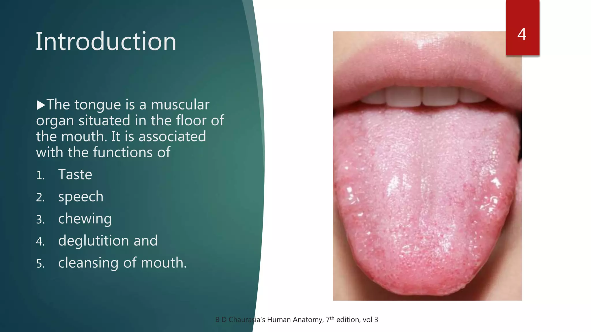
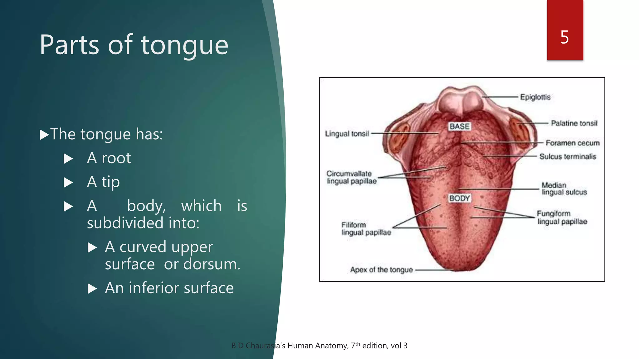
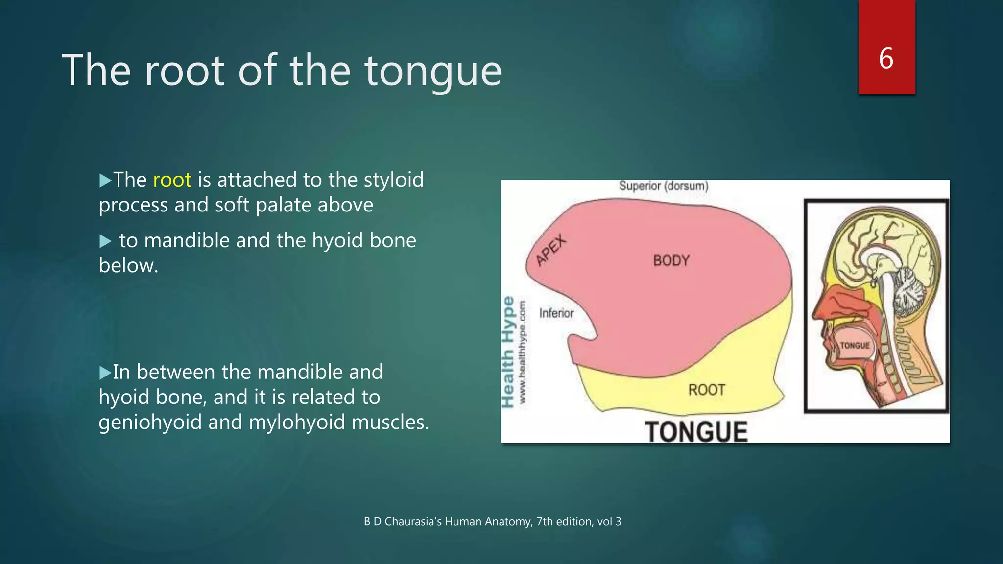
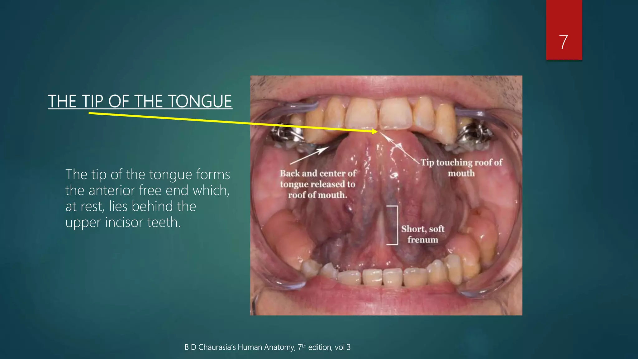
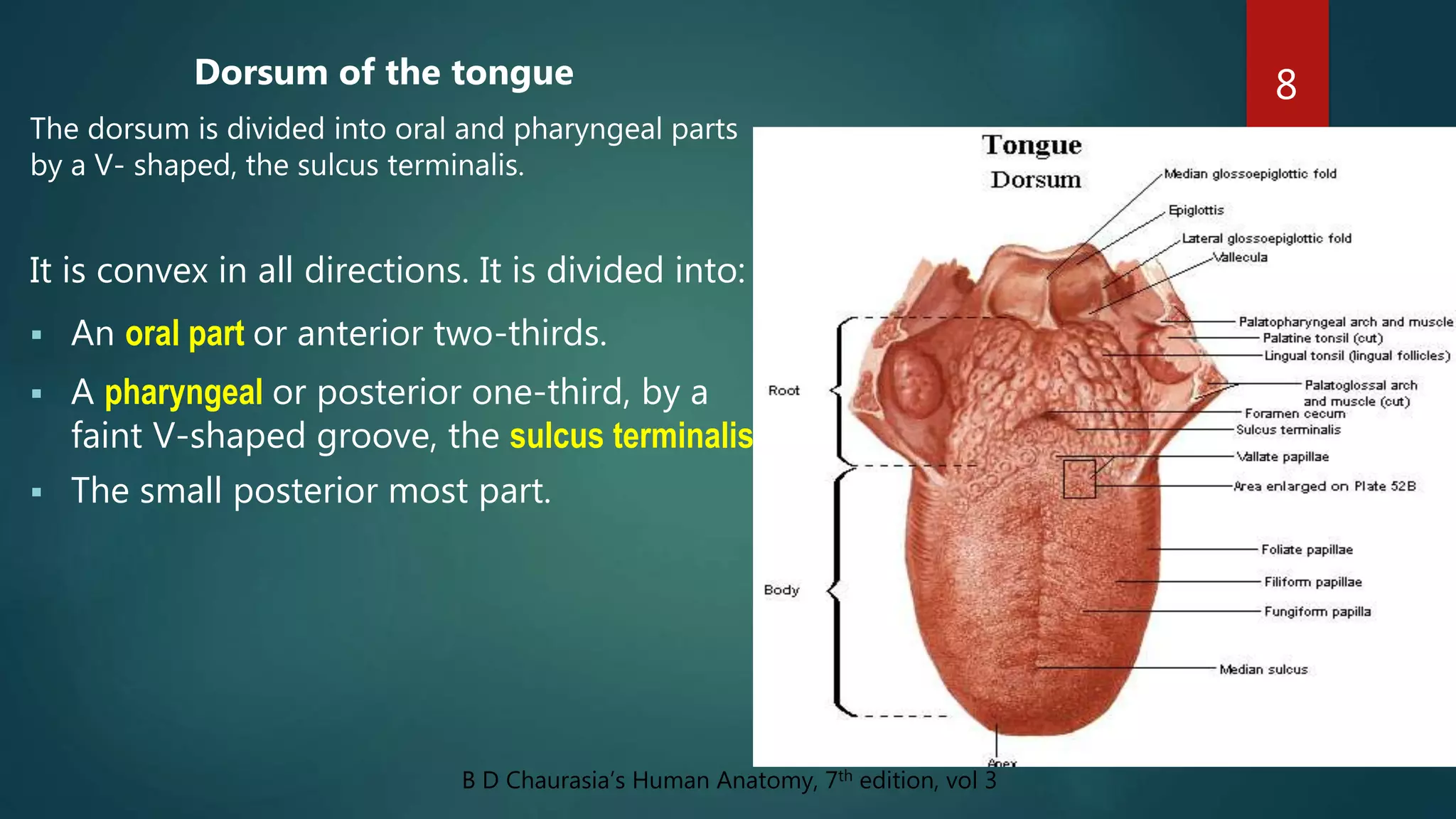
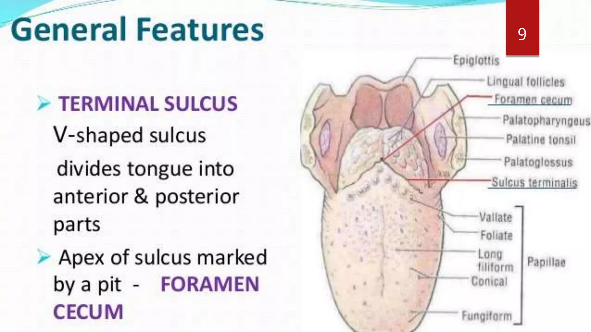
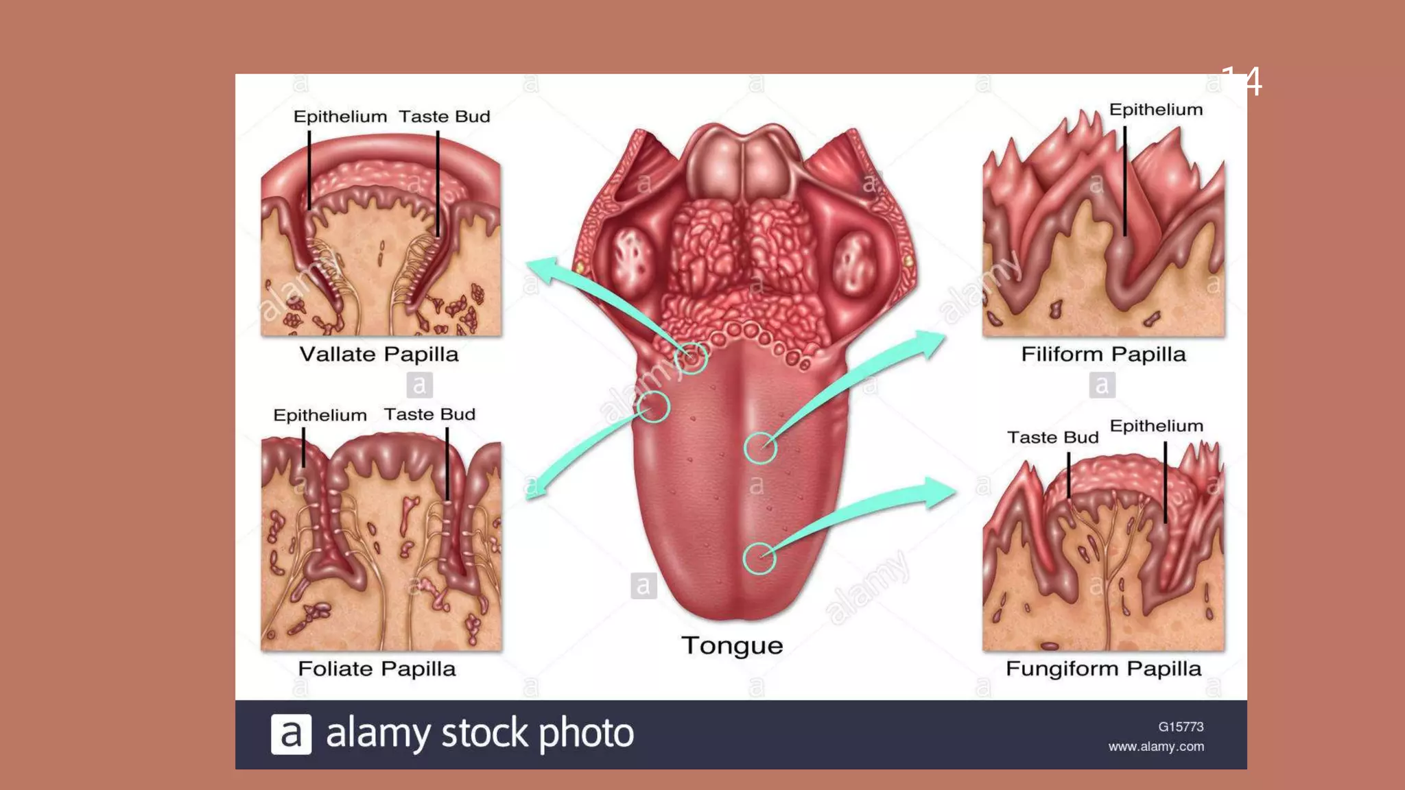
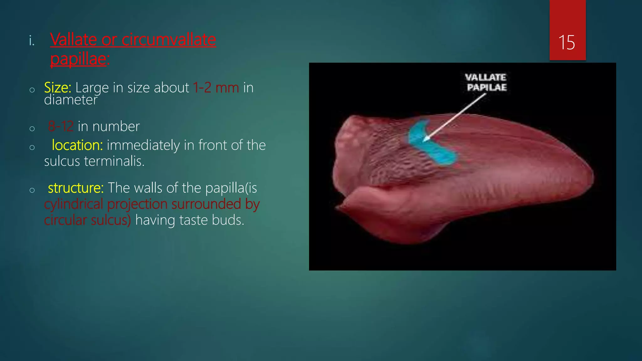
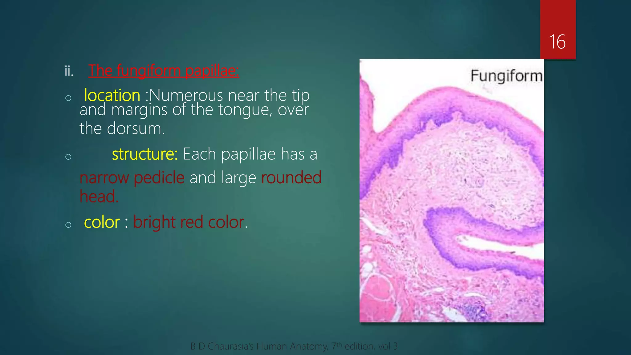
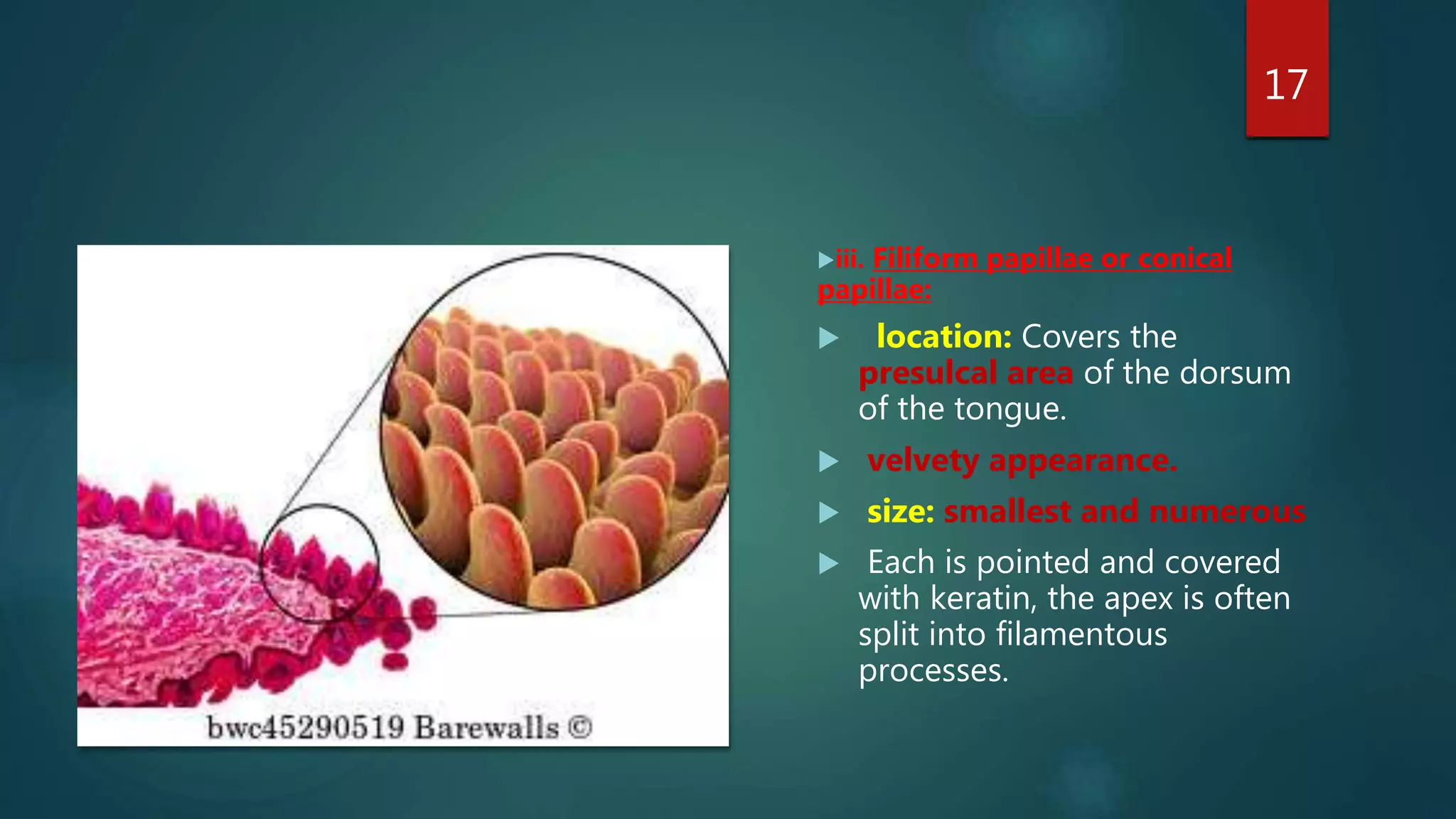
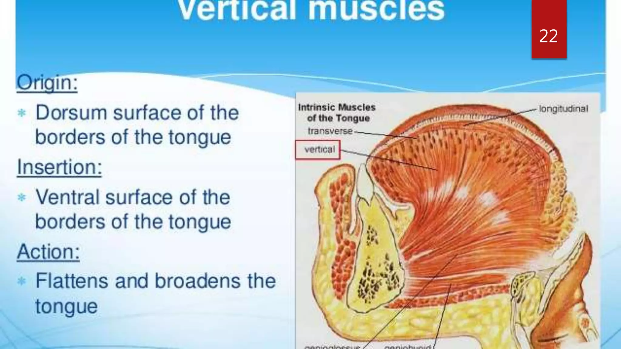
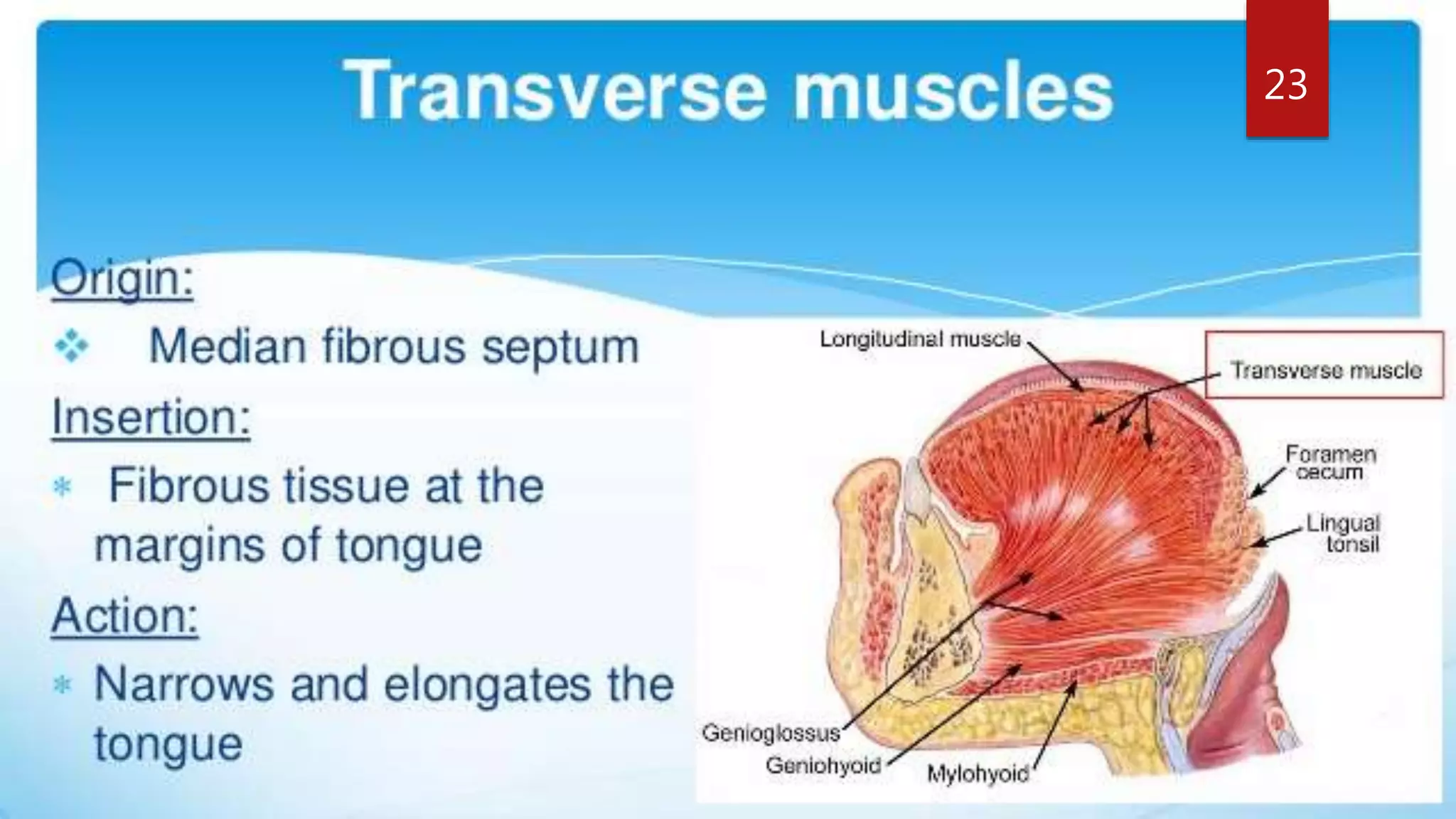
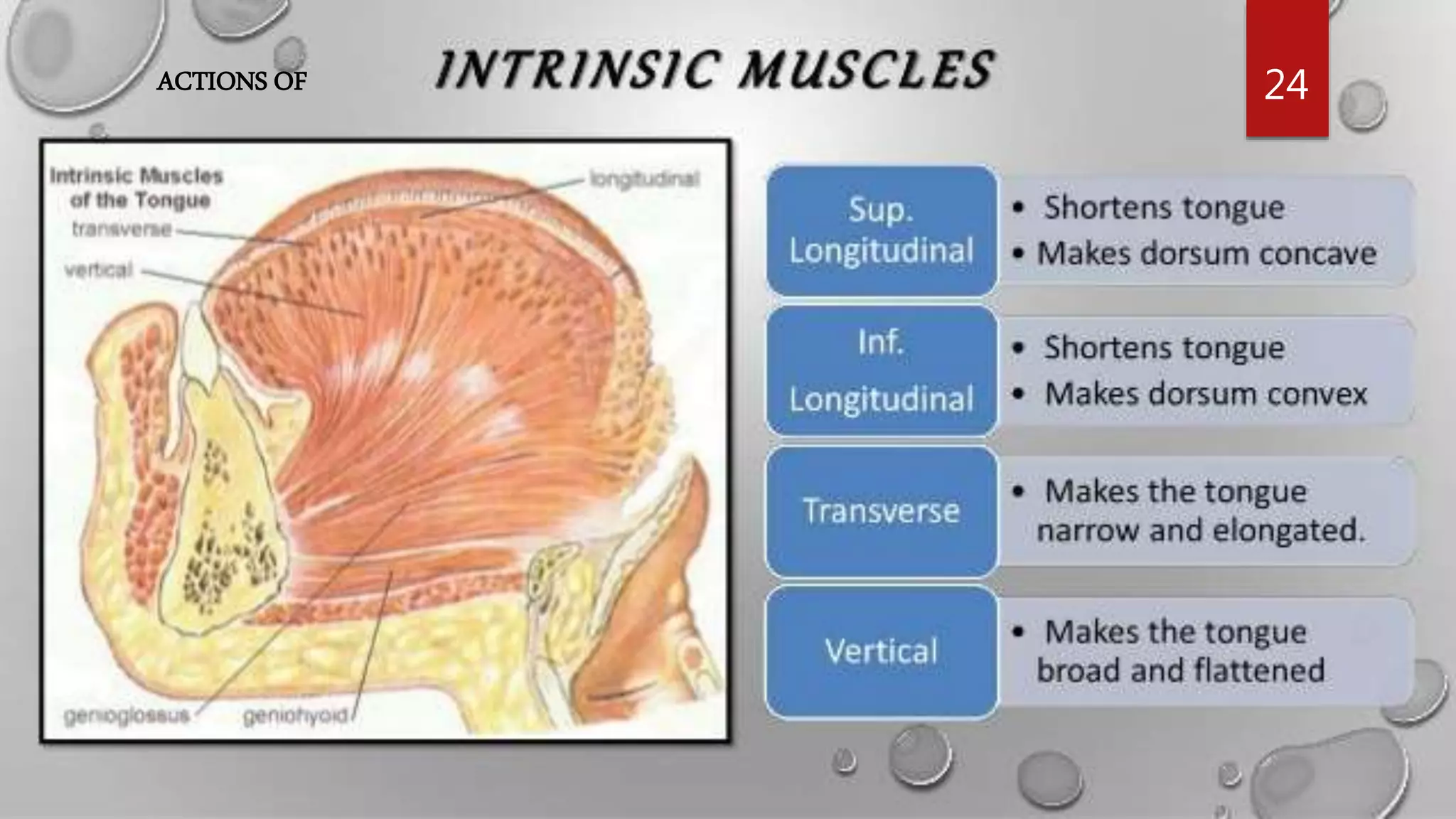
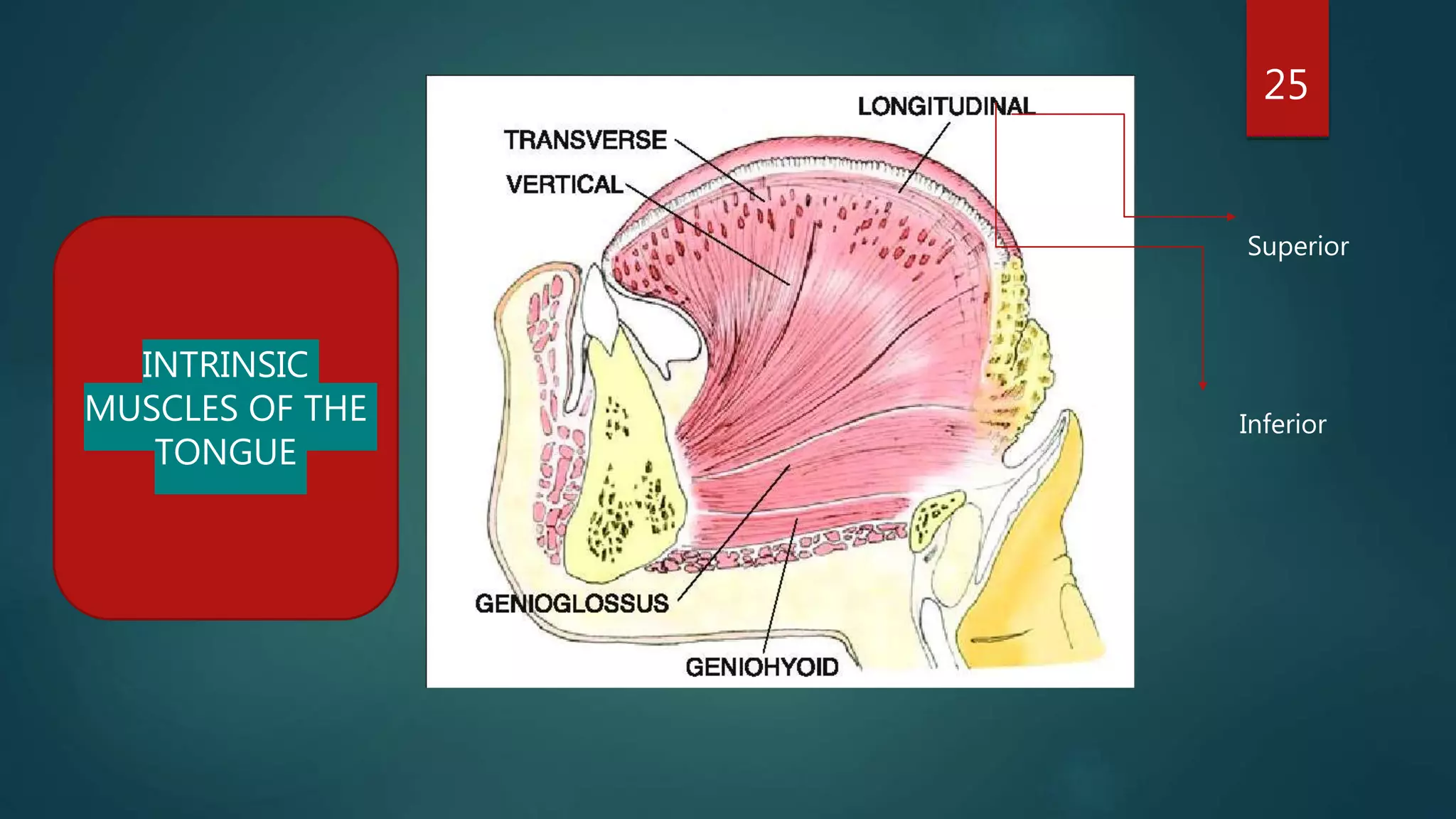
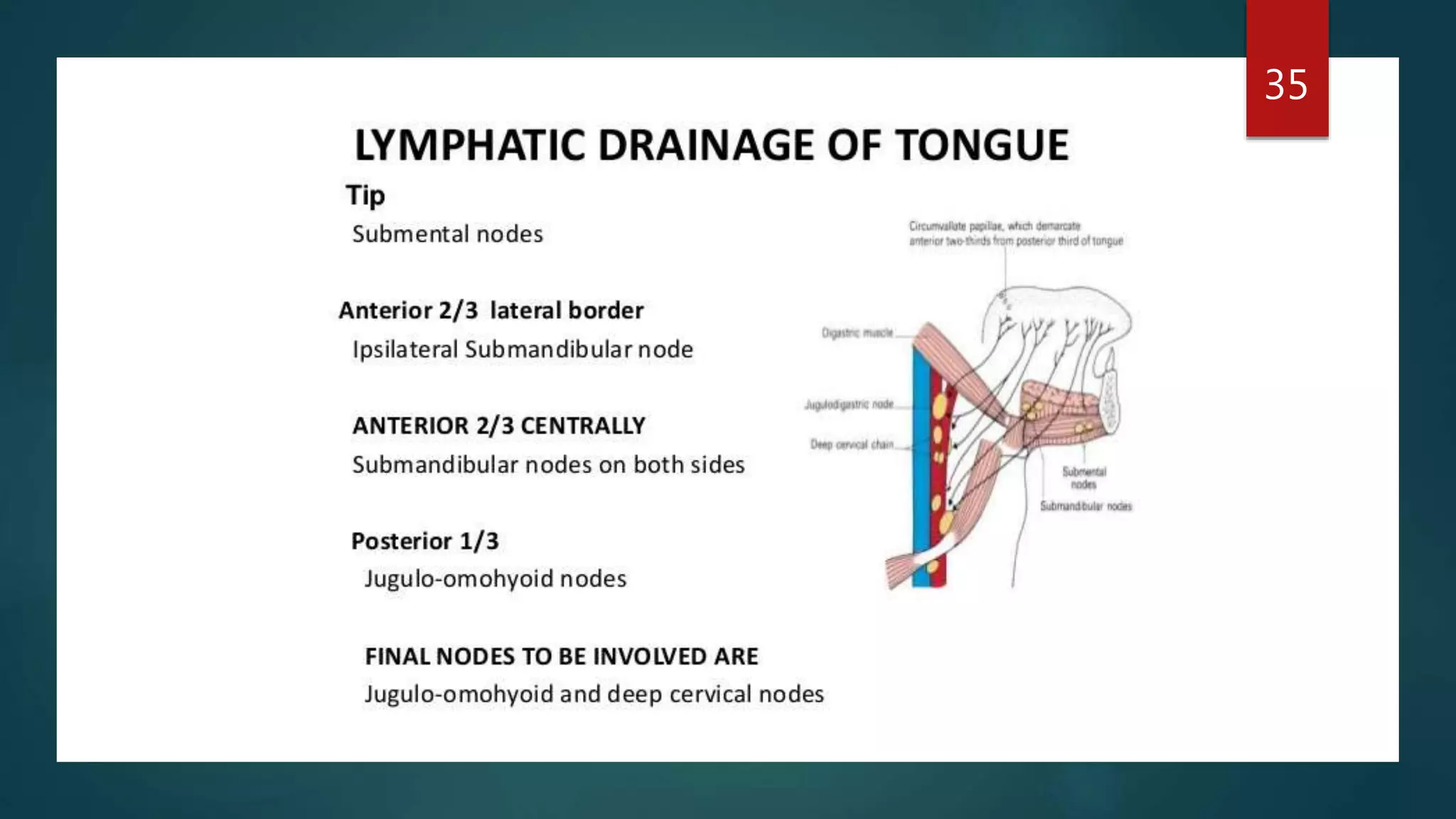
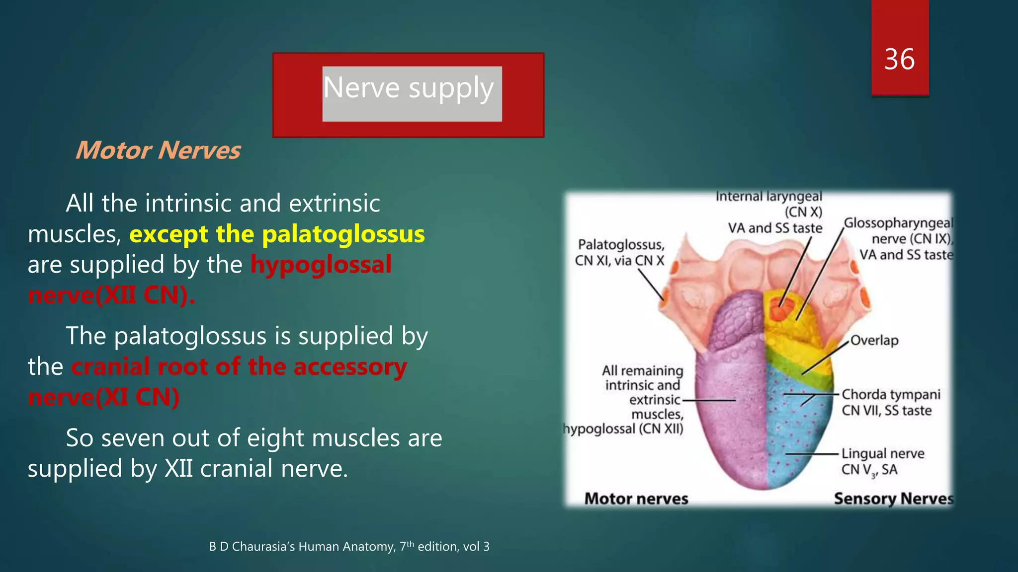
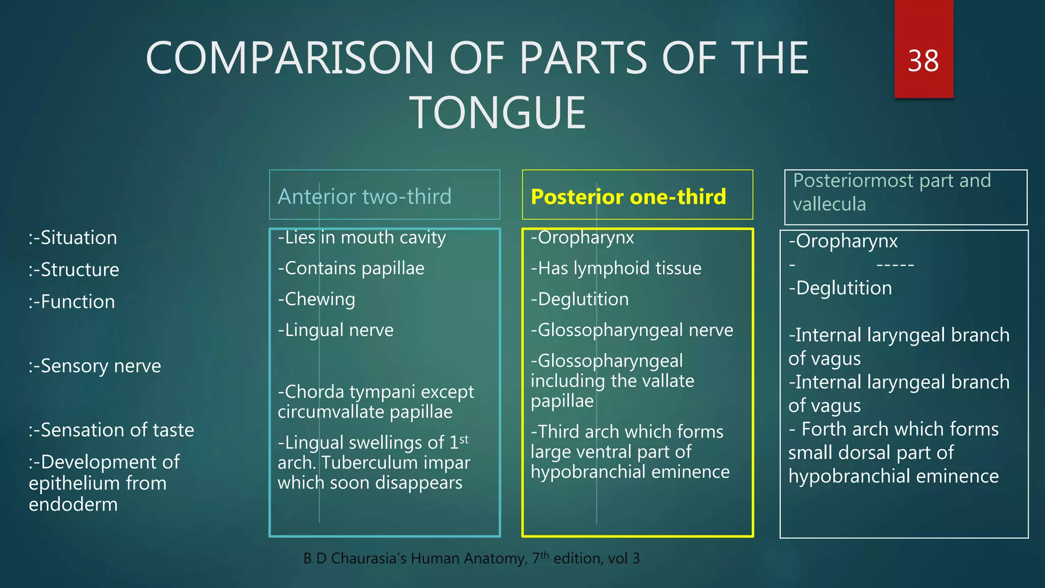
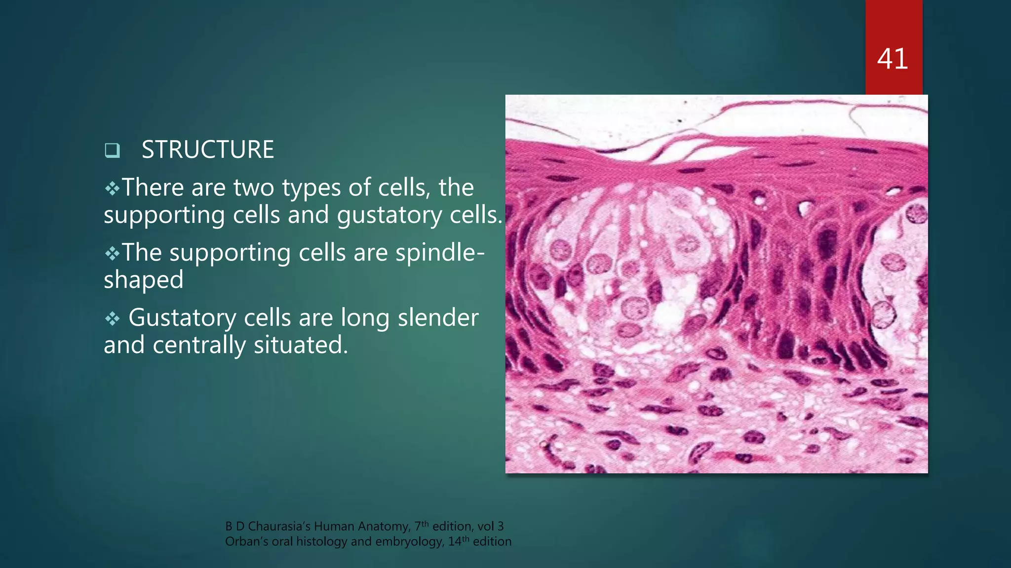
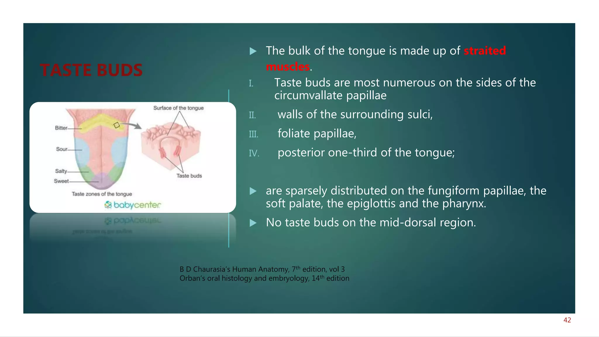
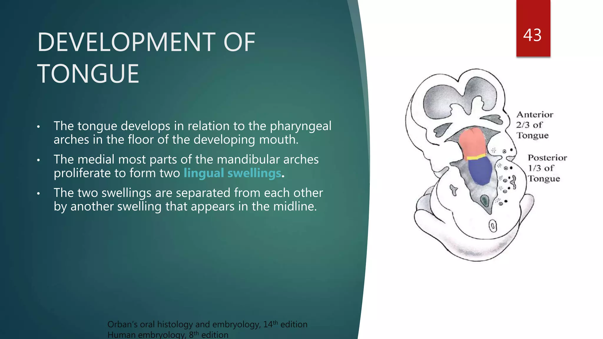
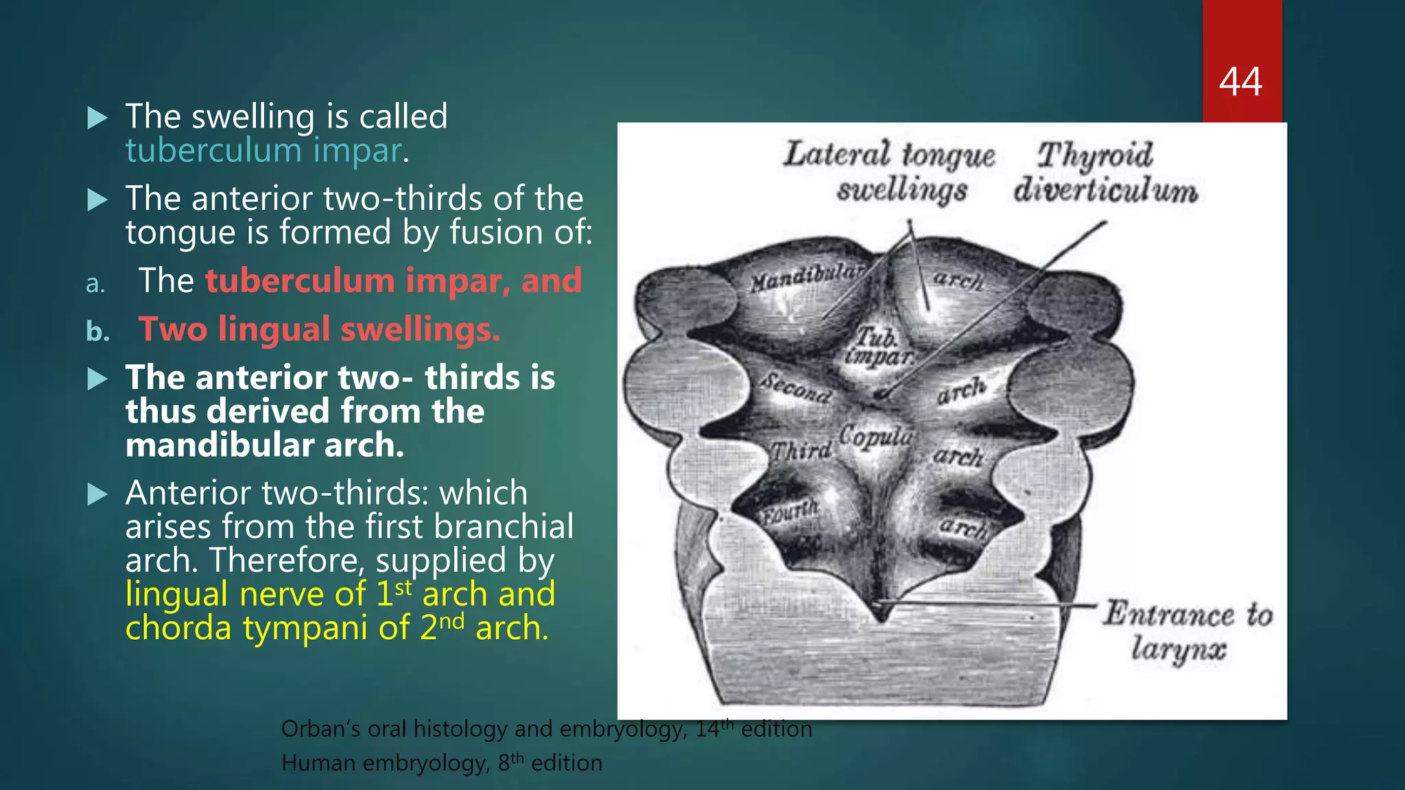
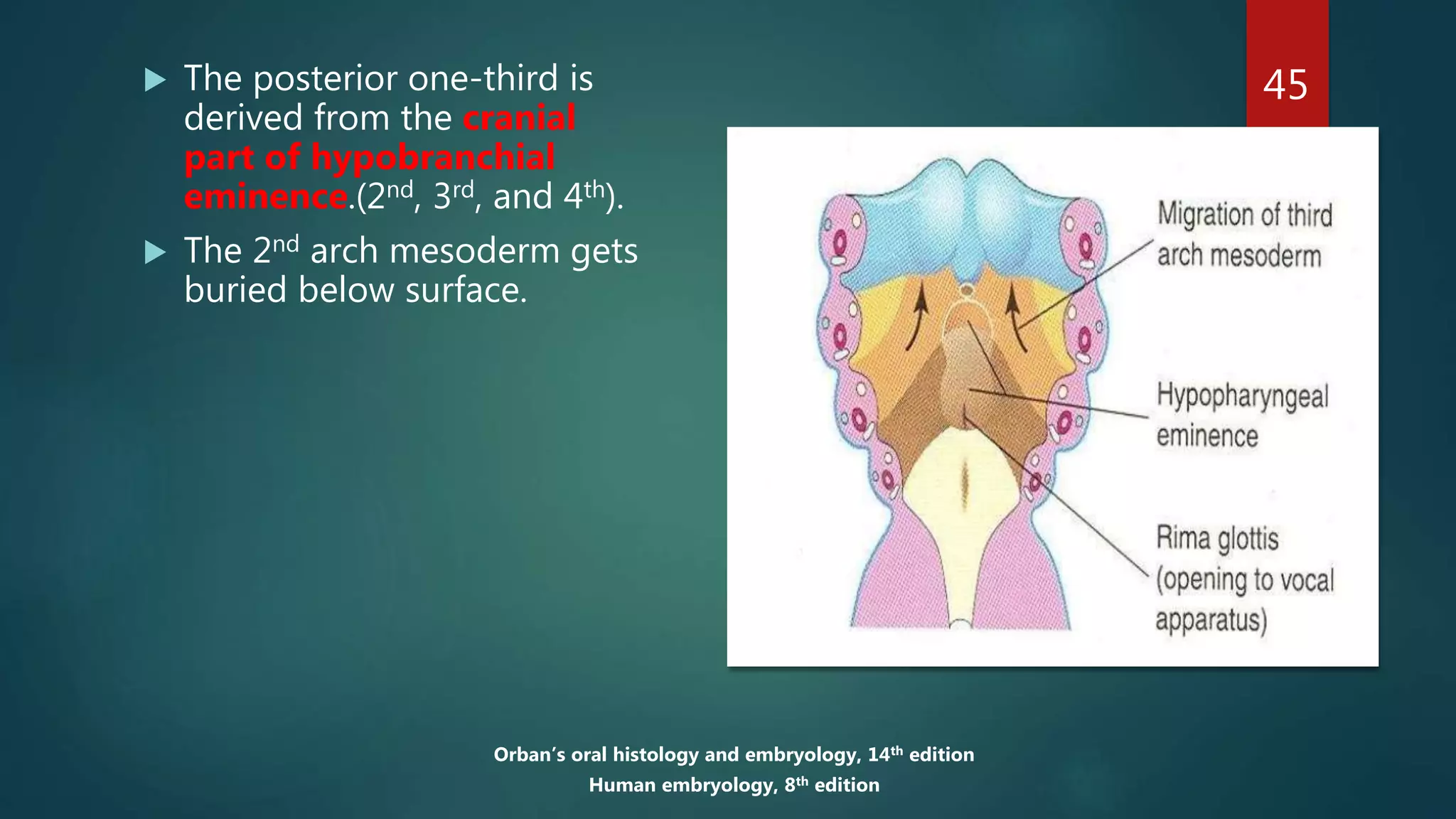
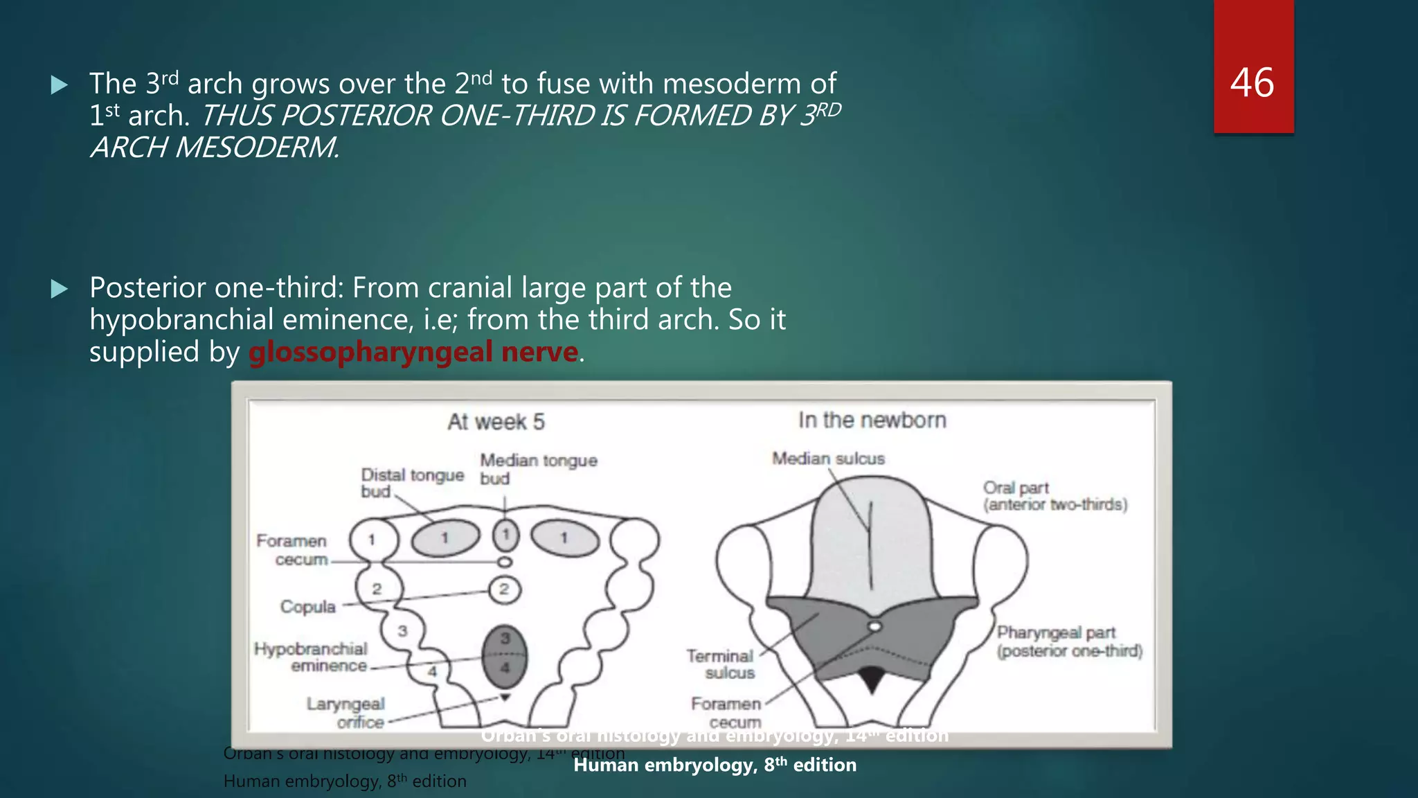
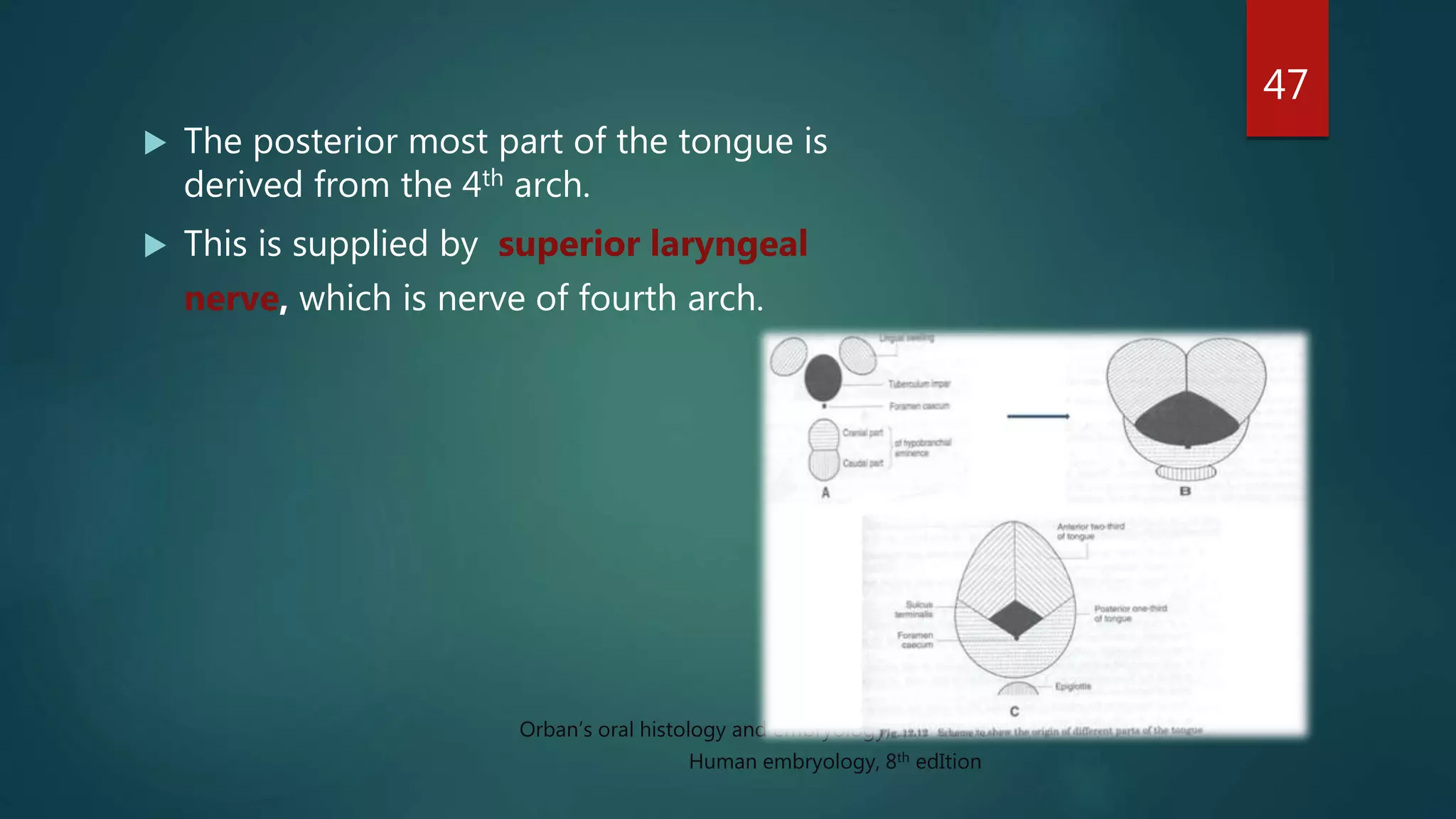
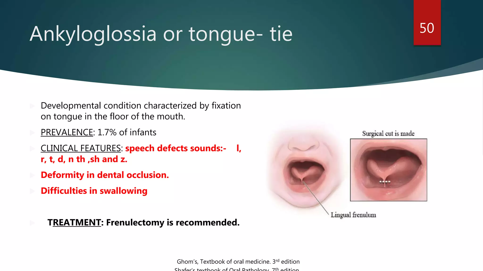
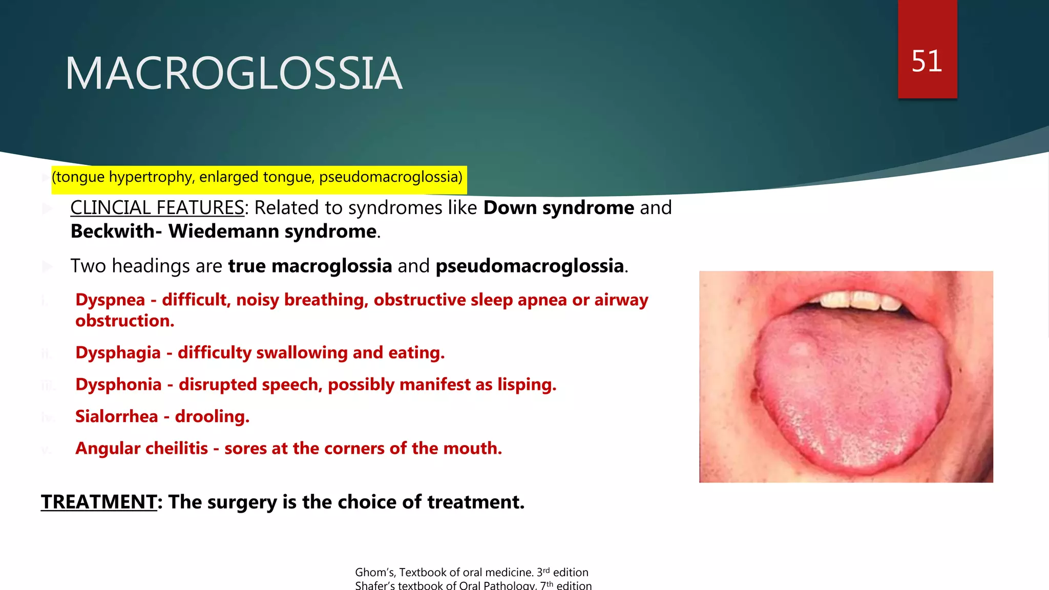
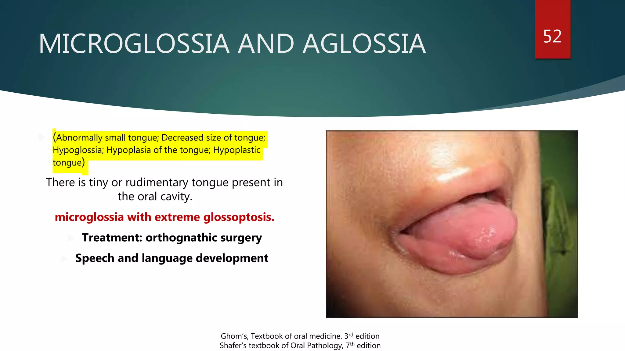
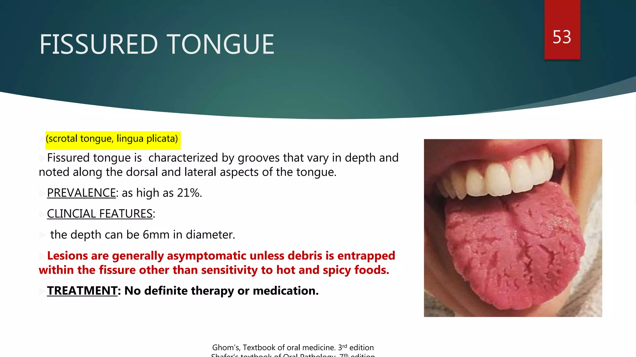
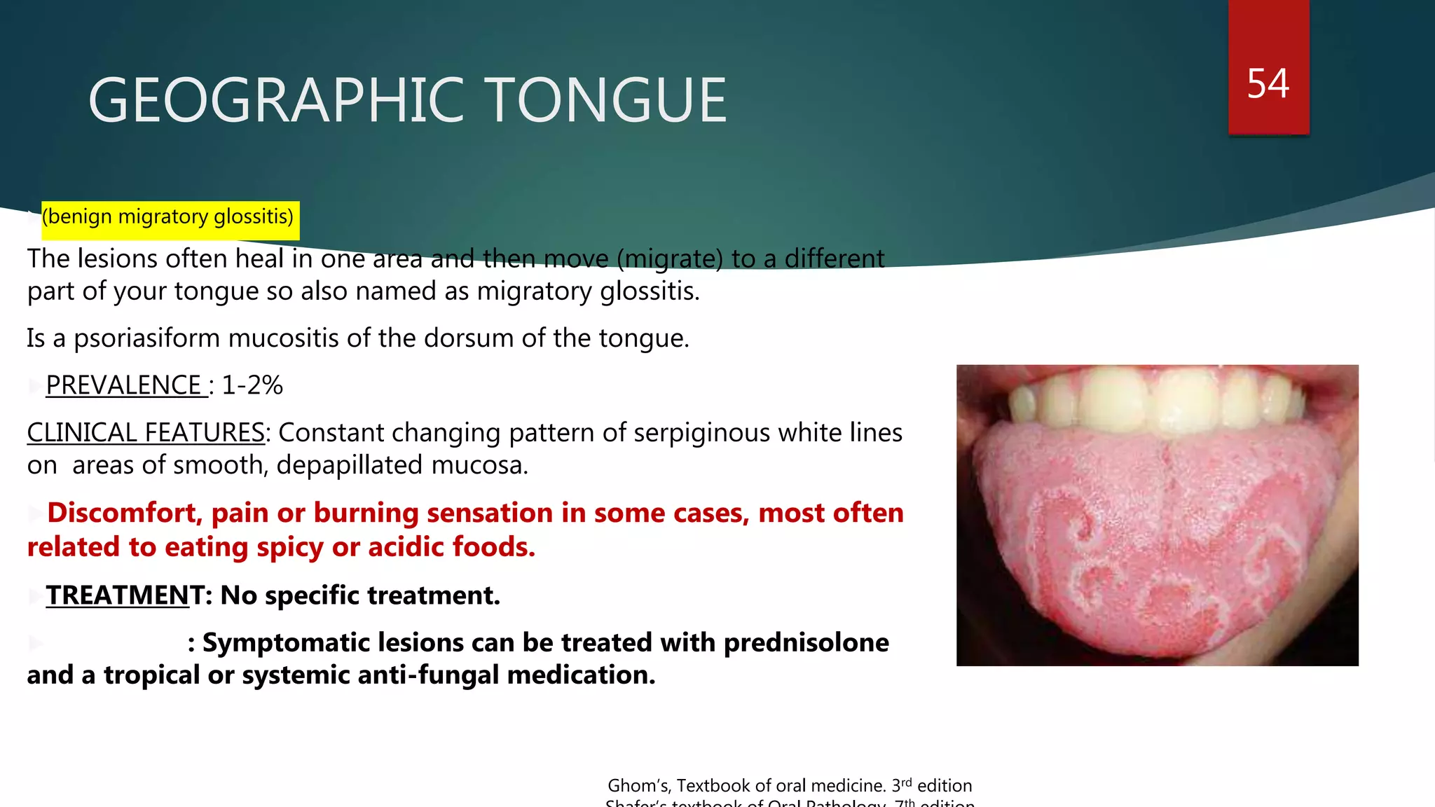
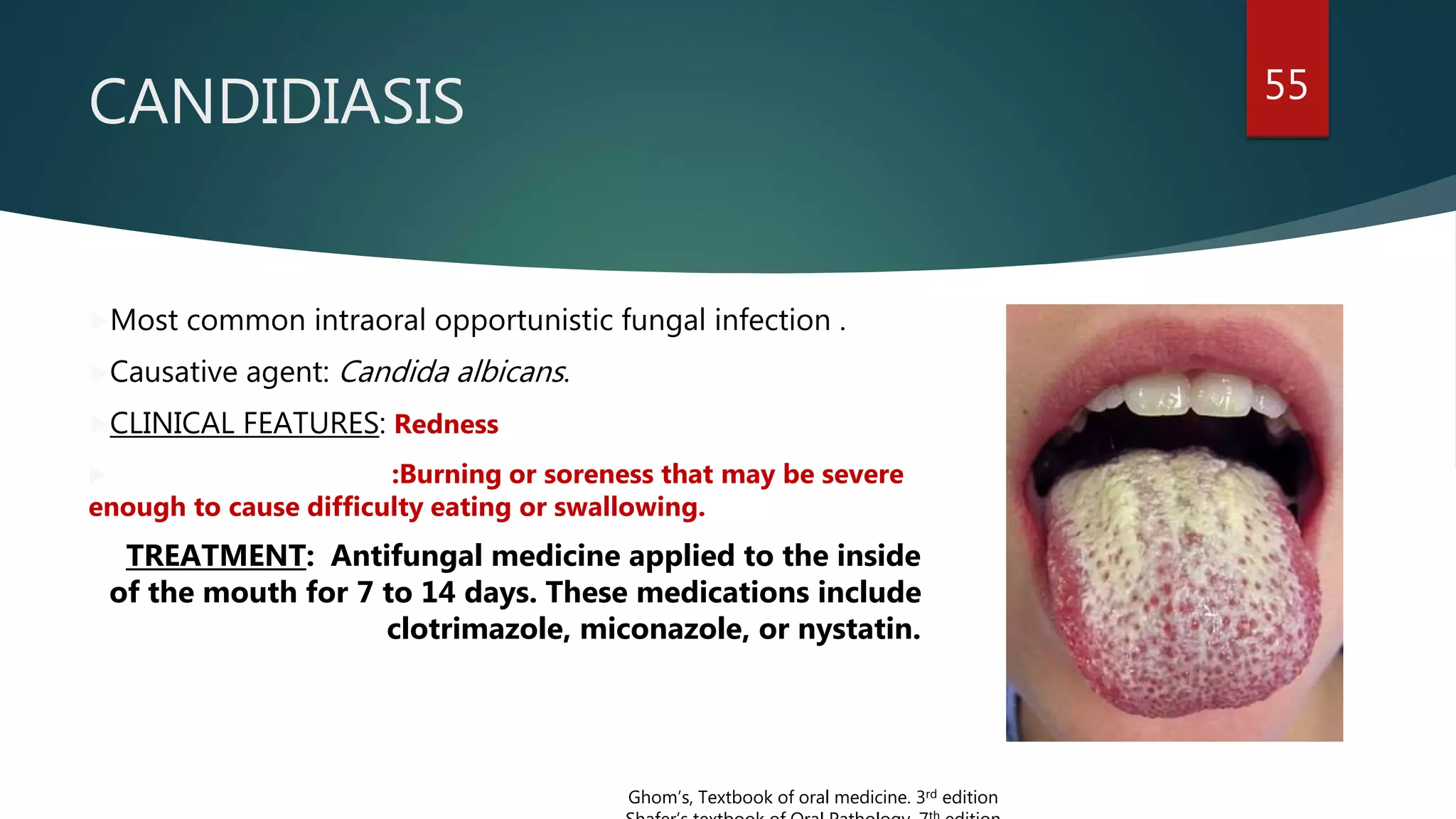
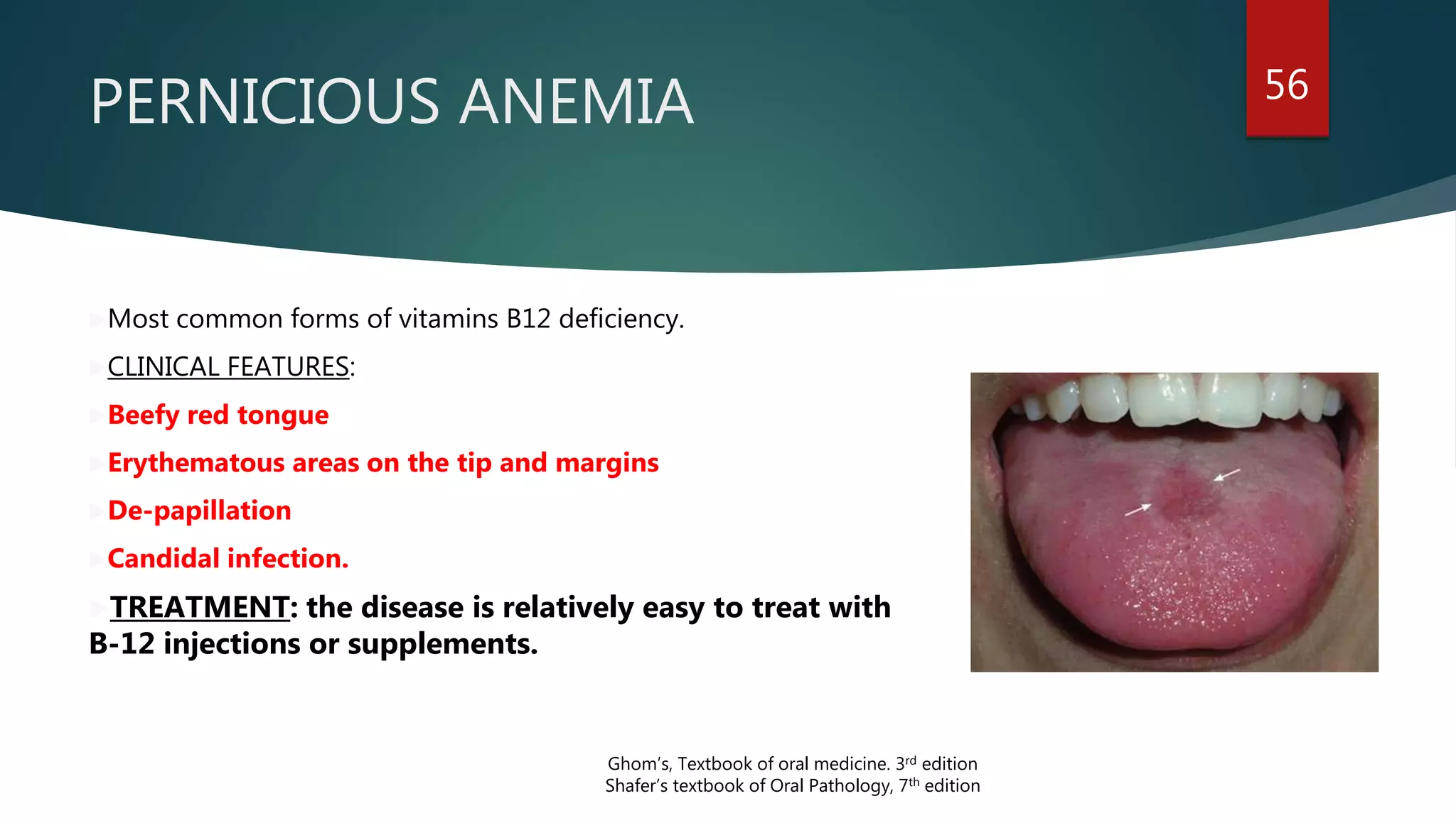
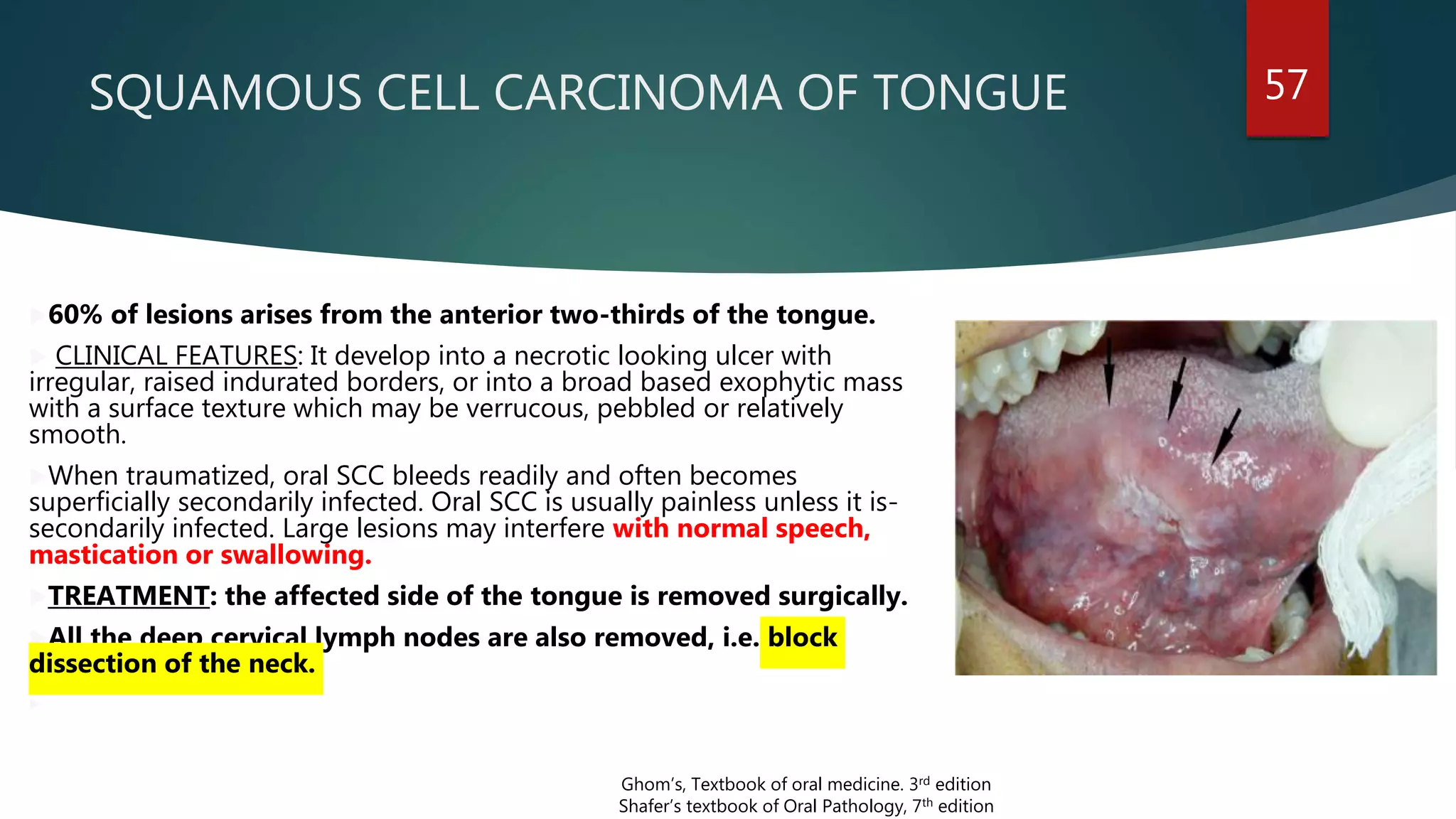
2. CONTENT
Introduction
Parts of Tongue
Muscles of the Tongue
Intrinsic muscles
Extrinsic muscles
Arterial supply of Tongue
Venous Drainage
2
3. Lymphatic Drainage
Nerve Supply
• Motor nerves
• Sensory nerves
Development of Tongue
Applied aspect : Anomalies, clinical features and its management
3
4. Introduction 4
The tongue is a muscular
organ situated in the floor of
the mouth. It is associated
with the functions of
1. Taste
2. speech
3. chewing
4. deglutition and
5. cleansing of mouth.
B D Chaurasia’s Human Anatomy, 7th edition, vol 3
5. Parts of tongue 5
The tongue has:
A root
A tip
A body, which is
subdivided into:
A curved upper
surface or dorsum.
An inferior surface
B D Chaurasia’s Human Anatomy, 7th edition, vol 3
6. The root of the tongue 6
The root is attached to the styloid
process and soft palate above
to mandible and the hyoid bone
below.
In between the mandible and
hyoid bone, and it is related to
geniohyoid and mylohyoid muscles.
B D Chaurasia’s Human Anatomy, 7th edition, vol 3
7. THE TIP OF THE TONGUE
The tip of the tongue forms
the anterior free end which,
at rest, lies behind the
upper incisor teeth.
7
B D Chaurasia’s Human Anatomy, 7th edition, vol 3
8. Dorsum of the tongue
The dorsum is divided into oral and pharyngeal parts
by a V- shaped, the sulcus terminalis.
It is convex in all directions. It is divided into:
An oral part or anterior two-thirds.
A pharyngeal or posterior one-third, by a
faint V-shaped groove, the sulcus terminalis.
The small posterior most part.
8
B D Chaurasia’s Human Anatomy, 7th edition, vol 3
10. 1. The oral part placed at floor
of the mouth.
2. Superior surface has
median furrow has papillae.
3. Inferior surface has smooth
mucous membrane
10
11. 11
B D Chaurasia’s Human Anatomy, 7th edition, vol 3
On other side of
frenulum, prominence
produced by deep
lingual veins.
Laterally a fold called
plica fimbriata goes
towards tip of tongue.
12. 2.The pharyngeal or lymphoid
part of the tongue lies behind
palatoglossal arches and the
sulcus terminalis.
3. The posterior most part of the
tongue is connected to
epiglottis by three-folds of
mucous membrane.
These are median
glossoepiglottic fold,
12
13. PAPILLAE OF TONGUE 13
WHAT IS A PAPILLAE?
These are projections of mucous membrane or corium
which gives the anterior two-thirds of the tongue, its
characteristic roughness. These are of four types:
Vallate or circumvallate
Fungiform
Foliate
Filiform
B D Chaurasia’s Human Anatomy, 7th edition, vol 3
Vallate
papillaeFoliate
Fungiform
Filiform
15. i. Vallate or circumvallate
papillae:
o Size: Large in size about 1-2 mm in
diameter
o 8-12 in number
o location: immediately in front of the
sulcus terminalis.
o structure: The walls of the papilla(is
cylindrical projection surrounded by
circular sulcus) having taste buds.
15
16. ii. The fungiform papillae:
o location :Numerous near the tip
and margins of the tongue, over
the dorsum.
o structure: Each papillae has a
narrow pedicle and large rounded
head.
o color : bright red color.
16
B D Chaurasia’s Human Anatomy, 7th edition, vol 3
17. 17
iii. Filiform papillae or conical
papillae:
location: Covers the
presulcal area of the dorsum
of the tongue.
velvety appearance.
size: smallest and numerous
Each is pointed and covered
with keratin, the apex is often
split into filamentous
processes.
19. MUSCLES OF THE
TONGUE
LONGITUDINAL : A. SUPERIOR
B. INFERIOR
TRANSVERE
VERTICAL
A middle fibrous septum divides tongue into right and
let halves. Each half contains 4 intrinsic and 4 extrinsic
muscles.
Intrinsic muscles:
They occupy the upper part of the tongue, and are
attached to the submucous fibrous layer and to the
median fibrous septum.
19
B D Chaurasia’s Human Anatomy, 7th edition, vol 3
26. Extrinsic muscles
1. Genioglossus
2. Hyoglossus
3. Styloglossus
4. Palatoglossus
The extrinsic muscles
connect the tongue to
the mandible via
genioglossus, to the
hyoid bone through
hyoglossus, to styloid
process via styloglossus,
and the palate via
palatoglossus.
26
B D Chaurasia’s Human Anatomy, 7th edition, vol 3
32. ARTERIAL SUPPY
OF TONGUE
32
It is derived from the
tortuous lingual artery, a
branch of the external
carotid artery.
a) The root of the
tongue by tonsillar
artery, a branch of
facial artery,
b) and ascending
pharyngeal branch of
external carotid artery.
B D Chaurasia’s Human Anatomy, 7th edition, vol 3
33. VENOUS DRAINAGE 33
Two venae comitantes supplement with
the lingual artery, and one vena comitant
accompanies the hyoglossal nerve.
I. The deep lingual vein is the largest
and principal vein of the tongue.
II. It is seen on the inferior surface of the
tongue.
B D Chaurasia’s Human Anatomy, 7th edition, vol 3
34. It runs backwards and crosses the genioglossus and the
hyoglossus below the hypoglossal nerve.
These veins unite at the posterior border of the hyoglossus to
form the lingual vein which ends in the internal jugular vein.
34
36. Motor Nerves
All the intrinsic and extrinsic
muscles, except the palatoglossus
are supplied by the hypoglossal
nerve(XII CN).
The palatoglossus is supplied by
the cranial root of the accessory
nerve(XI CN)
So seven out of eight muscles are
supplied by XII cranial nerve.
36
Nerve supply
B D Chaurasia’s Human Anatomy, 7th edition, vol 3
37. 37
Sensory nerve supply of tongue
The lingual nerve is the nerve of general sensation and the chorda tympani is the nerve for the
taste of the anterior two-thirds of the tongue except vallate papillae.
The glossopharyngeal nerve(IX CN) is the nerve for both general sensation and taste for
the posterior one-third of the tongue including the circumvallate papillae.
The posterior most part of the tongue is supplied by the vagus nerve(X CN) through the
internal laryngeal branch.
B D Chaurasia’s Human Anatomy, 7th edition, vol 3
38. COMPARISON OF PARTS OF THE
TONGUE
:-Situation
:-Structure
:-Function
:-Sensory nerve
:-Sensation of taste
:-Development of
epithelium from
endoderm
Anterior two-third
-Lies in mouth cavity
-Contains papillae
-Chewing
-Lingual nerve
-Chorda tympani except
circumvallate papillae
-Lingual swellings of 1st
arch. Tuberculum impar
which soon disappears
Posterior one-third
-Oropharynx
-Has lymphoid tissue
-Deglutition
-Glossopharyngeal nerve
-Glossopharyngeal
including the vallate
papillae
-Third arch which forms
large ventral part of
hypobranchial eminence
38
Posteriormost part and
vallecula
-Oropharynx
- -----
-Deglutition
-Internal laryngeal branch
of vagus
-Internal laryngeal branch
of vagus
- Forth arch which forms
small dorsal part of
hypobranchial eminence
B D Chaurasia’s Human Anatomy, 7th edition, vol 3
39. 39
HISTOLOGY
The tongue forms in the ventral floor of the pharynx after arrival of the
hypoglossal muscle cells.
It is known that the anterior two-thirds of the tongue is covered by
ectoderm whereas endoderm covers the posterior one-third.
A residual pit left in the epithelium at the site of invagination marks the
junction between the anterior two-thirds and posterior one-thirds of the
tongue.
B D Chaurasia’s Human Anatomy, 7th edition, vol 3
th
40. The mucous membrane consists of a layer of connective tissue, lined by stratified
squamous epithelium.
On the oral part of the dorsum, it is thin, forms papillae, and is adherent to the
muscles.
On the pharyngeal part of the dorsum, it is very rich in lymphoid follicles.
On the inferior surface, it is thin and smooth. Numerous gland, both serous and
mucous lie deep to the mucous membrane.
40
B D Chaurasia’s Human Anatomy, 7th edition, vol 3
Orban’s oral histology and embryology, 14th edition
41. STRUCTURE
There are two types of cells, the
supporting cells and gustatory cells.
The supporting cells are spindle-
shaped
Gustatory cells are long slender
and centrally situated.
41
B D Chaurasia’s Human Anatomy, 7th edition, vol 3
Orban’s oral histology and embryology, 14th edition
42. The bulk of the tongue is made up of straited
muscles.
I. Taste buds are most numerous on the sides of the
circumvallate papillae
II. walls of the surrounding sulci,
III. foliate papillae,
IV. posterior one-third of the tongue;
are sparsely distributed on the fungiform papillae, the
soft palate, the epiglottis and the pharynx.
No taste buds on the mid-dorsal region.
42
B D Chaurasia’s Human Anatomy, 7th edition, vol 3
Orban’s oral histology and embryology, 14th edition
TASTE BUDS
43. DEVELOPMENT OF
TONGUE
43
• The tongue develops in relation to the pharyngeal
arches in the floor of the developing mouth.
• The medial most parts of the mandibular arches
proliferate to form two lingual swellings.
• The two swellings are separated from each other
by another swelling that appears in the midline.
Orban’s oral histology and embryology, 14th edition
Human embryology, 8th edition
44. 44
The swelling is called
tuberculum impar.
The anterior two-thirds of the
tongue is formed by fusion of:
a. The tuberculum impar, and
b. Two lingual swellings.
The anterior two- thirds is
thus derived from the
mandibular arch.
Anterior two-thirds: which
arises from the first branchial
arch. Therefore, supplied by
lingual nerve of 1st arch and
chorda tympani of 2nd arch.
Orban’s oral histology and embryology, 14th edition
Human embryology, 8th edition
45. 45 The posterior one-third is
derived from the cranial
part of hypobranchial
eminence.(2nd, 3rd, and 4th).
The 2nd arch mesoderm gets
buried below surface.
Orban’s oral histology and embryology, 14th edition
Human embryology, 8th edition
46. The 3rd arch grows over the 2nd to fuse with mesoderm of
1st arch. THUS POSTERIOR ONE-THIRD IS FORMED BY 3RD
ARCH MESODERM.
Posterior one-third: From cranial large part of the
hypobranchial eminence, i.e; from the third arch. So it
supplied by glossopharyngeal nerve.
46
Orban’s oral histology and embryology, 14th edition
Human embryology, 8th edition
Orban’s oral histology and embryology, 14th edition
Human embryology, 8th edition
47. The posterior most part of the tongue is
derived from the 4th arch.
This is supplied by superior laryngeal
nerve, which is nerve of fourth arch.
47
Orban’s oral histology and embryology, 14th edition
Human embryology, 8th edItion
48. MUSCULATURE OF TONGUE
Derived from occipital myotomes.
nerve supply from hypoglossal
nerve.
EPITHELIUM OF TONGUE
Made firstly from a single layer of
cells.
Later, becomes stratified and
papilla becomes evident.
Taste buds are formed in relation
to the terminal branches of the
innervating nerve fibres.
48
Orban’s oral histology and embryology, 14th edition
Human embryology, 8th edition
49. Diseases of tongue
A. INHERITED, CONGENITAL, AND DEVELOPMENTAL ANOMALIES.
B. DISORDERS OF LINGUAL MUCOSA.
C. MALIGNANT TUMORS OF THE TONGUE.
50. Ankyloglossia or tongue- tie 50
Developmental condition characterized by fixation
on tongue in the floor of the mouth.
PREVALENCE: 1.7% of infants
CLINICAL FEATURES: speech defects sounds:- l,
r, t, d, n th ,sh and z.
Deformity in dental occlusion.
Difficulties in swallowing
TREATMENT: Frenulectomy is recommended.
Ghom’s, Textbook of oral medicine. 3rd edition
th
51. MACROGLOSSIA 51
(tongue hypertrophy, enlarged tongue, pseudomacroglossia)
CLINCIAL FEATURES: Related to syndromes like Down syndrome and
Beckwith- Wiedemann syndrome.
Two headings are true macroglossia and pseudomacroglossia.
i. Dyspnea - difficult, noisy breathing, obstructive sleep apnea or airway
obstruction.
ii. Dysphagia - difficulty swallowing and eating.
iii. Dysphonia - disrupted speech, possibly manifest as lisping.
iv. Sialorrhea - drooling.
v. Angular cheilitis - sores at the corners of the mouth.
TREATMENT: The surgery is the choice of treatment.
Ghom’s, Textbook of oral medicine. 3rd edition
Shafer’s textbook of Oral Pathology, 7th edition
52. MICROGLOSSIA AND AGLOSSIA 52
(Abnormally small tongue; Decreased size of tongue;
Hypoglossia; Hypoplasia of the tongue; Hypoplastic
tongue)
There is tiny or rudimentary tongue present in
the oral cavity.
microglossia with extreme glossoptosis.
Treatment: orthognathic surgery
Speech and language development
Ghom’s, Textbook of oral medicine. 3rd edition
Shafer’s textbook of Oral Pathology, 7th edition
53. FISSURED TONGUE 53
(scrotal tongue, lingua plicata)
Fissured tongue is characterized by grooves that vary in depth and
noted along the dorsal and lateral aspects of the tongue.
PREVALENCE: as high as 21%.
CLINCIAL FEATURES:
the depth can be 6mm in diameter.
Lesions are generally asymptomatic unless debris is entrapped
within the fissure other than sensitivity to hot and spicy foods.
TREATMENT: No definite therapy or medication.
Ghom’s, Textbook of oral medicine. 3rd edition
54. GEOGRAPHIC TONGUE 54
(benign migratory glossitis)
The lesions often heal in one area and then move (migrate) to a different
part of your tongue so also named as migratory glossitis.
Is a psoriasiform mucositis of the dorsum of the tongue.
PREVALENCE : 1-2%
CLINICAL FEATURES: Constant changing pattern of serpiginous white lines
on areas of smooth, depapillated mucosa.
Discomfort, pain or burning sensation in some cases, most often
related to eating spicy or acidic foods.
TREATMENT: No specific treatment.
: Symptomatic lesions can be treated with prednisolone
and a tropical or systemic anti-fungal medication.
Ghom’s, Textbook of oral medicine. 3rd edition
55. CANDIDIASIS 55
Most common intraoral opportunistic fungal infection .
Causative agent: Candida albicans.
CLINICAL FEATURES: Redness
:Burning or soreness that may be severe
enough to cause difficulty eating or swallowing.
TREATMENT: Antifungal medicine applied to the inside
of the mouth for 7 to 14 days. These medications include
clotrimazole, miconazole, or nystatin.
Ghom’s, Textbook of oral medicine. 3rd edition
56. PERNICIOUS ANEMIA 56
Most common forms of vitamins B12 deficiency.
CLINICAL FEATURES:
Beefy red tongue
Erythematous areas on the tip and margins
De-papillation
Candidal infection.
TREATMENT: the disease is relatively easy to treat with
B-12 injections or supplements.
Ghom’s, Textbook of oral medicine. 3rd edition
Shafer’s textbook of Oral Pathology, 7th edition
57. SQUAMOUS CELL CARCINOMA OF TONGUE 57
60% of lesions arises from the anterior two-thirds of the tongue.
CLINICAL FEATURES: It develop into a necrotic looking ulcer with
irregular, raised indurated borders, or into a broad based exophytic mass
with a surface texture which may be verrucous, pebbled or relatively
smooth.
When traumatized, oral SCC bleeds readily and often becomes
superficially secondarily infected. Oral SCC is usually painless unless it is-
secondarily infected. Large lesions may interfere with normal speech,
mastication or swallowing.
TREATMENT: the affected side of the tongue is removed surgically.
All the deep cervical lymph nodes are also removed, i.e. block
dissection of the neck.
Ghom’s, Textbook of oral medicine. 3rd edition
Shafer’s textbook of Oral Pathology, 7th edition
58. References
B D Chaurasia’s Human Anatomy, 7th edition, vol 3
Human embryology, 8th edItion
Ghom’s, Textbook of oral medicine. 3rd edition
Shafer’s textbook of Oral Pathology, 7th edition
58