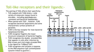
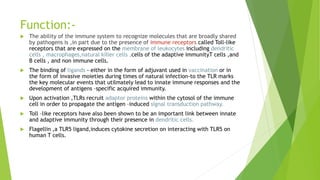
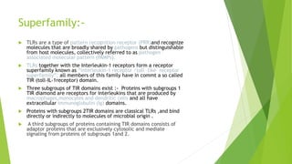
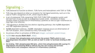
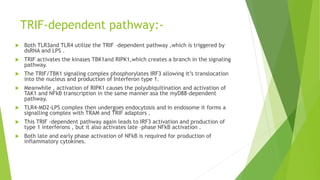
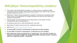
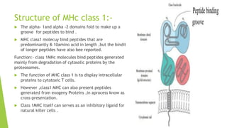
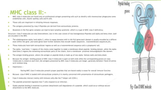
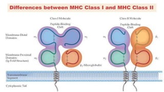
Toll like receptor
And MHC
Name-ritu
Roll-no – 14
MSC microbiology( first yr)
Toll like receptor-
 Toll Like receptors are a class of proteins that play a key role in innate
immune system.
 They are single pass membrane spanning receptors usually expressed
on sentinel cells such as macrophages and dendritic cells,that
recognize structurally conserved molecules derived from microbes.
 Once these microbes have breached physical barriers such as the skin
or intestinal tract mucosa they are recognized by TLRs which active
immune cell responses.
 The TLRs include
TLR1,TLR2,TLR3,TLR4,TLR5,TLR6,TLR7,TLR8,TLR9,TLR10,TLR11,TLR1
2,TLR13,though last three are not found in humans.
Discovery:-
 The protein Toll first attracted attention during the 1980s,when researchers in Germany found that
developing flies could not establish a proper dorsal –ventral axis without Toll.(Toll, referring to the
mutant flies bizarrely Scrambled anatomy,means “weird” in German slang.)
 Toll is transmembrane signal receptor protein ;related molecules with roles in innate immunity
came to be known as Toll-like receptors (TLRs)
 Three recent discoveries ignited an explosion of knowledge about the central role of TLRs in innate
immunity.
 The first observation came from fruit fly .In 1996,Jules Hoffman and Bruno Lemaitre found that
mutations in Toll, previously known to function in fly development ,made flies highly susceptible to
lethal infection with Aspergillus fumigatus,a fungus to which wild –type flies were immune.
 This landmark experiment convincingly demonstrated the importance of pathogen-triggerd immune
respoo in a no vertebrate organism.
 In 1997,Ruslan medzhitov and Charles Janeway discovered that a certain human protein, identified
by homology between its cytoplasmic domain and that of Toll,activated the expression of immune
response genes when transfected into a human experimental cell line.
 This human protein was subsequently named TLR4.
 This was the first evidence that an immune response pathway was conserved between fruit flies and
humans .
 In 1998 ,proof that TLRs are part of the normal immune physiology of
mammals came from studies with mutant mice conducted in the laboratory of
Bruce Beutler.
 Mice homozygous for the lps locus were resistant to lipopolysaccharide (LPS
),also known as endotoxin , which comes from the cell walls of gram –
negative bacteria .
 In humans ,a buildup of endotoxin from severe bacterial infection can cause
septic shock ,a life threatening condition in which vital organs such as the
brain ,heart ,kidney ,liver may fail.
 DNA sequencing revealed that the mouse lps Gene encoded a mutant form of
a Toll-like receptor,TLR,which differed from the normal form by a single
amino acid.
Structure :-
 Toll- like receptors are membrane –spanning proteins
that share a common structural element in their
extracellular region, repeating segments of 24to 29
amino acids containing the sequence xLxxLxLxx(x is
any amino acids and L is leucine ).
 These structural motifs are called leucine –rich repeats
(LRRS)
 All TLRs contain several LRRs, and a subset of the LRRs
make up the extracellular ligand –binding region of
molecules .
 TLR domains have three regions,highly conserved
among all members of the TLR family ,called boxes
1,2,3 that serve as binding sites for intracellular
proteins participating in the signaling pathways
mediated by TLRs.
Toll-like receptors and their ligands:-
The pairing of TLRs affects their specificity.
• TLR coupled with TLR6 binds a wide
variety of molecular classes found in
microbes , including peptidoglycans
,zymosans and bacterial lipopeptides .
• When paired with TLR1 , however ,TLR2
recognizes bacterial lipoproteins and some
characteristic surface proteins of
parasites.
• TLR4 is the key receptor for most bacterial
lipopolysaccharides.
• TLR5 recognize flagellin,the major
structural component of bacterial flagella.
• TLR3 recognizes the double –stranded RNA
that appears in cells after infection by
RNA viruses,and viral single-stranded RNA
is the ligand for TLR8and TLR7.
• TLR9 recognizes and initiates a response
to the DNA sequence CpG (unmethylated
cytosine linked to guanine ).
Function:-
 The ability of the immune system to recognize molecules that are broadly shared
by pathogens is ,in part due to the presence of immune receptors called Toll-like
receptors that are expressed on the membrane of leukocytes including dendritic
cells , macrophages,natural killer cells ,cells of the adaptive immunityT cells ,and
B cells , and non immune cells.
 The binding of ligands - either in the form of adjuvant used in vaccination or in
the form of invasive moieties during times of natural infection-to the TLR marks
the key molecular events that utilmately lead to innate immune responses and the
development of antigens –specific acquired immunity.
 Upon activation ,TLRs recruit adaptor proteins within the cytosol of the immune
cell in order to propagate the antigen –induced signal transduction pathway.
 Toll –like receptors have also been shown to be an important link between innate
and adaptive immunity through their presence in dendritic cells.
 Flagellin ,a TLR5 ligand,induces cytokine secretion on interacting with TLR5 on
human T cells.
Superfamily:-
 TLRs are a type of pattern recognition receptor (PRR)and recognize
molecules that are broadly shared by pathogens but distinguishable
from host molecules, collectively referred to as pathogen –
associated molecular pattern (PAMPs).
 TLRs together with the Interleukin-1 receptors form a receptor
superfamily known as “interleukin-1 receptor /toll –like receptor
superfamily”; all members of this family have in commt a so called
TIR (toll-IL-1receptor) domain.
 Three subgroups of TIR domains exist :- Proteins with subgroups 1
TIR diamond are receptors for interleukins that are produced by
macrophages,monocytes and dendritic cells and all have
extracellular immunoglobulin (Ig) domains.
 Proteins with subgroups 2TIR domains are classical TLRs ,and bind
directly or indirectly to molecules of microbial origin .
 A third subgroups of proteins containing TIR domains consists of
adaptor proteins that are exclusively cytosolic and mediate
signaling from proteins of subgroups 1and 2.
Signaling :-
 TLRs believed to function as dimers .TLRs forms and heterodimers with TLR1 or TLR6.
 TLRs may also depend on other co-receptors for full ligand sensitivity, such as in the
case of TLR4‘s recognition of LPS ,which requires MD-2 .
 A set of endosomal TLRs comprising TLR3,TLR7,TLR8,TLR9 recognize nucleic acid
derived from viruses as well as endogenous nucleic acids . Activation of these
receptor leads to production of inflammatory cytokines as well as type 1interferons to
help fight viral infection.
 TLR signaling is divided into two distinct signaling pathways :the myD88-dependent
and TRIF-dependent pathway .
 MyD88-dependent pathway :-MyD88- dependent response occurs on dimerization of
the TLR receptor ,and is utilized by every TLR except TLR3.
 Its primary effect is activation of NFkB and mitogen –activated protein kinase.
 MyD88 then recruits IRAK4.IRAK1 ,IRAK2.
 IRAK kinases then phosphorylate and activate the protein TRAF6.which in turn
polyubiquinates the protein TAK1,as well as itself in order to facilitate binding to IKK-
beta .
 On binding ,TAK1 phosphorylate IKK-beta ,which then phosphorylates IkB causing it’s
degradation and allowing NFkB to diffuse into the cell nucleus and activate
transcription and consequent induction of inflammatory cytokines.
TRIF-dependent pathway:-
 Both TLR3and TLR4 utilize the TRIF –dependent pathway ,which is triggered by
dsRNA and LPS .
 TRIF activates the kinases TBK1and RIPK1,which creates a branch in the signaling
pathway.
 The TRIF/TBK1 signaling complex phosphorylates IRF3 allowing it’s translocation
into the nucleus and production of Interferon type 1.
 Meanwhile , activation of RIPK1 causes the polyubiquitination and activation of
TAK1 and NFkB transcription in the same manner asa the myD88-dependent
pathway.
 TLR4-MD2-LPS complex then undergoes endocytosis and in endosome it forms a
signalling complex with TRAM and TRIF adaptors .
 This TRIF -dependent pathway again leads to IRF3 activation and production of
type 1 interferons , but it also activates late –phase NFkB activation .
 Both late and early phase activation of NFkB is required for production of
inflammatory cytokines.
MHC(Major Histocompatibility complex)
 The major Histocompatibility complex is a large locus on vertebrates DNA
containing a set of closely linked polymorphic genes that code for cell surface
proteins essential for the adaptive immune system.
 Definition:-”Major Histocompatibility complex is membrane attached protein
which work on recognization of antigen between self and non self body and
antigen presentation”.
 This locus got its name because it was discovered in the study of tissue
compatibility upon transplantation.
 Its products play role in discriminating self /non-self.
 Participant in both humoral and cell- mediated immunity.
 MHC act as antigen presenting structures.
 In human MHC is found in chromosome 6 referred to as HLA complex
 In mice MHC is found on chromosome 17 referred to as H-2 complex.
 MHC molecules always recognize only T lymphocytes .The two types of MHC are
worked in immunity.T helper cell recognized by MHC molecules 2,and T cytotoxic
cells are recognized by MHC 1 molecules.
History:-
 In the mid -1930 ,Peter Gorer who was using inbred strains of mice to identify blood –group antigens, identified four groups of
genes , designantes 1throug 4 ,that encoded blood – cell antigens.
 Snell was awarded the Nobel prize in1980 for their contribution to the discovery of MHC molecules.
 Classes of MHC moecules:-
class 1 MHC molecules
Class II MHC moecules
Class III MHC molecules
 Class 1MHC genes encoded glycoproteins expressed on the surface of nearly all nucleated cells ; the major function of the class 1
Gene products is presentation of peptide antigens to Tc cells .
 Class II MHC Gene encoded glycoproteins expressed primarily on antigen- presenting cells (macrophages , dendritic ND B cells
),where they present processed antigenic peptides to TH cells
 Class III MHC Gene encoded,in addition to other prody, various secreted proteins that have immune functions, including
components of the complement system and molecuy involved in inflammation.
 MHC molecules mediate interactions of leukocytes ,also called white blood cells ,which are immune cells,with other leukocytes or
with body cells.
 The MHC determines compatibility of donors for organ transplant ,as well as one’s susceptibility to an autoimmune disease via
cross-reacting immunization.
 Each MHC molecule on the cell surface displays a small peptide , molecular fraction of a protein,called an epitope.
 Diversity of antigen presentation , mediated by MHC antigens ,is attained in at least three ways:-
 1. an organism ‘s MHC repertoire is poygenic
 2.MHc expression is codominant
 3.MHC Gene variants are highly polymorphic varying from organism to organism within a species.
MHC class 1:-
 MHC class 1 molecules are found on the cell surface of all nucleated cells in the bodies of
vertebrates.
 They also occur on platelets ,but not on red blood cells.their function is to display peptide
fragments of Proteins from within the cell to cytotoxic T cells.; this will trigger an immediate respoy
from the immune system against a particular non-self antigen displayed with the help of an MHC
class 1 Protein.
 Because MHC class 1 molecules preset peptides derived from cytosoly proteins the pathway of MHC
class 1 presentation is often called cytosolic or endogey pathway.
 In humans ,the HLAs corresponding to MHc class 1are HLA-A ,HLA-B,HLA-C.
Structure:-MHc class1 molecules are heterodimers that consist of two polypeptide chains,alpha and beta
-2microglobulin .
 The two Chains are linked noncovalently via interaction of B2M and the alpha 3 domain.
 Only the alpha chain is poymorphic and encoded by a HLA gene while the B2M subunit is not
polymorphic and encoded by beta-2microglobulin gene.
 The alpha -3 domain is plasma membrane spanning and interacts with the CD8 co –receptor of T –
cells.
 The alpha 3-CD8interaction holds the MHC 1 molecule in place while the T cell receptor on the
surface of the cytotoxic T cell bind it’s alpha 1,alpha-2heterodimer ligand and checks the coupled
peptide for antigenicity..
Structure of MHc class 1:-
 The alpha- 1and alpha -2 domains fold to make up a
groove for peptides to bind .
 MHC class1 molecuy bind peptides that are
predominantly 8-10amino acid in length ,but the bindit
of longer peptides have also bee reported.
Function:- class 1MHc molecules bind peptides generated
mainly from degradation of cytosolic proteins by the
proteosomes.
 The function of MHC class 1 is to display intracellular
proteins to cytotoxic T cells.
 However ,class1 MHC can also present peptides
generated from exogeny Proteins ,in aprocess know as
cross-presentation.
 Class 1MHC itself can serves as an inhibitory ligand for
natural killer cells .
MHC class II:-
 MHC class II molecules are found only on professional antigen presenting cells such as dendriy cells mononuclear phagocytes some
endothelial cells ,thymic epithey cells and B cells
 These cells are important in initiating immune responses.
 The antigens presented by class II Peptides are derived from extracellular proteins.
 Mutations in the HLA gene complex can lead to bare lymphoy syndrome ,which is a type of MHC class II deficiency.
Structure:-class II molecules are also heterodimers ,but in this case consist of two homogeneous Peptides and alpha and beta chain ,both
are encoded in the MHC
 The subdesignation alpha 1and alpha 2.,refers to sepay domains with in the HLA gene;each domain is usually encoded by a different
exon within the gene ,and some genes have further domains that encode leader sequences , transmembrane sequences,etc.
 These molecules have both extracellular regions as well as a transmembrane sequence and a cytoplasmic tail.
 The alpha -1and beta -1 regions of the chains come together to make a membrane-distal peptide –binding domain ,while the alpha
2and beta 2 regions,the remaining extracellular parts of the chains ,form a membrane –proximal immunoglobulin-like domain.
 The antigen binding groove ,where the antigen or peptide binds,is made up of two alpha –helixes walls and beta sheet.
 Because the antigen –binding groove of MHC class II molecules is open at both ends while the corresponding groove on class
1molecules is closed at each end ,the antigens presented by MHC class II molecules are longer, generally between 15and 24 amino
acid residues long.
 Importance :-
 Having MHC class II molecules present proper peptides that are bound stably is esseo for overall immune function.
 Because class II MHC is loaded with extracellular proteins,it is mainly concerned with presentation of extracellular pathogens.
 Class II molecules interact mainly with immune cells,like the T helper cell (CD4+).
 The peptide presented regulates how T cells respond to an infection .
 Stable peptide binding is essential to prevent detachment and degradation of a peptide ,which could occur without secure
attachment to the MHC molecule.
Toll like receptor and MHC

Toll like receptor and MHC

  • 1.
    Toll like receptor AndMHC Name-ritu Roll-no – 14 MSC microbiology( first yr)
  • 2.
    Toll like receptor- Toll Like receptors are a class of proteins that play a key role in innate immune system.  They are single pass membrane spanning receptors usually expressed on sentinel cells such as macrophages and dendritic cells,that recognize structurally conserved molecules derived from microbes.  Once these microbes have breached physical barriers such as the skin or intestinal tract mucosa they are recognized by TLRs which active immune cell responses.  The TLRs include TLR1,TLR2,TLR3,TLR4,TLR5,TLR6,TLR7,TLR8,TLR9,TLR10,TLR11,TLR1 2,TLR13,though last three are not found in humans.
  • 3.
    Discovery:-  The proteinToll first attracted attention during the 1980s,when researchers in Germany found that developing flies could not establish a proper dorsal –ventral axis without Toll.(Toll, referring to the mutant flies bizarrely Scrambled anatomy,means “weird” in German slang.)  Toll is transmembrane signal receptor protein ;related molecules with roles in innate immunity came to be known as Toll-like receptors (TLRs)  Three recent discoveries ignited an explosion of knowledge about the central role of TLRs in innate immunity.  The first observation came from fruit fly .In 1996,Jules Hoffman and Bruno Lemaitre found that mutations in Toll, previously known to function in fly development ,made flies highly susceptible to lethal infection with Aspergillus fumigatus,a fungus to which wild –type flies were immune.  This landmark experiment convincingly demonstrated the importance of pathogen-triggerd immune respoo in a no vertebrate organism.  In 1997,Ruslan medzhitov and Charles Janeway discovered that a certain human protein, identified by homology between its cytoplasmic domain and that of Toll,activated the expression of immune response genes when transfected into a human experimental cell line.  This human protein was subsequently named TLR4.  This was the first evidence that an immune response pathway was conserved between fruit flies and humans .
  • 4.
     In 1998,proof that TLRs are part of the normal immune physiology of mammals came from studies with mutant mice conducted in the laboratory of Bruce Beutler.  Mice homozygous for the lps locus were resistant to lipopolysaccharide (LPS ),also known as endotoxin , which comes from the cell walls of gram – negative bacteria .  In humans ,a buildup of endotoxin from severe bacterial infection can cause septic shock ,a life threatening condition in which vital organs such as the brain ,heart ,kidney ,liver may fail.  DNA sequencing revealed that the mouse lps Gene encoded a mutant form of a Toll-like receptor,TLR,which differed from the normal form by a single amino acid.
  • 5.
    Structure :-  Toll-like receptors are membrane –spanning proteins that share a common structural element in their extracellular region, repeating segments of 24to 29 amino acids containing the sequence xLxxLxLxx(x is any amino acids and L is leucine ).  These structural motifs are called leucine –rich repeats (LRRS)  All TLRs contain several LRRs, and a subset of the LRRs make up the extracellular ligand –binding region of molecules .  TLR domains have three regions,highly conserved among all members of the TLR family ,called boxes 1,2,3 that serve as binding sites for intracellular proteins participating in the signaling pathways mediated by TLRs.
  • 6.
    Toll-like receptors andtheir ligands:- The pairing of TLRs affects their specificity. • TLR coupled with TLR6 binds a wide variety of molecular classes found in microbes , including peptidoglycans ,zymosans and bacterial lipopeptides . • When paired with TLR1 , however ,TLR2 recognizes bacterial lipoproteins and some characteristic surface proteins of parasites. • TLR4 is the key receptor for most bacterial lipopolysaccharides. • TLR5 recognize flagellin,the major structural component of bacterial flagella. • TLR3 recognizes the double –stranded RNA that appears in cells after infection by RNA viruses,and viral single-stranded RNA is the ligand for TLR8and TLR7. • TLR9 recognizes and initiates a response to the DNA sequence CpG (unmethylated cytosine linked to guanine ).
  • 7.
    Function:-  The abilityof the immune system to recognize molecules that are broadly shared by pathogens is ,in part due to the presence of immune receptors called Toll-like receptors that are expressed on the membrane of leukocytes including dendritic cells , macrophages,natural killer cells ,cells of the adaptive immunityT cells ,and B cells , and non immune cells.  The binding of ligands - either in the form of adjuvant used in vaccination or in the form of invasive moieties during times of natural infection-to the TLR marks the key molecular events that utilmately lead to innate immune responses and the development of antigens –specific acquired immunity.  Upon activation ,TLRs recruit adaptor proteins within the cytosol of the immune cell in order to propagate the antigen –induced signal transduction pathway.  Toll –like receptors have also been shown to be an important link between innate and adaptive immunity through their presence in dendritic cells.  Flagellin ,a TLR5 ligand,induces cytokine secretion on interacting with TLR5 on human T cells.
  • 8.
    Superfamily:-  TLRs area type of pattern recognition receptor (PRR)and recognize molecules that are broadly shared by pathogens but distinguishable from host molecules, collectively referred to as pathogen – associated molecular pattern (PAMPs).  TLRs together with the Interleukin-1 receptors form a receptor superfamily known as “interleukin-1 receptor /toll –like receptor superfamily”; all members of this family have in commt a so called TIR (toll-IL-1receptor) domain.  Three subgroups of TIR domains exist :- Proteins with subgroups 1 TIR diamond are receptors for interleukins that are produced by macrophages,monocytes and dendritic cells and all have extracellular immunoglobulin (Ig) domains.  Proteins with subgroups 2TIR domains are classical TLRs ,and bind directly or indirectly to molecules of microbial origin .  A third subgroups of proteins containing TIR domains consists of adaptor proteins that are exclusively cytosolic and mediate signaling from proteins of subgroups 1and 2.
  • 9.
    Signaling :-  TLRsbelieved to function as dimers .TLRs forms and heterodimers with TLR1 or TLR6.  TLRs may also depend on other co-receptors for full ligand sensitivity, such as in the case of TLR4‘s recognition of LPS ,which requires MD-2 .  A set of endosomal TLRs comprising TLR3,TLR7,TLR8,TLR9 recognize nucleic acid derived from viruses as well as endogenous nucleic acids . Activation of these receptor leads to production of inflammatory cytokines as well as type 1interferons to help fight viral infection.  TLR signaling is divided into two distinct signaling pathways :the myD88-dependent and TRIF-dependent pathway .  MyD88-dependent pathway :-MyD88- dependent response occurs on dimerization of the TLR receptor ,and is utilized by every TLR except TLR3.  Its primary effect is activation of NFkB and mitogen –activated protein kinase.  MyD88 then recruits IRAK4.IRAK1 ,IRAK2.  IRAK kinases then phosphorylate and activate the protein TRAF6.which in turn polyubiquinates the protein TAK1,as well as itself in order to facilitate binding to IKK- beta .  On binding ,TAK1 phosphorylate IKK-beta ,which then phosphorylates IkB causing it’s degradation and allowing NFkB to diffuse into the cell nucleus and activate transcription and consequent induction of inflammatory cytokines.
  • 10.
    TRIF-dependent pathway:-  BothTLR3and TLR4 utilize the TRIF –dependent pathway ,which is triggered by dsRNA and LPS .  TRIF activates the kinases TBK1and RIPK1,which creates a branch in the signaling pathway.  The TRIF/TBK1 signaling complex phosphorylates IRF3 allowing it’s translocation into the nucleus and production of Interferon type 1.  Meanwhile , activation of RIPK1 causes the polyubiquitination and activation of TAK1 and NFkB transcription in the same manner asa the myD88-dependent pathway.  TLR4-MD2-LPS complex then undergoes endocytosis and in endosome it forms a signalling complex with TRAM and TRIF adaptors .  This TRIF -dependent pathway again leads to IRF3 activation and production of type 1 interferons , but it also activates late –phase NFkB activation .  Both late and early phase activation of NFkB is required for production of inflammatory cytokines.
  • 11.
    MHC(Major Histocompatibility complex) The major Histocompatibility complex is a large locus on vertebrates DNA containing a set of closely linked polymorphic genes that code for cell surface proteins essential for the adaptive immune system.  Definition:-”Major Histocompatibility complex is membrane attached protein which work on recognization of antigen between self and non self body and antigen presentation”.  This locus got its name because it was discovered in the study of tissue compatibility upon transplantation.  Its products play role in discriminating self /non-self.  Participant in both humoral and cell- mediated immunity.  MHC act as antigen presenting structures.  In human MHC is found in chromosome 6 referred to as HLA complex  In mice MHC is found on chromosome 17 referred to as H-2 complex.  MHC molecules always recognize only T lymphocytes .The two types of MHC are worked in immunity.T helper cell recognized by MHC molecules 2,and T cytotoxic cells are recognized by MHC 1 molecules.
  • 12.
    History:-  In themid -1930 ,Peter Gorer who was using inbred strains of mice to identify blood –group antigens, identified four groups of genes , designantes 1throug 4 ,that encoded blood – cell antigens.  Snell was awarded the Nobel prize in1980 for their contribution to the discovery of MHC molecules.  Classes of MHC moecules:- class 1 MHC molecules Class II MHC moecules Class III MHC molecules  Class 1MHC genes encoded glycoproteins expressed on the surface of nearly all nucleated cells ; the major function of the class 1 Gene products is presentation of peptide antigens to Tc cells .  Class II MHC Gene encoded glycoproteins expressed primarily on antigen- presenting cells (macrophages , dendritic ND B cells ),where they present processed antigenic peptides to TH cells  Class III MHC Gene encoded,in addition to other prody, various secreted proteins that have immune functions, including components of the complement system and molecuy involved in inflammation.  MHC molecules mediate interactions of leukocytes ,also called white blood cells ,which are immune cells,with other leukocytes or with body cells.  The MHC determines compatibility of donors for organ transplant ,as well as one’s susceptibility to an autoimmune disease via cross-reacting immunization.  Each MHC molecule on the cell surface displays a small peptide , molecular fraction of a protein,called an epitope.  Diversity of antigen presentation , mediated by MHC antigens ,is attained in at least three ways:-  1. an organism ‘s MHC repertoire is poygenic  2.MHc expression is codominant  3.MHC Gene variants are highly polymorphic varying from organism to organism within a species.
  • 13.
    MHC class 1:- MHC class 1 molecules are found on the cell surface of all nucleated cells in the bodies of vertebrates.  They also occur on platelets ,but not on red blood cells.their function is to display peptide fragments of Proteins from within the cell to cytotoxic T cells.; this will trigger an immediate respoy from the immune system against a particular non-self antigen displayed with the help of an MHC class 1 Protein.  Because MHC class 1 molecules preset peptides derived from cytosoly proteins the pathway of MHC class 1 presentation is often called cytosolic or endogey pathway.  In humans ,the HLAs corresponding to MHc class 1are HLA-A ,HLA-B,HLA-C. Structure:-MHc class1 molecules are heterodimers that consist of two polypeptide chains,alpha and beta -2microglobulin .  The two Chains are linked noncovalently via interaction of B2M and the alpha 3 domain.  Only the alpha chain is poymorphic and encoded by a HLA gene while the B2M subunit is not polymorphic and encoded by beta-2microglobulin gene.  The alpha -3 domain is plasma membrane spanning and interacts with the CD8 co –receptor of T – cells.  The alpha 3-CD8interaction holds the MHC 1 molecule in place while the T cell receptor on the surface of the cytotoxic T cell bind it’s alpha 1,alpha-2heterodimer ligand and checks the coupled peptide for antigenicity..
  • 14.
    Structure of MHcclass 1:-  The alpha- 1and alpha -2 domains fold to make up a groove for peptides to bind .  MHC class1 molecuy bind peptides that are predominantly 8-10amino acid in length ,but the bindit of longer peptides have also bee reported. Function:- class 1MHc molecules bind peptides generated mainly from degradation of cytosolic proteins by the proteosomes.  The function of MHC class 1 is to display intracellular proteins to cytotoxic T cells.  However ,class1 MHC can also present peptides generated from exogeny Proteins ,in aprocess know as cross-presentation.  Class 1MHC itself can serves as an inhibitory ligand for natural killer cells .
  • 15.
    MHC class II:- MHC class II molecules are found only on professional antigen presenting cells such as dendriy cells mononuclear phagocytes some endothelial cells ,thymic epithey cells and B cells  These cells are important in initiating immune responses.  The antigens presented by class II Peptides are derived from extracellular proteins.  Mutations in the HLA gene complex can lead to bare lymphoy syndrome ,which is a type of MHC class II deficiency. Structure:-class II molecules are also heterodimers ,but in this case consist of two homogeneous Peptides and alpha and beta chain ,both are encoded in the MHC  The subdesignation alpha 1and alpha 2.,refers to sepay domains with in the HLA gene;each domain is usually encoded by a different exon within the gene ,and some genes have further domains that encode leader sequences , transmembrane sequences,etc.  These molecules have both extracellular regions as well as a transmembrane sequence and a cytoplasmic tail.  The alpha -1and beta -1 regions of the chains come together to make a membrane-distal peptide –binding domain ,while the alpha 2and beta 2 regions,the remaining extracellular parts of the chains ,form a membrane –proximal immunoglobulin-like domain.  The antigen binding groove ,where the antigen or peptide binds,is made up of two alpha –helixes walls and beta sheet.  Because the antigen –binding groove of MHC class II molecules is open at both ends while the corresponding groove on class 1molecules is closed at each end ,the antigens presented by MHC class II molecules are longer, generally between 15and 24 amino acid residues long.  Importance :-  Having MHC class II molecules present proper peptides that are bound stably is esseo for overall immune function.  Because class II MHC is loaded with extracellular proteins,it is mainly concerned with presentation of extracellular pathogens.  Class II molecules interact mainly with immune cells,like the T helper cell (CD4+).  The peptide presented regulates how T cells respond to an infection .  Stable peptide binding is essential to prevent detachment and degradation of a peptide ,which could occur without secure attachment to the MHC molecule.