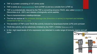
Tumour Necrosis Factor
PRESENTED BY
CHANDANA DINAKARAN
AAH-PB0-01
PHD FIRST YEAR
ICAR-CIFE
 The regulation of immune responses occurs through cytokines which help in growth, differentiation, and
activation function to regulate the immune responses (Muñoz-Carrillo et al. 2018).
 These cytokines are proteins - inducing the defense activity in the host body, generation of T-cells and
production of antibodies.
 These secreted proteins are produced from the immune cells such as macrophages, lymphocytes,
(Reyes-Cerpa et al. 2012).
 Tumor necrosis factor superfamily (TNFSF) members are considered as essential cytokines in signaling
cascade ( Sivangala and Sumanlatha 2015).
 TNFSF comprises of 19 members, which signals through 29 different receptors.
INTRODUCTION
Tumor Necrosis Factor (TNF)
 TNF also known as cachexin or cachectin
 Tumor necrosis factor (TNF, also referred to as TNFα) was identified in the late1970s as a
cytokine produced by immune cells having a capacity to suppress tumor cell proliferation and
induce tumor regression
 Fish tumor necrosis factor (TNF) ligands are important cytokines which play pivotal roles in
regulating immune functions, metabolism and morphological development.
 The first two members of the family to be identified were: Tumor Necrosis Factor Alpha (TNF
Alpha) and Tumor necrosis factor-beta (TNF-β), also known as Lymphotoxin-alpha
 TNFSF2 (TNF-α)
 TNF-α (TNFSF2) was the first member of the TNFSF identified in fish
 TNF-α mediates a broad range of biological functions in regulating
inflammation, apoptosis, fat metabolism and organ regeneration in fish (Zou
and Secombes, 2016).
 Fish TNF-α are type II membrane proteins and share a conserved TNF
homology domain (THD)
 Activation - binding to the respective receptor(s).
 The THD of fish TNF- α is located in the extracellular region and is
composed of 10 β-strands forming a compact “jellyroll” topology.
 TNF is a protein consisting of 157 amino acids
 TNF-α exists as a transmembrane form (mTNF-α) and as a soluble form (sTNF-α).
 TNF-α is proteolytically cleaved by the TNF-α converting enzyme (TACE, also called ADAM17)
(Secombes et al., 2001) and release a biologically active peptide.
 Tace is transmembrane metalloproteinase
 The first aa residue at the protease cleavage site (threonine or serine) is important and remains
conserved in fish (Kim et al., 2009).
 The secretion of TNF-α and TACE-like activity induced by lipopolysaccharide (LPS) and zymosan
 TACE-like activity can be selectively inhibited by the TAPI-1 inhibitor (Roher et al., 2011).
 In fish, high basal levels of tnf-α expression are detected in a wide range of immune organs and cell
types
 Monocytes/macrophages are the primary leukocyte population capable of producing a large quantity of
TNF-α (Ronza et al., 2015a), and TNF-α has been detected in adipocytes (Bou et al., 2014).
 Administration of immunogen, poly I:C or β-glucan into fish can upregulate tnf-α gene expression (Milne et al.,
2017).
 TNF-α plays a central role in regulating the inflammatory process.
 Fish tnf-α genes are up-regulated during the early stages of infection
 It was found that tnf-α was significantly increased during early acute infection, whereas it was surprisingly
down-regulated during chronic infection
 Parasite infection alters tnf-α expression in fish hosts.
 Proinflammatory cytokines such as interleukin (IL)-1β and IFN-γ have been shown to activate tnf-α
expression.
 The bmal1 and clock1 gene, which play important roles in controlling circadian rhythm, have also been
shown to activate the tnf-α gene when overexpressed in medaka embryos. This suggests circadian rhythms
can influence tnf-α functions (Onoue et al., 2019).
Functions of TNF-α are
conserved in fish
 Fish TNF-α augments the respiratory burst response and stimulates nitric oxide production
 Fish TNF-α enhances migration of immune cells.
 Intraperitoneal injection of TNF-α increased phagocyte mobilization and induced
granulopoiesis in the head kidney of sea bream (Ordás et al., 2007), and enhanced phagocytic
activity and the migration of leukocytes in other fish species such as Bluefin tuna and rainbow
trout
 TNF-α plays a central role in mediating apoptosis and necrotic cell death in fish
 TNF-α enhances replication of the spring viremia of carp virus (SVCV) by inhibiting the host
autophagic antiviral response
 TNF-α is a key factor in the regulation of lipid metabolism in fish(Zhou et al., 2017).
 Treatment with TNF-α also led to lowered plasma levels of triglyceride, total cholesterol, low
density lipoprotein cholesterol, nonesterified fatty acid, and glycerol (Liu et al., 2016a)---- TNF-α is
involved in the regulation of hepatic lipid metabolism and fat distribution.
 On the other hand, TNF-α can induce hepatic steatosis and lipid accumulation
 TNF-α is involved in tissue development and regeneration in fish.
 TNF-α affects basal testosterone production and ovarian functions
 In zebrafish, TNF-α mediated signaling pathways are crucial for the homeostasis of skin and fin
regeneration (Nguyen-Chi et al., 2017).
 TNF-α deficiency leads to the inhibition of the infiltration of neutrophils into the skin, increased
proliferation of keratinocytes and local activation of the NF-κB signaling pathway (Candel et al., 2014).
 Knockdown of tnf-α expression inhibits the proliferation of Müller glia, indicating Müller glia-derived
TNF-α is required for recruiting additional Müller glia to enter the cell cycle of the retinal
regeneration process in zebrafish.
TNFSF5 (CD40L, CD154)
 The TNFSF member CD40L (also known as CD154) plays critical roles in the survival, proliferation
and maturation of T and B cells and also functions as a co-stimulatory molecule for T cells to regulate
antibody production (Lu et al., 2018).
 The CD40L homologue is present in teleost fish and is predicted to be a type II membrane-bound
protein
 The cd40l gene has recently been identified in a non-teleost fish, the small spotted catshark
(Scyliorhinus canicula)
TNFSF6 (FASL)
 Two forms of FASL(First Apototic Signal Ligand) homologues, a membrane-bound form and a soluble
form, have been reported in fish.
 FASL is a death receptor controlling programmed cell death, apoptosis and cytotoxic processes (Wang et
al., 2019).
 The FASL-FasR system is hinted to activate anti-parasite innate immunity
TNFSF10 (TRAIL)
 The TNF-related apoptosis-inducing ligand (TRAIL), also termed TNFSF10, regulates many biological
processes including cell proliferation and apoptotic cell death.
 It is predominantly expressed in the spleen, kidney and intestine, but at moderate levels in the gills and muscle
 The expression of trail can be modulated by various stimuli.
TNFSF11 (RANKL)
 The receptor activator of the nuclear factor κB ligand (RANKL, TNFSF11) plays key roles in
osteoclastogenesis and bone resorption.
 RANKL stimulates bone resorption via the osteoclast surface receptor RANK.
TNFSF12 (TWEAK)
 The tumor necrosis factor-like weak inducer of apoptosis (TWEAK, TNFSF12) is a multifunctional cytokine in
regulating angiogenesis, inflammation and apoptosis.
 Expression is detected mainly in immune organs such as the spleen, gills, and kidney and can be up-regulated
by bacterial and viral pathogens
TNFSF13 (APRIL)
 A proliferation-inducing ligand (APRIL) is involved in immune regulation, tumor cell proliferation and
apoptosis (Dillon et al., 2006).
 In grass carp, the april transcripts are predominantly detected in the skin, spleen, and head kidney, and
are significantly up-regulated after infection of A. hydrophila and Aquareovirus
TNFSF13B (BAFF)
 B-cell activating factor (BAFF) is essential for the survival of B cells (Liu et al., 2016b) and duplicated copies of
Baff genes have been reported in teleost fish species
 Ammonia and environmental pollutants also induce baff expression in pufferfish.
 BAFF plays a central role in regulating B cell homeostasis and can trigger specific signals controlling
differentiation of peritoneal B cell subsets
TNFSF14 (LIGHT)
 Lymphotoxin-related inducible ligand (LIGHT) serves as one of the receptors for the entry of the herpes
into humans and exists in fish.
EDA ( Ectodysplasin A)
 EDA is an ancient TNFSF member and is functionally conserved.
 In fish, it is essential for the development and patterning of teeth, scales and fins (Pujolar et al., 2017).
TNFSF New (TNF-N)
 A novel TNF homologue (designated as tnf-new, tnf-n, lymphotoxin-beta (LT-β)) has been
described in rainbow trout, Japanese pufferfish and zebrafish
TNF receptors
 The TNF receptor superfamily (TNFRSF) members possess at least one conserved cysteine-rich
domain (CRD) (Locksley et al., 2001).
 The CRDs are considered as the hallmark of the TNFRSF members and dictate ligand specificity.
 The intracellular region of TNFRSF can be divided into three types, those containing a death
domain (DD), those lacking a cytosolic domain, and those containing a cytosolic region but lacking
the DD.
 The former are central in regulating programmed cell death or apoptosis whilst those lacking a
cytosolic domain function as decoy receptors.
TNFRSF1a (TNFR1)
 TNFR1 (TNFRSF1a) is a DD containing receptor and serves as the receptor for TNF-α and LT-α.
 Activation of TNFR1 by TNF-α initiates a cascade of downstream signaling pathways involving adapter
proteins such as FADD, RIP and TRADD, and caspases to regulate cell differentiation, apoptosis and
cytotoxicity (Chen and Goeddel, 2002).
 TNFR1 mediated signaling pathway is also important in balancing local tissue macrophage populations
and tissue regeneration (Nguyen-Chi et al., 2017)
TNFRSF1b (TNFR2)
 TNFR2 (TNFRSF1b) binds to TNF-α.
 TNFR2 is thought to primarily promote cell survival.
 TNFR2 provides a co-stimulatory signal for T cell proliferation and survival (Ward-Kavanagh et al., 2016).
 Deficiency of tnfr2 causes apoptosis of embryo endothelial cells in zebrafish, which involves caspase-8,
caspase-2 and P53 (Espín et al., 2013).
TNFRSF5 (CD40)
 CD40 (TNFRSF5) is primarily expressed in B cells, macrophages, dendritic cells (DCs)
 The CD40-CD40L interaction is vital for the survival and proliferation of B cells, antibody production
and isotype switching
TNFRSF6 (FAS/CD95)
 The death receptor FAS/CD95 (TNFRSF6) is the sole receptor of FASL (CD95L) and contains a DD in
the cytoplasmic tail.
 Interaction between FAS and FASL triggers a conformational alteration of FAS
 The apoptosis pathway mediated by FAS/FASL is conserved in fish.
EDAR
 EDAR (ectodysplasin A receptor) is a DD-containing TNF receptor and is paired with EDA
 The EDA/EDAR pathway is conserved in fish and has been shown to be important for the formation of
teeth and scales which are believed to have evolved from a common ancestor
Fish viral TNFRSF homologues
 The TNFRSFs are one group of host receptors that can be exploited by viruses to gain entry into cells
to establish infection (Kinkade and Ware, 2006).
 Viruses generate TNFRSF homologues in order to manipulate host immune responses, likely through
interaction with their host
Cell signaling
 TNF can bind two receptors, TNFR1 (TNF receptor type 1) and TNFR2
 TNFR1 is expressed in most tissues, whereas TNFR2 is found typically in cells of the immune
system
 TNFR-1 is a death domain (DD)-containing receptor with an extracellular domain (ECD), a
transmembrane domain (TMD), and an intracellular domain (ICD)- capability of inducing apoptotic
cell death.
 The initial step in TNF signaling- the binding of the TNF to the extracellular domain of TNF-R1 and
the release of the inhibitory protein silencer of death domains (SODD) from TNF-R1’s intracellular
domain (ICD).
 TNF-R1 ICD is recognized by the adaptor protein TNF receptor–associated death domain
(TRADD), which recruits additional adaptor proteins receptor-interacting protein (RIP), TNF-R–
associated factor 2 (TRAF2), and Fas-associated death domain (FADD). These latter proteins
recruit key enzymes to TNF-R1 that are responsible for initiating signaling events
 Following TRADD binding, three pathways can be initiated
Fig b--TNFR receptor have a
molecule attached to its
intracellular side – SODD(silencer
of Death domain (DD)), SODD
keeps the DD of TNFR in its
inactive stage.
Fig a– when TNF binds to TNFR –
results in conformational change in
TNFR- dissociation of SODD
 Three pathways are activated
 Activation of NF-kB pathway
 activation of MAPK pathway
 Induction of death signaling– apoptosis
 apoptosis pathway 2 types-extrinsic and intrinsic
 extrinsic pathway –driven by extracellular signal
 intrinsic pathway – driven by intracellular signal
NF-κB pathway
NF-κB
pathway
 Activation of NF-κB:
 Following the Binding of TNF to TNF receptor 1 (TNF-R1),DD is activated and recruits TNF-R associated
death domain (TRADD), TNF-R associated factor 2 (TRAF2), and receptor interacting protein 1 (serine-
threonine kinase -RIP1) are recruited to form the so called complex I. TRADD recruits TRAF2 and RIP.
 NF-κBs are normally sequestered in the cytoplasm as inactive complexes bound by a family of inhibitory
proteins, inhibitory κBs (IκBs)--- which normally retain NF-kB within the cytoplasm of unstimulated cells
 IkB kinase (IKK) complex that mediates phosphorylation of IkB
 The core of the IKK complex consists of two catalytic subunits, IKKa and IKKb, and a regulatory subunit,
NFkB essential modulator (NEMO, or IKKg). Activation of IKK is typically mediated by transforming growth
factor beta-activated kinase 1 (TAK1), a MAPK kinase kinase (MAP3K) that responds to various immune
receptor signals and relies on ubiquitination for its catalytic activation and signaling function
 Phosphorylation of IkB leads to its subsequent ubiquitination and proteasomal degradation, thereby
allowing NF-kB to translocate to the nucleus where it activates the transcription of NF-kBresponsive genes-
NF-κB's target genes, cIAP-1, cIAP-2, Bcl-xL, XIAP, and IEX-1L are found to have anti-apoptotic properties
 Mediates the transcription of a vast array of proteins involved in cell survival and proliferation, inflammatory
response, and anti-apoptotic factors
 Activation of the MAPK pathways:
 Mitogen-activated protein kinases form a large family of serine/ threonine kinases that respond to diverse
extracellular and intracellular stimuli and mediate multiple biological processes
 The mammalian MAPK family includes three subfamilies: the extracellular signal-regulated kinases (ERKs),
the c-Jun N-terminal kinases (JNKs), the p38 MAP kinases
 TNF induces a strong activation of the stress-related JNK group, evokes moderate response of the p38-
MAPK, and is responsible for minimal activation of the classical ERKs
 TRAF2 is also thought to activate a mitogen activated protein kinase kinase kinase (MAPKKK), such as
extracellular signal-regulated kinase kinase kinase 1 (MEKK1) or apoptosis-stimulated kinase 1 (ASK1),
thereby activating a cascade of kinases resulting in the activation of c-Jun NH2-terminal kinase ( JNK), a
kinase that phosphorylates c-Jun and increases its transcriptional activity
 MAP3K MAP2 MAPK
 MKK4 and MKK7 for JNKs
 The two JNK kinases (JNKKs) phosphorylate JNK at Thr183 and Tyr185, leading to its activation
 JNK translocates to the nucleus and activates transcription factors such as c-Jun and ATF2.
The JNK pathway is involved in cell differentiation, proliferation, and is generally pro-apoptotic
Activation of the MAPK pathway
MAPK kinase cascade activate each other
in a sequential manner via phosporylation
 Induction of death signaling:
 Pathway is initiated by TNFR1 internally signaling Complex I - form Complex II that consists of
TRADD, RIP, FADD, and caspase-8.
 The death receptor-mediated apoptosis pathway is also called the extrinsic apoptosis pathway
 Depending on the signaling molecule and receptor – are of two types extrinsic pathway
 TNF path and FAS path
 Interaction with membrane-bound FasL reorganizes these complexes and allows the formation of a
death inducing signaling complex (DISC). The Fas DISC contains the adaptor protein Fas-
associated death domain protein (FADD) and caspases 8 and 10, which can initiate the process of
apoptosis
 Caspase-8 is auto-activated to trigger activation of the executor capsases-3, and -7, and the
endonucleases, resulting in destruction of cell component proteins, fragmentation of DNA, and,
eventually, apoptotic cell death.
TNF pathway of extrinsic apoptosis
 TNF a binds to TNFR --- Activate DD---- activated DD recruit TRAD---
which inturn recruit----FADD ----cleaves procaspase 8-------- caspase 8---
-----------caspase cascade----- cell death
TNF path
FAS pathway of extrinsic apoptosis
 This involves 2 different cell
 a) signaling cell- kill the target cell by singal molecule- FasL
 b) target cell– killed by apoptosis
 FasL- FasR binding produce first apptotic signal-Activate DD---- activated
DD recruit TRAD---which inturn recruit----FADD ----recruits procaspase 8
 FADD+ procaspase 8 form DISC(death inducing signaling complex)
 Caspase 8 is released from DISC ---cleaves procaspase 3---caspase
cascade—act on inactive apoptotic substrates(caspase dependent
DNAase, proteolytic enzymes)-----degrade DNA and cytosolic protein -----
cell death
FAS PATH
Intrinsic apoptotic pathway
 3 stage
 Initiation– initiated by intracellular signal( DNA damage, hypoxia, stress)
 Execution
 Phagocytosis
 DNA lesion(act as apoptotic signal)----activate serine /threonine kinase -------P53 ---
--PUMA----Activate BAX(Bcell lymphoma associated x protein)- BAX become
integrated into Mitochondrial membrane – open Voltage dependent anionic channel
--- form MAC(mitochondrial induced apoptotic channel)
 Cyt c+ APAF1( apoptotic protease activating factor)+ procaspase 9==== form
Apoptosome
 Apoptosome --release caspase 9 -----caspase cascade--- cell death
 TNF-induced apoptosis also uses the mitochondria-mediated (intrinsic) apoptosis pathway.
 This is achieved by caspase-8 activating BCL-2 interacting domain (Bid), a BH3-only Bcl2 family
member
 caspase-8 is activated through auto-catalytic cleavage. Active caspase-8 sets the cell death machinery
in motion by cleaving Bid.
 The resulting tBid fragment translocates to the mitochondria where it causes permeabilization of the
mitochondrial outermembrane.
 This leads to the release of cytochrome c and other mitochondrial apoptogenic factors(second
mitochondria-derived activator of caspases (SMAC, / direct IAP binding protein with low pIaso called
Diablo from mt to cytosol)), which cause activation of other caspases and ultimately cell death
 Cytochrome c binds to apoptotic protease activating factor 1(Apaf-1) and pro-caspase-9 to form
apoptosome, resulting in caspase-9-mediated activation of the executor caspases (caspase-3)
 Smac binds to and inhibits the inhibitor of apoptosis proteins (IAP, including c-IAP1, c-IAP2, X-linked
Inhibitor of Apoptosis Protein [XIAP], and survivin)
Reference
 Chen, G. and Goeddel, D.V., 2002. TNF-R1 signaling: a beautiful
pathway. Science, 296(5573), pp.1634-1635.
 Gaur, U. and Aggarwal, B.B., 2003. Regulation of proliferation, survival and apoptosis by
members of the TNF superfamily. Biochemical pharmacology, 66(8), pp.1403-1408.
 Li, Y., Xiao, T. and Zou, J., 2020. Fish TNF and TNF receptors. Science China Life Sciences,
pp.1-25.
 Shi, J.H. and Sun, S.C., 2018. Tumor necrosis factor receptor-associated factor regulation of
nuclear factor κB and mitogen-activated protein kinase pathways. Frontiers in
immunology, 9, p.1849.
 Salomon, B.L., Leclerc, M., Tosello, J., Ronin, E., Piaggio, E. and Cohen, J.L., 2018. Tumor
necrosis factor α and regulatory T cells in oncoimmunology. Frontiers in immunology, 9,
p.444.
 Banerjee, R., Samanta, M. and Das, S., 2020. Regulation of tumor necrosis superfamily
molecules, BAFF and APRIL, in response to pathogenic exposure and ligand stimulation in
freshwater carp, Catla catla. Aquaculture International, 28(5), pp.2139-2159.
 Wullaert, A., Heyninck, K. and Beyaert, R., 2006. Mechanisms of crosstalk between TNF-
induced NF-κB and JNK activation in hepatocytes. Biochemical pharmacology, 72(9),
pp.1090-1101.
 Sabio, G. and Davis, R.J., 2014, June. TNF and MAP kinase signalling pathways.
In Seminars in immunology (Vol. 26, No. 3, pp. 237-245). Academic Press.
 Wang, X. and Lin, Y., 2008. Tumor necrosis factor and cancer, buddies or foes? 1. Acta
Pharmacologica Sinica, 29(11), pp.1275-1288.
THANK YOU

TNF

  • 1.
    Tumour Necrosis Factor PRESENTEDBY CHANDANA DINAKARAN AAH-PB0-01 PHD FIRST YEAR ICAR-CIFE
  • 2.
     The regulationof immune responses occurs through cytokines which help in growth, differentiation, and activation function to regulate the immune responses (Muñoz-Carrillo et al. 2018).  These cytokines are proteins - inducing the defense activity in the host body, generation of T-cells and production of antibodies.  These secreted proteins are produced from the immune cells such as macrophages, lymphocytes, (Reyes-Cerpa et al. 2012).  Tumor necrosis factor superfamily (TNFSF) members are considered as essential cytokines in signaling cascade ( Sivangala and Sumanlatha 2015).  TNFSF comprises of 19 members, which signals through 29 different receptors. INTRODUCTION
  • 3.
    Tumor Necrosis Factor(TNF)  TNF also known as cachexin or cachectin  Tumor necrosis factor (TNF, also referred to as TNFα) was identified in the late1970s as a cytokine produced by immune cells having a capacity to suppress tumor cell proliferation and induce tumor regression  Fish tumor necrosis factor (TNF) ligands are important cytokines which play pivotal roles in regulating immune functions, metabolism and morphological development.  The first two members of the family to be identified were: Tumor Necrosis Factor Alpha (TNF Alpha) and Tumor necrosis factor-beta (TNF-β), also known as Lymphotoxin-alpha
  • 4.
     TNFSF2 (TNF-α) TNF-α (TNFSF2) was the first member of the TNFSF identified in fish  TNF-α mediates a broad range of biological functions in regulating inflammation, apoptosis, fat metabolism and organ regeneration in fish (Zou and Secombes, 2016).  Fish TNF-α are type II membrane proteins and share a conserved TNF homology domain (THD)  Activation - binding to the respective receptor(s).  The THD of fish TNF- α is located in the extracellular region and is composed of 10 β-strands forming a compact “jellyroll” topology.
  • 5.
     TNF isa protein consisting of 157 amino acids  TNF-α exists as a transmembrane form (mTNF-α) and as a soluble form (sTNF-α).  TNF-α is proteolytically cleaved by the TNF-α converting enzyme (TACE, also called ADAM17) (Secombes et al., 2001) and release a biologically active peptide.  Tace is transmembrane metalloproteinase  The first aa residue at the protease cleavage site (threonine or serine) is important and remains conserved in fish (Kim et al., 2009).  The secretion of TNF-α and TACE-like activity induced by lipopolysaccharide (LPS) and zymosan  TACE-like activity can be selectively inhibited by the TAPI-1 inhibitor (Roher et al., 2011).  In fish, high basal levels of tnf-α expression are detected in a wide range of immune organs and cell types
  • 7.
     Monocytes/macrophages arethe primary leukocyte population capable of producing a large quantity of TNF-α (Ronza et al., 2015a), and TNF-α has been detected in adipocytes (Bou et al., 2014).  Administration of immunogen, poly I:C or β-glucan into fish can upregulate tnf-α gene expression (Milne et al., 2017).  TNF-α plays a central role in regulating the inflammatory process.  Fish tnf-α genes are up-regulated during the early stages of infection  It was found that tnf-α was significantly increased during early acute infection, whereas it was surprisingly down-regulated during chronic infection  Parasite infection alters tnf-α expression in fish hosts.  Proinflammatory cytokines such as interleukin (IL)-1β and IFN-γ have been shown to activate tnf-α expression.  The bmal1 and clock1 gene, which play important roles in controlling circadian rhythm, have also been shown to activate the tnf-α gene when overexpressed in medaka embryos. This suggests circadian rhythms can influence tnf-α functions (Onoue et al., 2019).
  • 8.
    Functions of TNF-αare conserved in fish
  • 9.
     Fish TNF-αaugments the respiratory burst response and stimulates nitric oxide production  Fish TNF-α enhances migration of immune cells.  Intraperitoneal injection of TNF-α increased phagocyte mobilization and induced granulopoiesis in the head kidney of sea bream (Ordás et al., 2007), and enhanced phagocytic activity and the migration of leukocytes in other fish species such as Bluefin tuna and rainbow trout  TNF-α plays a central role in mediating apoptosis and necrotic cell death in fish  TNF-α enhances replication of the spring viremia of carp virus (SVCV) by inhibiting the host autophagic antiviral response  TNF-α is a key factor in the regulation of lipid metabolism in fish(Zhou et al., 2017).  Treatment with TNF-α also led to lowered plasma levels of triglyceride, total cholesterol, low density lipoprotein cholesterol, nonesterified fatty acid, and glycerol (Liu et al., 2016a)---- TNF-α is involved in the regulation of hepatic lipid metabolism and fat distribution.
  • 10.
     On theother hand, TNF-α can induce hepatic steatosis and lipid accumulation  TNF-α is involved in tissue development and regeneration in fish.  TNF-α affects basal testosterone production and ovarian functions  In zebrafish, TNF-α mediated signaling pathways are crucial for the homeostasis of skin and fin regeneration (Nguyen-Chi et al., 2017).  TNF-α deficiency leads to the inhibition of the infiltration of neutrophils into the skin, increased proliferation of keratinocytes and local activation of the NF-κB signaling pathway (Candel et al., 2014).  Knockdown of tnf-α expression inhibits the proliferation of Müller glia, indicating Müller glia-derived TNF-α is required for recruiting additional Müller glia to enter the cell cycle of the retinal regeneration process in zebrafish.
  • 11.
    TNFSF5 (CD40L, CD154) The TNFSF member CD40L (also known as CD154) plays critical roles in the survival, proliferation and maturation of T and B cells and also functions as a co-stimulatory molecule for T cells to regulate antibody production (Lu et al., 2018).  The CD40L homologue is present in teleost fish and is predicted to be a type II membrane-bound protein  The cd40l gene has recently been identified in a non-teleost fish, the small spotted catshark (Scyliorhinus canicula) TNFSF6 (FASL)  Two forms of FASL(First Apototic Signal Ligand) homologues, a membrane-bound form and a soluble form, have been reported in fish.  FASL is a death receptor controlling programmed cell death, apoptosis and cytotoxic processes (Wang et al., 2019).  The FASL-FasR system is hinted to activate anti-parasite innate immunity
  • 12.
    TNFSF10 (TRAIL)  TheTNF-related apoptosis-inducing ligand (TRAIL), also termed TNFSF10, regulates many biological processes including cell proliferation and apoptotic cell death.  It is predominantly expressed in the spleen, kidney and intestine, but at moderate levels in the gills and muscle  The expression of trail can be modulated by various stimuli. TNFSF11 (RANKL)  The receptor activator of the nuclear factor κB ligand (RANKL, TNFSF11) plays key roles in osteoclastogenesis and bone resorption.  RANKL stimulates bone resorption via the osteoclast surface receptor RANK. TNFSF12 (TWEAK)  The tumor necrosis factor-like weak inducer of apoptosis (TWEAK, TNFSF12) is a multifunctional cytokine in regulating angiogenesis, inflammation and apoptosis.  Expression is detected mainly in immune organs such as the spleen, gills, and kidney and can be up-regulated by bacterial and viral pathogens
  • 13.
    TNFSF13 (APRIL)  Aproliferation-inducing ligand (APRIL) is involved in immune regulation, tumor cell proliferation and apoptosis (Dillon et al., 2006).  In grass carp, the april transcripts are predominantly detected in the skin, spleen, and head kidney, and are significantly up-regulated after infection of A. hydrophila and Aquareovirus TNFSF13B (BAFF)  B-cell activating factor (BAFF) is essential for the survival of B cells (Liu et al., 2016b) and duplicated copies of Baff genes have been reported in teleost fish species  Ammonia and environmental pollutants also induce baff expression in pufferfish.  BAFF plays a central role in regulating B cell homeostasis and can trigger specific signals controlling differentiation of peritoneal B cell subsets TNFSF14 (LIGHT)  Lymphotoxin-related inducible ligand (LIGHT) serves as one of the receptors for the entry of the herpes into humans and exists in fish.
  • 14.
    EDA ( EctodysplasinA)  EDA is an ancient TNFSF member and is functionally conserved.  In fish, it is essential for the development and patterning of teeth, scales and fins (Pujolar et al., 2017). TNFSF New (TNF-N)  A novel TNF homologue (designated as tnf-new, tnf-n, lymphotoxin-beta (LT-β)) has been described in rainbow trout, Japanese pufferfish and zebrafish
  • 15.
    TNF receptors  TheTNF receptor superfamily (TNFRSF) members possess at least one conserved cysteine-rich domain (CRD) (Locksley et al., 2001).  The CRDs are considered as the hallmark of the TNFRSF members and dictate ligand specificity.  The intracellular region of TNFRSF can be divided into three types, those containing a death domain (DD), those lacking a cytosolic domain, and those containing a cytosolic region but lacking the DD.  The former are central in regulating programmed cell death or apoptosis whilst those lacking a cytosolic domain function as decoy receptors.
  • 16.
    TNFRSF1a (TNFR1)  TNFR1(TNFRSF1a) is a DD containing receptor and serves as the receptor for TNF-α and LT-α.  Activation of TNFR1 by TNF-α initiates a cascade of downstream signaling pathways involving adapter proteins such as FADD, RIP and TRADD, and caspases to regulate cell differentiation, apoptosis and cytotoxicity (Chen and Goeddel, 2002).  TNFR1 mediated signaling pathway is also important in balancing local tissue macrophage populations and tissue regeneration (Nguyen-Chi et al., 2017) TNFRSF1b (TNFR2)  TNFR2 (TNFRSF1b) binds to TNF-α.  TNFR2 is thought to primarily promote cell survival.  TNFR2 provides a co-stimulatory signal for T cell proliferation and survival (Ward-Kavanagh et al., 2016).  Deficiency of tnfr2 causes apoptosis of embryo endothelial cells in zebrafish, which involves caspase-8, caspase-2 and P53 (Espín et al., 2013).
  • 17.
    TNFRSF5 (CD40)  CD40(TNFRSF5) is primarily expressed in B cells, macrophages, dendritic cells (DCs)  The CD40-CD40L interaction is vital for the survival and proliferation of B cells, antibody production and isotype switching TNFRSF6 (FAS/CD95)  The death receptor FAS/CD95 (TNFRSF6) is the sole receptor of FASL (CD95L) and contains a DD in the cytoplasmic tail.  Interaction between FAS and FASL triggers a conformational alteration of FAS  The apoptosis pathway mediated by FAS/FASL is conserved in fish.
  • 18.
    EDAR  EDAR (ectodysplasinA receptor) is a DD-containing TNF receptor and is paired with EDA  The EDA/EDAR pathway is conserved in fish and has been shown to be important for the formation of teeth and scales which are believed to have evolved from a common ancestor Fish viral TNFRSF homologues  The TNFRSFs are one group of host receptors that can be exploited by viruses to gain entry into cells to establish infection (Kinkade and Ware, 2006).  Viruses generate TNFRSF homologues in order to manipulate host immune responses, likely through interaction with their host
  • 19.
    Cell signaling  TNFcan bind two receptors, TNFR1 (TNF receptor type 1) and TNFR2  TNFR1 is expressed in most tissues, whereas TNFR2 is found typically in cells of the immune system  TNFR-1 is a death domain (DD)-containing receptor with an extracellular domain (ECD), a transmembrane domain (TMD), and an intracellular domain (ICD)- capability of inducing apoptotic cell death.  The initial step in TNF signaling- the binding of the TNF to the extracellular domain of TNF-R1 and the release of the inhibitory protein silencer of death domains (SODD) from TNF-R1’s intracellular domain (ICD).  TNF-R1 ICD is recognized by the adaptor protein TNF receptor–associated death domain (TRADD), which recruits additional adaptor proteins receptor-interacting protein (RIP), TNF-R– associated factor 2 (TRAF2), and Fas-associated death domain (FADD). These latter proteins recruit key enzymes to TNF-R1 that are responsible for initiating signaling events  Following TRADD binding, three pathways can be initiated
  • 20.
    Fig b--TNFR receptorhave a molecule attached to its intracellular side – SODD(silencer of Death domain (DD)), SODD keeps the DD of TNFR in its inactive stage. Fig a– when TNF binds to TNFR – results in conformational change in TNFR- dissociation of SODD
  • 21.
     Three pathwaysare activated  Activation of NF-kB pathway  activation of MAPK pathway  Induction of death signaling– apoptosis  apoptosis pathway 2 types-extrinsic and intrinsic  extrinsic pathway –driven by extracellular signal  intrinsic pathway – driven by intracellular signal
  • 22.
  • 23.
  • 24.
     Activation ofNF-κB:  Following the Binding of TNF to TNF receptor 1 (TNF-R1),DD is activated and recruits TNF-R associated death domain (TRADD), TNF-R associated factor 2 (TRAF2), and receptor interacting protein 1 (serine- threonine kinase -RIP1) are recruited to form the so called complex I. TRADD recruits TRAF2 and RIP.  NF-κBs are normally sequestered in the cytoplasm as inactive complexes bound by a family of inhibitory proteins, inhibitory κBs (IκBs)--- which normally retain NF-kB within the cytoplasm of unstimulated cells  IkB kinase (IKK) complex that mediates phosphorylation of IkB  The core of the IKK complex consists of two catalytic subunits, IKKa and IKKb, and a regulatory subunit, NFkB essential modulator (NEMO, or IKKg). Activation of IKK is typically mediated by transforming growth factor beta-activated kinase 1 (TAK1), a MAPK kinase kinase (MAP3K) that responds to various immune receptor signals and relies on ubiquitination for its catalytic activation and signaling function  Phosphorylation of IkB leads to its subsequent ubiquitination and proteasomal degradation, thereby allowing NF-kB to translocate to the nucleus where it activates the transcription of NF-kBresponsive genes- NF-κB's target genes, cIAP-1, cIAP-2, Bcl-xL, XIAP, and IEX-1L are found to have anti-apoptotic properties  Mediates the transcription of a vast array of proteins involved in cell survival and proliferation, inflammatory response, and anti-apoptotic factors
  • 25.
     Activation ofthe MAPK pathways:  Mitogen-activated protein kinases form a large family of serine/ threonine kinases that respond to diverse extracellular and intracellular stimuli and mediate multiple biological processes  The mammalian MAPK family includes three subfamilies: the extracellular signal-regulated kinases (ERKs), the c-Jun N-terminal kinases (JNKs), the p38 MAP kinases  TNF induces a strong activation of the stress-related JNK group, evokes moderate response of the p38- MAPK, and is responsible for minimal activation of the classical ERKs  TRAF2 is also thought to activate a mitogen activated protein kinase kinase kinase (MAPKKK), such as extracellular signal-regulated kinase kinase kinase 1 (MEKK1) or apoptosis-stimulated kinase 1 (ASK1), thereby activating a cascade of kinases resulting in the activation of c-Jun NH2-terminal kinase ( JNK), a kinase that phosphorylates c-Jun and increases its transcriptional activity  MAP3K MAP2 MAPK  MKK4 and MKK7 for JNKs  The two JNK kinases (JNKKs) phosphorylate JNK at Thr183 and Tyr185, leading to its activation  JNK translocates to the nucleus and activates transcription factors such as c-Jun and ATF2. The JNK pathway is involved in cell differentiation, proliferation, and is generally pro-apoptotic
  • 26.
    Activation of theMAPK pathway MAPK kinase cascade activate each other in a sequential manner via phosporylation
  • 27.
     Induction ofdeath signaling:  Pathway is initiated by TNFR1 internally signaling Complex I - form Complex II that consists of TRADD, RIP, FADD, and caspase-8.  The death receptor-mediated apoptosis pathway is also called the extrinsic apoptosis pathway  Depending on the signaling molecule and receptor – are of two types extrinsic pathway  TNF path and FAS path  Interaction with membrane-bound FasL reorganizes these complexes and allows the formation of a death inducing signaling complex (DISC). The Fas DISC contains the adaptor protein Fas- associated death domain protein (FADD) and caspases 8 and 10, which can initiate the process of apoptosis  Caspase-8 is auto-activated to trigger activation of the executor capsases-3, and -7, and the endonucleases, resulting in destruction of cell component proteins, fragmentation of DNA, and, eventually, apoptotic cell death.
  • 28.
    TNF pathway ofextrinsic apoptosis  TNF a binds to TNFR --- Activate DD---- activated DD recruit TRAD--- which inturn recruit----FADD ----cleaves procaspase 8-------- caspase 8--- -----------caspase cascade----- cell death
  • 29.
  • 30.
    FAS pathway ofextrinsic apoptosis  This involves 2 different cell  a) signaling cell- kill the target cell by singal molecule- FasL  b) target cell– killed by apoptosis  FasL- FasR binding produce first apptotic signal-Activate DD---- activated DD recruit TRAD---which inturn recruit----FADD ----recruits procaspase 8  FADD+ procaspase 8 form DISC(death inducing signaling complex)  Caspase 8 is released from DISC ---cleaves procaspase 3---caspase cascade—act on inactive apoptotic substrates(caspase dependent DNAase, proteolytic enzymes)-----degrade DNA and cytosolic protein ----- cell death
  • 31.
  • 32.
    Intrinsic apoptotic pathway 3 stage  Initiation– initiated by intracellular signal( DNA damage, hypoxia, stress)  Execution  Phagocytosis  DNA lesion(act as apoptotic signal)----activate serine /threonine kinase -------P53 --- --PUMA----Activate BAX(Bcell lymphoma associated x protein)- BAX become integrated into Mitochondrial membrane – open Voltage dependent anionic channel --- form MAC(mitochondrial induced apoptotic channel)  Cyt c+ APAF1( apoptotic protease activating factor)+ procaspase 9==== form Apoptosome  Apoptosome --release caspase 9 -----caspase cascade--- cell death
  • 35.
     TNF-induced apoptosisalso uses the mitochondria-mediated (intrinsic) apoptosis pathway.  This is achieved by caspase-8 activating BCL-2 interacting domain (Bid), a BH3-only Bcl2 family member  caspase-8 is activated through auto-catalytic cleavage. Active caspase-8 sets the cell death machinery in motion by cleaving Bid.  The resulting tBid fragment translocates to the mitochondria where it causes permeabilization of the mitochondrial outermembrane.  This leads to the release of cytochrome c and other mitochondrial apoptogenic factors(second mitochondria-derived activator of caspases (SMAC, / direct IAP binding protein with low pIaso called Diablo from mt to cytosol)), which cause activation of other caspases and ultimately cell death  Cytochrome c binds to apoptotic protease activating factor 1(Apaf-1) and pro-caspase-9 to form apoptosome, resulting in caspase-9-mediated activation of the executor caspases (caspase-3)  Smac binds to and inhibits the inhibitor of apoptosis proteins (IAP, including c-IAP1, c-IAP2, X-linked Inhibitor of Apoptosis Protein [XIAP], and survivin)
  • 37.
    Reference  Chen, G.and Goeddel, D.V., 2002. TNF-R1 signaling: a beautiful pathway. Science, 296(5573), pp.1634-1635.  Gaur, U. and Aggarwal, B.B., 2003. Regulation of proliferation, survival and apoptosis by members of the TNF superfamily. Biochemical pharmacology, 66(8), pp.1403-1408.  Li, Y., Xiao, T. and Zou, J., 2020. Fish TNF and TNF receptors. Science China Life Sciences, pp.1-25.  Shi, J.H. and Sun, S.C., 2018. Tumor necrosis factor receptor-associated factor regulation of nuclear factor κB and mitogen-activated protein kinase pathways. Frontiers in immunology, 9, p.1849.  Salomon, B.L., Leclerc, M., Tosello, J., Ronin, E., Piaggio, E. and Cohen, J.L., 2018. Tumor necrosis factor α and regulatory T cells in oncoimmunology. Frontiers in immunology, 9, p.444.
  • 38.
     Banerjee, R.,Samanta, M. and Das, S., 2020. Regulation of tumor necrosis superfamily molecules, BAFF and APRIL, in response to pathogenic exposure and ligand stimulation in freshwater carp, Catla catla. Aquaculture International, 28(5), pp.2139-2159.  Wullaert, A., Heyninck, K. and Beyaert, R., 2006. Mechanisms of crosstalk between TNF- induced NF-κB and JNK activation in hepatocytes. Biochemical pharmacology, 72(9), pp.1090-1101.  Sabio, G. and Davis, R.J., 2014, June. TNF and MAP kinase signalling pathways. In Seminars in immunology (Vol. 26, No. 3, pp. 237-245). Academic Press.  Wang, X. and Lin, Y., 2008. Tumor necrosis factor and cancer, buddies or foes? 1. Acta Pharmacologica Sinica, 29(11), pp.1275-1288.
  • 39.