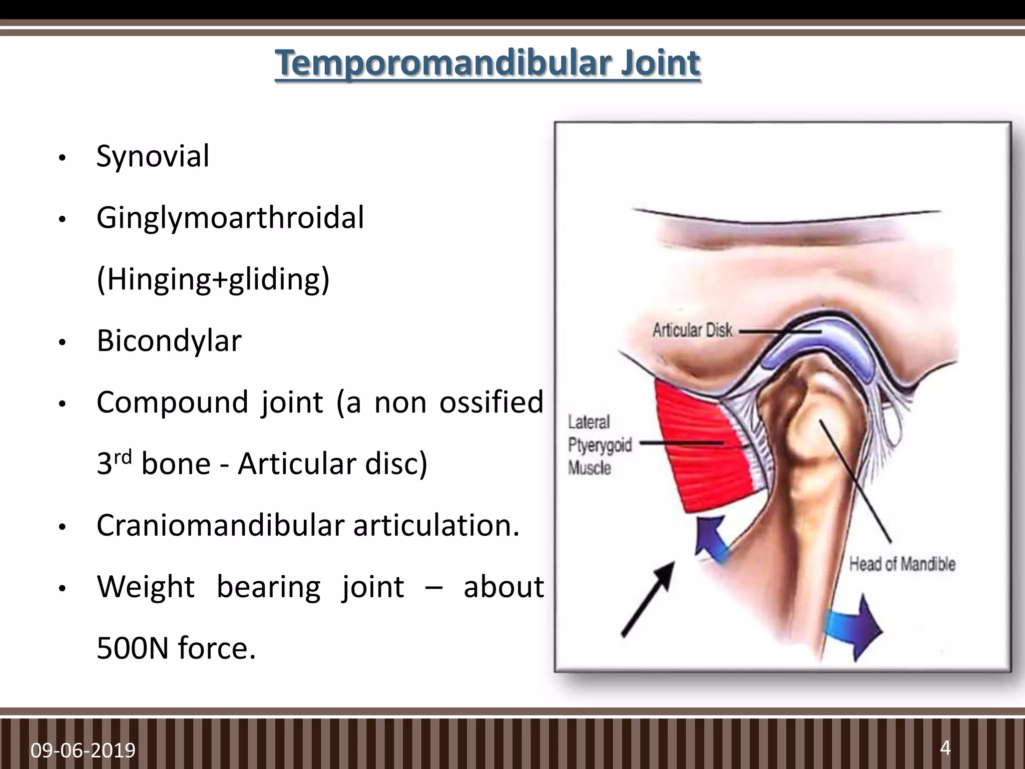
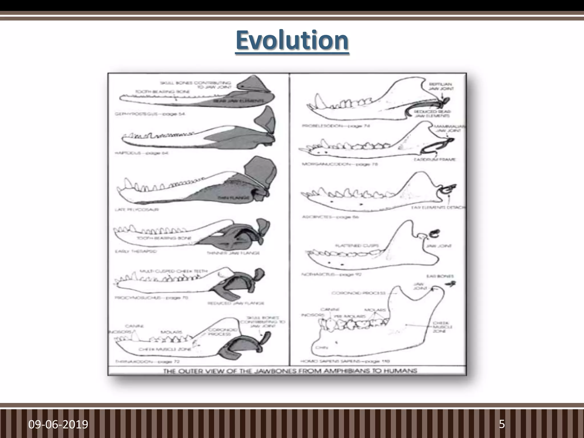
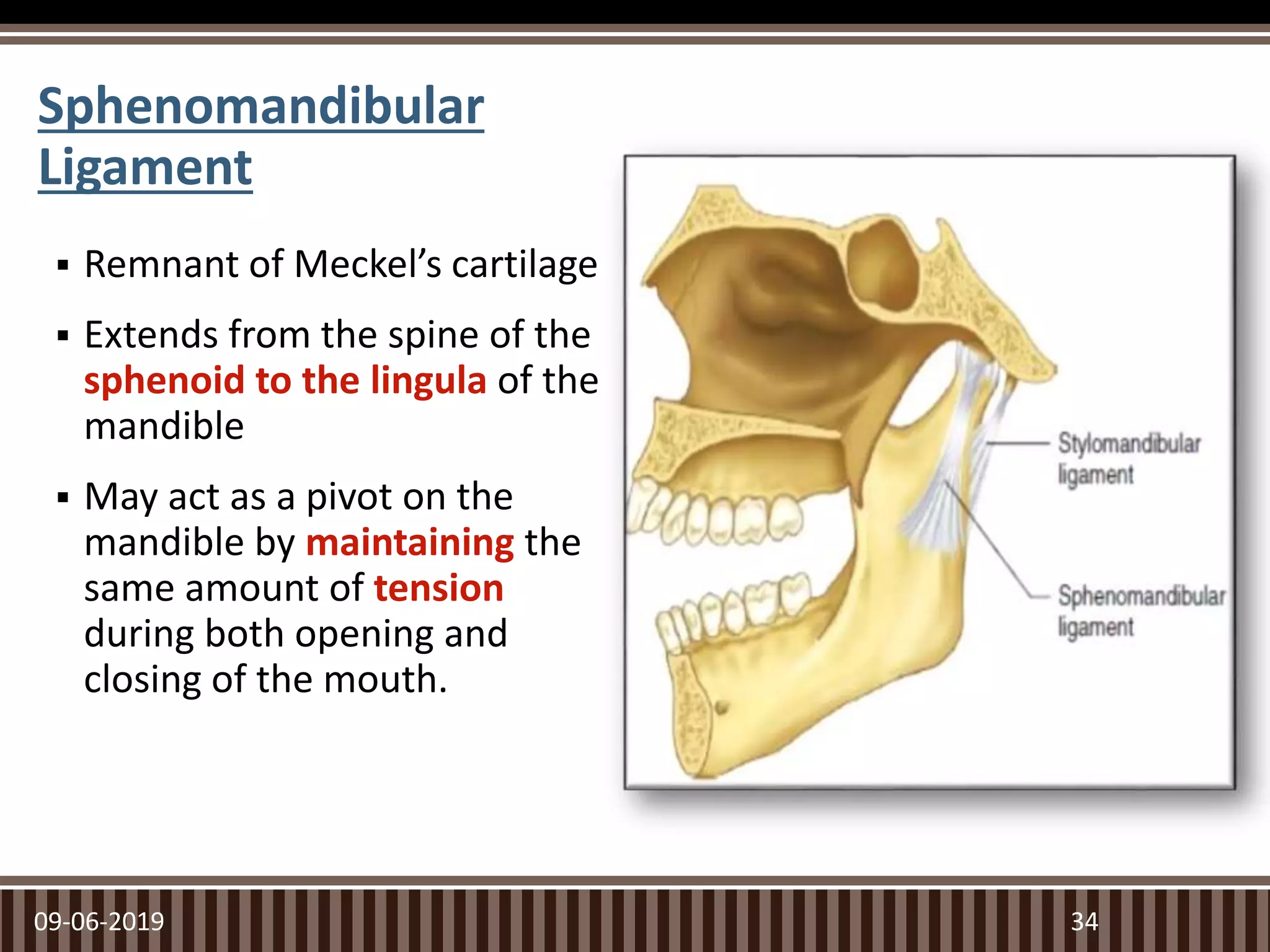
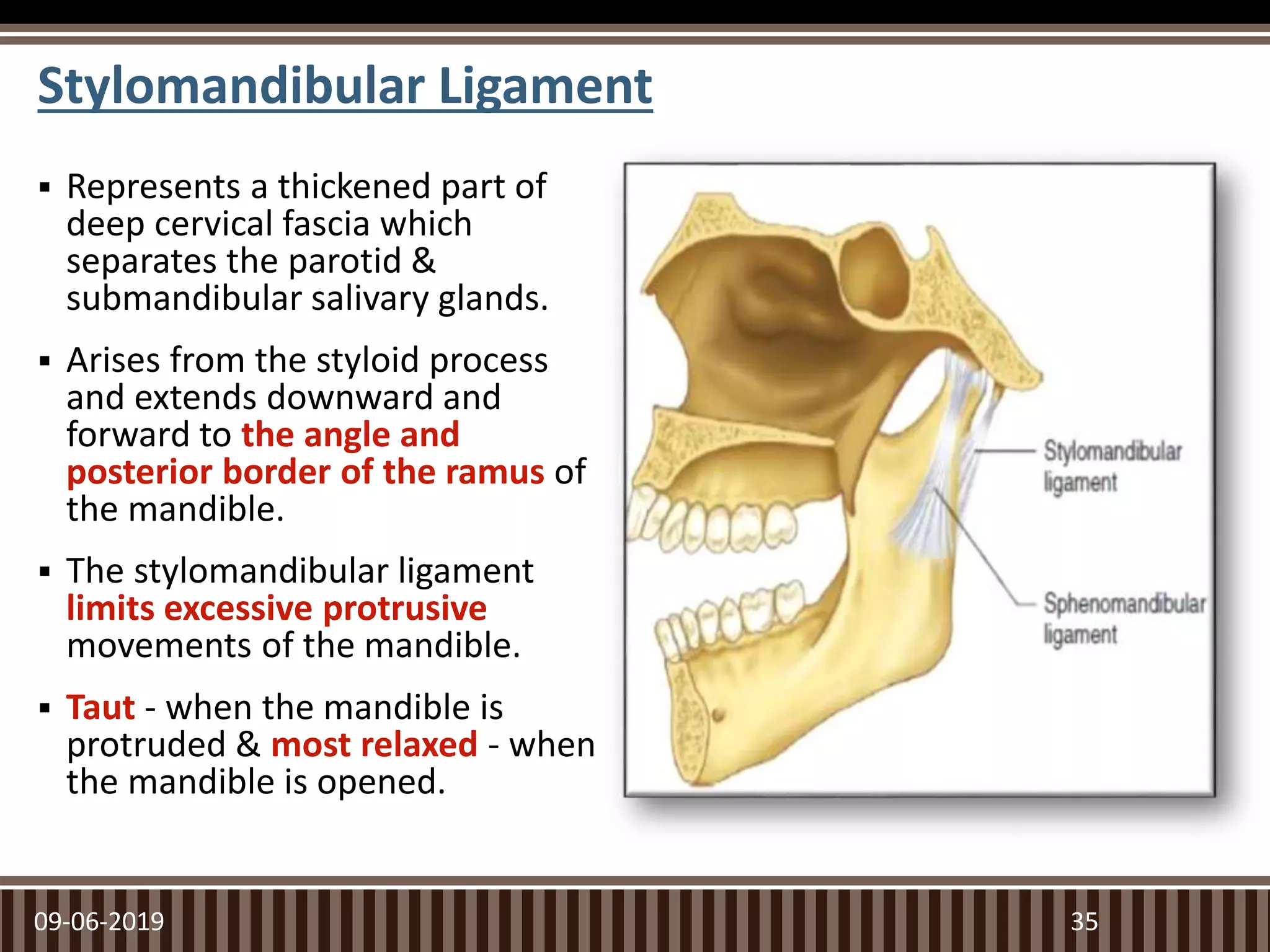
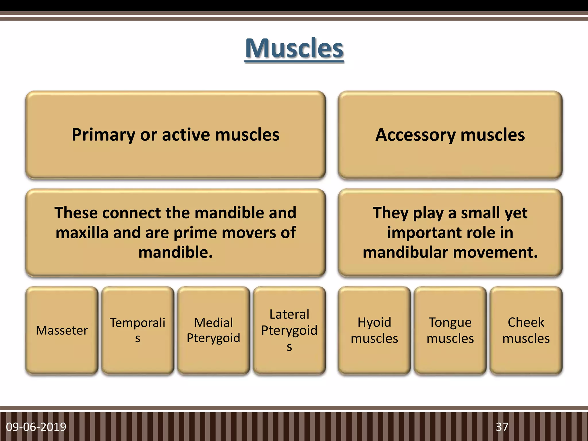
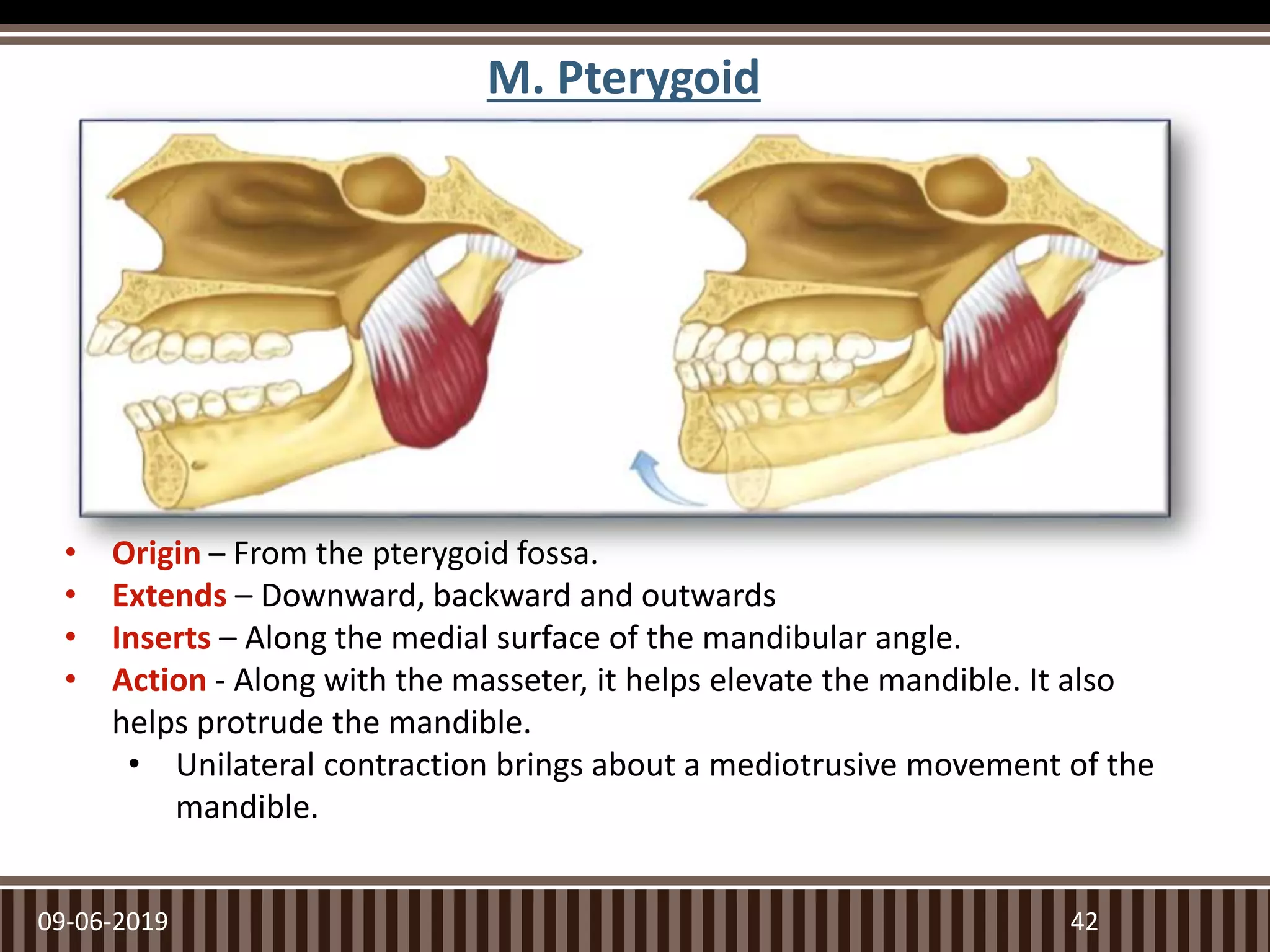
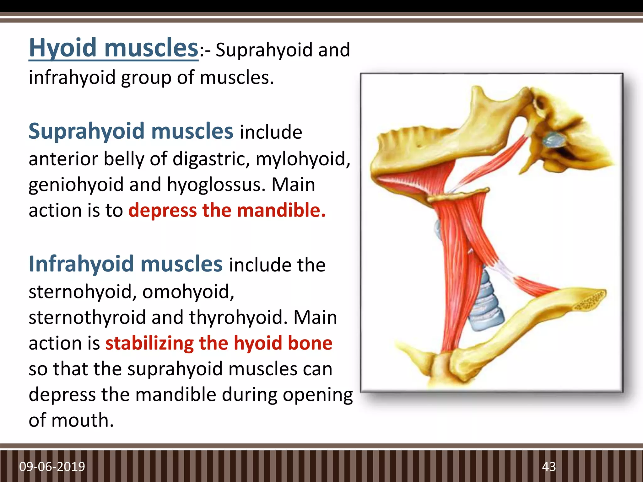
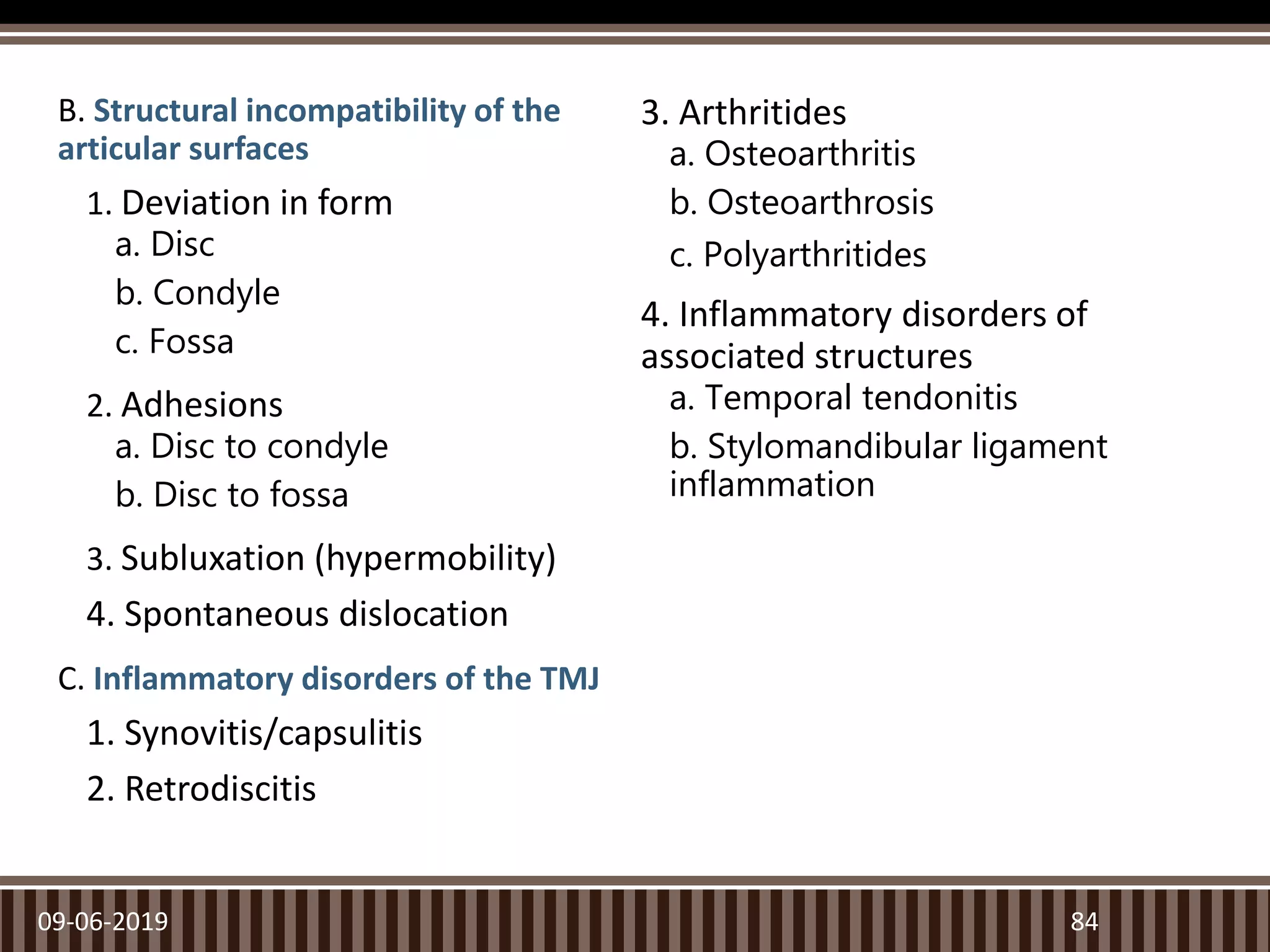
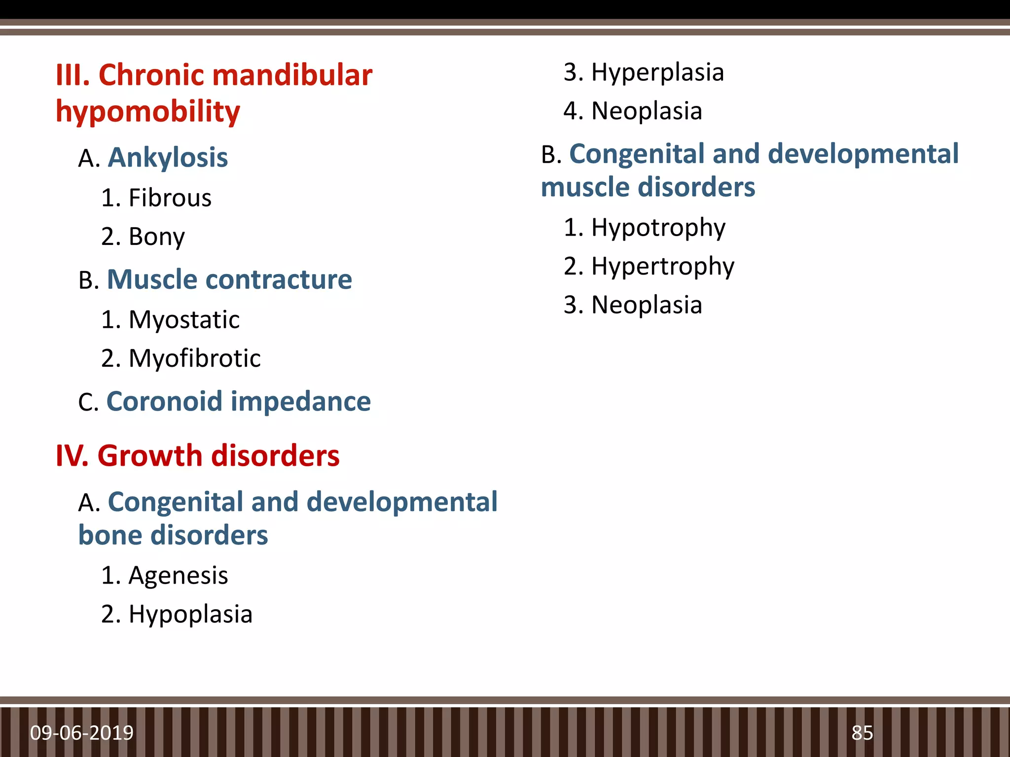
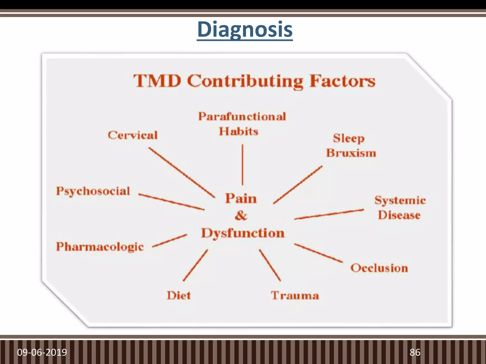
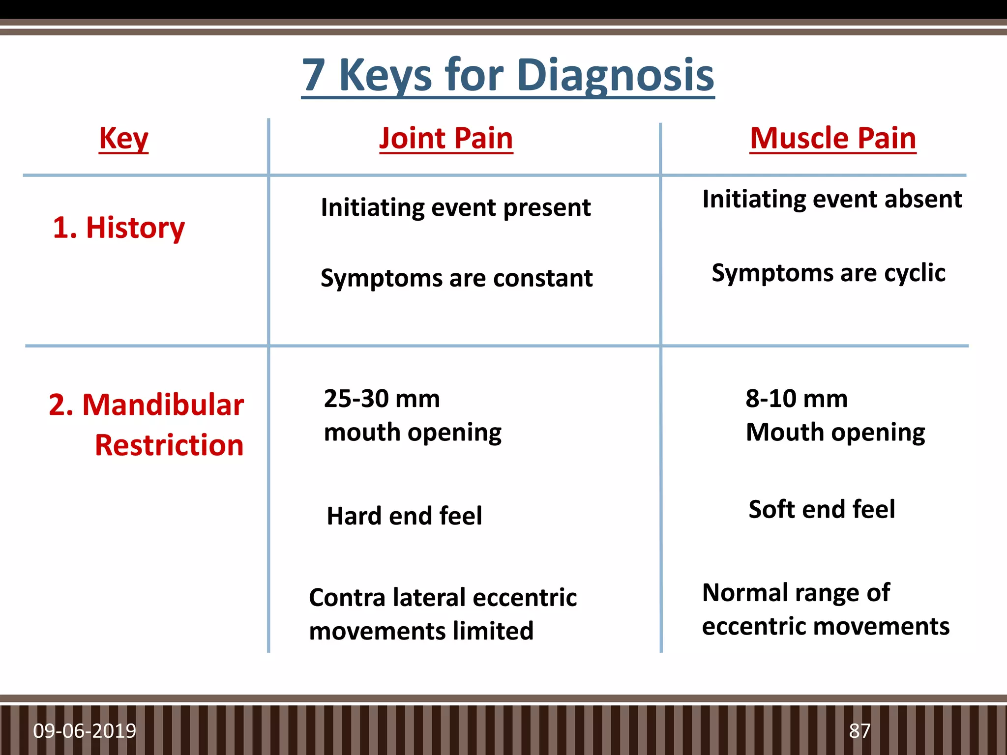
Seminar-
TMJ And Its Applied
Aspects In
Prosthodontics
Presented by
Dr Tanmay Popat
Ist Year PG
09-06-2019 1
CONTENTS:
● Introduction
● Evolution
● Growth & Development
● Age Changes
● Functional Anatomy
○ Bony Components
○ Soft Tissue Components
○ Muscles
● Innervations and Blood
Supply
● Biomechanics of TMJ
○ Functional Movements
○ Mechanism of Disc
Control
○ Border Movements
● Examination of TMJ
● Classification Of TMJ
Disorders
● Role of occlusion in TMJ
Disorders
● Mounting of Diagnostic
Casts
● Treatment of TMJ Disorders
● Conclusion
● References
09-06-2019 2
Introduction
 Temporomandibular joint is one of the most complex
joints in the body and a major component of
masticatory system which is responsible for chewing,
speaking and swallowing.
 TMJ co-ordinates with the other components of
Masticatory system in performing various masticatory
functions.
09-06-2019 3
• Synovial
• Ginglymoarthroidal
(Hinging+gliding)
• Bicondylar
• Compound joint (a non ossified
3rd bone - Articular disc)
• Craniomandibular articulation.
• Weight bearing joint – about
500N force.
4
Temporomandibular Joint
09-06-2019
Evolution
509-06-2019
 There are 3 stages that define the normal embryologic
development of the TMJ:
–Blastemic Stage
–Cavitation Stage
–Maturation Stage
09-06-2019 6
Growth and Development
 Acc to Baume, temporomandibular articulation
originate from two different blastema.
09-06-2019 7
Growth and Development
Condylar
blastema –
(primodium
of the
mandible)
condylar
cartilage
aponeurosis
of the external
pterygoid
muscle
the disc
the capsular
elements of
the lower
joint.
Temporal
Blastema
Articular
structures of
the upper level.
09-06-2019 8
Bender et al. Oral Maxillofacial Surg Clin N Am 30 (2018) 1–9
Growth and Development – Joint Innervations
 4th fetal month – nerve fibers may be observed in the
articular capsule
 5th month – appear to reach the disc.
 6th month – widest distribution over the condyle & within
the disc.
 Nerve fibers in capsule innervate the synovial membrane
of the joint as well.
 Acc. to Kitamura;
- branches of Auriculotemporal N., masseter N., & the
posterior deep temporal N. Branches of Mandibular
portion of Trigeminal N.
09-06-2019 9
 The growth of face and cranium involves two basic
types of growth changes:
1)Displacement
2)Remodelling
 Both these process, together constitute the growth
mechanism of craniofacial skeleton.
 As mandibular moves forward and downward, it grows
upward and backward at the same time by an equal
amount
09-06-2019 10
Growth and Development
09-06-2019 11
Growth and Development
09-06-2019 12
Growth and Development – Age Changes
A – At Birth
B – At 6 Years
C – In an Adult
1st Decade of Life:
 Due to decreased vascularization entire cartilage layer
becomes significantly thinner. This continues upto the
third year.
 By 2 ½ years the articular eminence increase from 2 to
4mm.
 This is due to resorption of the bone in the roof of the
mandibular fossa and bone deposition anterior and
posterior to the fossa leading to formation of ‘S’ shape
curve.
09-06-2019 13
2nd Decade of Life
 Characterized by progressive slowing of growth process.
 By 13-15 years decreased thickness of cartilage layer.
 Presence of proliferative layer at least till age of 18 years.
 A cortical bone cap coalescing with subchondral trabecular
bone by 10-12 years of age. This increases in thickness upto
3rd decade of life.
 Bone cap is completed by 20 years of age although cartilage
and sparse cartilage cells remain.
09-06-2019 14
Adult TMJ
 Deep to the articular layer in the region where
subchondral growth cartilage was located a chondroid type
bone may be found which directly overlies the bone cap.
 This marks the end of active growth of the condyle.
 Up through the 5th decade mandibular fossa became more
deep and articular eminence becomes more prominent.
 As age progresses further there is flattening of the
articular fossa and decrease in prominence of the articular
eminence
09-06-2019 15
09-06-2019 16
Functional Anatomy – Bony Components
09-06-2019 17
• Condylar Head
• Glenoid Fossa
• Articular
Eminence
CONDYLAR HEAD
09-06-2019 18
‘Rugby ball’ or ‘Date-
stone’ shape
•Composed of cancellous bone
covered by a thin layer of compact
bone
•During the period of growth a layer
of hyaline cartilage lies
underneath the fibrous covering of
the condyle.
•Hyaline cartilage of the condyle is
hence referred to as SECONDARY
CARTILAGE
• Articulating surface is more convex anteroposteriorly & slightly
convex mediolaterally.
• 15-20mm mediolaterally
• 8-10mm anteroposteriorly
• The main load-bearing areas are on the lateral aspect
09-06-2019 19
 The medial pole extends farther beyond the condylar
neck than the lateral pole does and is positioned more
posteriorly so that the long axis of the condyle deviates
posteriorly and meets a similar axis drawn from the
opposite condyle at the anterior border of the foramen
magnum.
09-06-2019 20
Glenoid Fossa
09-06-2019 21
• Concavity within temporal bone
that houses mandibular condyle.
• Covered by hard layer of bone.
• Single layer of cortical bone
separates glenoid fossa from
middle cranial fossa.
• It is covered with thin fibrous
layer.
• Anterior wall- Articular
eminence
• Posterior wall- SquamoTympanic
plate
 Anterior to the Glenoid fossa
there is a convex bony
prominence i.e. - “Articular
Eminence/Articular Tubercle”.
 Composed of spongy bone
covered by a thin layer of
compact bone
 The degree of convexity i.e.,
the steepness dictates the
pathway of condyle when
mandible is positioned
anteriorly.
09-06-2019 22
Articular Eminence
Functional Anatomy – Soft Tissue Components
09-06-2019 23
• Articular Disc
• Synovial Membrane
• Ligaments
• Muscles
 Firm,fibrous coonective tissue,
biconcave with a thick periphery
and thin central part.
 In sagittal plane it can be divided
into 3 parts
09-06-2019 24
Articular Disc
ARTICULAR DISC LOCATION
Ant. band Under the articular
tubercle
Central thin zone b/w condyle & post.
part of articular
tubercle
Post. band Over the condyle
Articular Disc
09-06-2019 25
ANTERIOR ATTACHMENT
(CAPSULAR LIGAMENT)
ATTACHES ARTICULAR DISC TO TYPE OF FIBERS
1. Superior Attchment Ant. Margin of Articular Surface of
Temporal Bone
Collagen fibers
2. Inferior Attachment Ant. Margin of Articulating Surface of
Condyle
Collagen fibers
POSTERIOR ATTACHMENT
(RETRODISCAL TISSUE/
BILAMINAR ZONE)
ATTACHES ARTICULAR DISC TO TYPE OF FIBERS
1. Superior Retrodiscal lamina Tympanic plate Elastic fibers
2. Inferior Retrodiscal lamina Post. surface of articulating surface of
condyle
Collagen fibers
The remaining portion of the retrodiscal tissue is
attached to a large venous plexus called the A-V shunt
or the Vascular Knee
09-06-2019 26
Synovial
Membrane and
Fluid
 A specialized layer of endothelial cells surrounding the internal
surfaces of joint cavity.
 This layer along with the synovial fringe at the anterior border of
retrodiscal tissues produces synovial fluid which fills both the
superior & inferior joint cavities.
 2 main Purposes –
–Medium for providing metabolic requirements. Exchange
exists between vessels of capsule, synovial fluid and articular
tissues.
–Serves as a lubricant during function
09-06-2019 27
09-06-2019 28
Boundary
Lubrication Weeping
Lubrication
Mechanism of Lubrication
The Ligaments
09-06-2019 29
Functional Ligaments
• The Capsular Ligament
• The Collateral (Discal)
Ligaments
• The temporomandibular
Ligament
Accessory Ligaments
• The Sphenomandibular
Ligament
• The Stylomandibular
Ligament
The Capsular Ligament or The Joint capsule
• Surrounds and encompasses the
entire TMJ.
• The Fibers
• Superiorly - temporal bone
along the borders of the
articular surfaces of the
mandibular fossa & articular
eminence.
• Inferiorly – neck of the condyle
• Resist any medial, lateral / inferior
forces that tend to separate /
dislocate the articular surfaces.
• Helps to retain synovial fluid &
provides proprioceptive feedback.
09-06-2019 30
The Collateral (Discal) Ligaments
09-06-2019 31
• Attaches the medial & lateral
borders of the articular disc to
the poles of the condyles.
• Divides the joint mediolaterally
into the superior & inferior
cavities.
• True ligaments - do not stretch &
restricts movement of the disc
away from condyle.
• Responsible for hinging
movement of TMJ.
• Strain on these ligaments
produces pain.
The Temporomandibular Ligament
09-06-2019 32
Outer Oblique Portion - resists
excessive dropping of the condyle,
limiting the extent of mouth opening.
Inner Horizontal Portion – limits
posterior movement of the condyle &
disc, hence protects the retrodiscal
tissues from trauma.
– Also protects the lateral pterygoid
muscle from over-lengthening or
extension
09-06-2019 33
 Remnant of Meckel’s cartilage
 Extends from the spine of the
sphenoid to the lingula of the
mandible
 May act as a pivot on the
mandible by maintaining the
same amount of tension
during both opening and
closing of the mouth.
09-06-2019 34
Sphenomandibular
Ligament
 Represents a thickened part of
deep cervical fascia which
separates the parotid &
submandibular salivary glands.
 Arises from the styloid process
and extends downward and
forward to the angle and
posterior border of the ramus of
the mandible.
 The stylomandibular ligament
limits excessive protrusive
movements of the mandible.
 Taut - when the mandible is
protruded & most relaxed - when
the mandible is opened.
09-06-2019 35
Stylomandibular Ligament
09-06-2019 36
Muscles
09-06-2019 37
Primary or active muscles
These connect the mandible and
maxilla and are prime movers of
mandible.
Masseter
Temporali
s
Medial
Pterygoid
Lateral
Pterygoid
s
Accessory muscles
They play a small yet
important role in
mandibular movement.
Hyoid
muscles
Tongue
muscles
Cheek
muscles
Masseter
09-06-2019 38
• Origin – Zygomatic Arch
• Superficial portion (SP) -- Runs downward and slightly backward to get
inserted in the posterior part of the lateral surface of the mandible.
• Deep portion (DP) -- Runs in a predominantly vertical direction to get inserted
in upper part of the ramus and some area on the coronoid process
• Action – Elevates the mandible and brings teeth into contact
SP -- Aids in protruding the mandible.
DP -- Pulls the mandible into its retruded position in relation to the
maxilla.
09-06-2019
Temporalis
39
• Origin – Temporal Fossa
• Insertion -- on the coronoid process and the anterior border of the ascending
ramus.
• Anterior Portion (AP) – Fibers directed almost vertically.
• Middle Portion (MP) – Fibers run obliquely forward.
• Posterior portion (PP) – Fibers directed almost horizontally
• Action – AP contracts the mandible is raised vertically.
• MP contracts it will elevate and retrude the mandible.
• PP fibres are relatively weak and they pull the condyle back into CR.
09-06-2019
L. Pterygoid
40
• SLP – originates at the infratemporal surface of the greater sphenoid wing, - Runs
horizontally, backward, and outward – Inserts on the articular capsule, the disc, and
the neck of the condyle
• ILP – originates at the outer surface of the lateral pterygoid plate – Runs backward,
upward, and outward – Inserts primarily on the neck of the condyle
• Action – SLP It becomes active only in conjugation with elevator muscles especially
when the teeth are held together and power stroke (i.e. closure against resistance).
• ILP - When both right and left ILP contract simultaneously, the condyles are pulled
down the articular eminence. Unilateral contraction – mediotrusive movement of
that condyle and lateral movement of the mandible to the opposite side. Along with
the mandibular depressors,it helps depress the mandible.
09-06-2019 41
09-06-2019
M. Pterygoid
42
• Origin – From the pterygoid fossa.
• Extends – Downward, backward and outwards
• Inserts – Along the medial surface of the mandibular angle.
• Action - Along with the masseter, it helps elevate the mandible. It also
helps protrude the mandible.
• Unilateral contraction brings about a mediotrusive movement of the
mandible.
09-06-2019 43
Hyoid muscles:- Suprahyoid and
infrahyoid group of muscles.
Suprahyoid muscles include
anterior belly of digastric, mylohyoid,
geniohyoid and hyoglossus. Main
action is to depress the mandible.
Infrahyoid muscles include the
sternohyoid, omohyoid,
sternothyroid and thyrohyoid. Main
action is stabilizing the hyoid bone
so that the suprahyoid muscles can
depress the mandible during opening
of mouth.
09-06-2019 44
09-06-2019 45
TEMPORALIS MASSETER
MEDIAL PTERYGOID
PROTRACTION
LATERAL PTERYGOID
DIGASTRIC
GENIOHYOID
RETRACTION
MYLOHYOID
DEPRESSION
ELEVATION
Innervations And Blood Supply
Innervations
 The trigeminal nerve – provides both motor &
sensory innervation to the muscles that control it.
 Afferent innervation – branches of the mandibular
nerve.
 Also by auriculotemporal nerve as it leaves the
mandibular nerve behind the joint & ascends
laterally & superior to wrap around the posterior
region of the joint.
 Additional nerves – temporal & masseteric .
09-06-2019 46
Innervations And Blood Supply
Blood Supply
The blood supply to TMJ is majorly superficial, there is no
blood supply inside the capsule.
 Posteriorly: Superficial temporal artery
 Anteriorly: middle meningeal artery
 Inferiorly: internal maxillary artery
Other arteries are: deep auricular, anterior tympanic and
ascending pharyngeal artery.
Condyle receives its supply through the marrow spaces by
inferior alveolar artery and feeder vesels that enter
directly into condylar head from larger vessels.
09-06-2019 47
Biomechanics of TMJ
09-06-2019 48
• Functional Movements
of TMJ
• Disc Control
• Mandibular Movements
The TMJ is a compound joint – can be divided into two
distinct systems:-
 One joint system - the tissues that surround the inferior
synovial cavity(the condyle and the articular disc). The disc
and its attachment to the condyle is called the condyle-disc
complex, this joint system is responsible for rotational
movement in the TMJ.
 Second system – the condyle – disc complex functioning
against the surface of the mandibular fossa. Translation
occurs between the superior surface of the articular disc
and the mandibular fossa. Thus the articular disc functions
as a nonossified bone contributing to both joint systems.
Hence justifying TMJ as a true compound joint.
09-06-2019 49
09-06-2019 50
Mechanism of Disc Control
09-06-2019 51
Mandibular Movements
 Acc. to Okeson – 2 types of Movements
–Rotational/hinge
 Occurs mainly between the disc and condyle in the
lower joint compartment.
–Translational/sliding
 Occurs mainly between the articular eminence and
disc (and mandible) in the upper compartment.
 Based on Plane of border movements:
–horizontal, vertical (frontal) & sagittal plane
movements
09-06-2019 52
09-06-2019 53
Rotation around
horizontal axis
Rotation around
sagittal axis
Rotation around Vertical axis Translation
09-06-2019 54
 By combining the mandibular border movements in the three
planes a three dimensional envelope of motion can be produced
that represents the maximum range of movement of the
mandible. Although it has a characteristic shape it varies from
person to person.
09-06-2019 55
Eccentric Mandibular Movements
 Any movement of the mandible from centric occlusal
position that results in tooth contact is called as
eccentric movement.
 Types of eccentric movements:
– Protrusive movements.
– Retrusive movements.
– Laterotrusive movements
09-06-2019 56
Protrusive Movements
 Consists mainly of condylar translations.
 Sagittal Protrusive condylar path: 5-55o and mean is 30o
 Sagittal protrusive incisal path: range 50-70o
09-06-2019 57
Lateral Movements
 Sagittal lateral condylar path:
–When lateral movement is
executed, the working condyle
rotates and moves outward
while other, non-working
condyle translates forward,
medially downward orbiting
around the rotating working
condyle.
–This orbiting condylar path of
working condyle is called as
BENNETT MOVEMENT
09-06-2019 58
Examination of TMJ
 Clinical examination of TMJ:
–Inspection of TMJ
–Auscultation of TMJ
–Palpation of TMJ & Musculature associated with mandibular
movements
–Functional analysis of mandibular movements
 Radiographic examination of TMJ
–Transcranial & Transpharyngeal projections
–Panaromic projection -Transorbital projection
–Submentovertex projection -Computed tomography(CT)
–Arthrography -Magnetic resonance imaging(MRI)
09-06-2019 59
Inspection Of TMJ
 Area surrounding TMJ is inspected for any signs of
inflammation.
 Any gross asymmetry.
 Any Swelling/Growth
 Depression
 Discharge
 Colour change of skin over TMJ
 Surface of the overlying skin in the preauricular area.
 Mouth opening – Normal/Resricted
 Deviation/Deflection
09-06-2019 60
Ausculatation
 Sounds made by the TMJ can be examined with a
stethoscope. Also the timing of clicking during opening
and closure can be noted .
09-06-2019 61
Crepitations
 A grating or scalping noise that occurs on jaw
movements . Sound like when sand paper is rubbed
against a surface.
 Crepitation is very uncommon in asymptomatic joint
and may be an early sign of degenerative joint
disease.
 Crepitus is caused by roughened, irregular anterior
surface.
09-06-2019 62
Clicking
CLICK- single sound of short
duration
POP- loud click
Occurs due to the uncoordinated
movement of condylar head and
T.M.J disc.
 Joint clicking is differentiated as:
Initial
Intermediate
Terminal
Reciprocal
09-06-2019 63
 Initial clicking : It is a sign of retruded condyle
 Intermediate clicking : Is a sign of unevenness of the
condylar surfaces and articular disc
 Terminal clicking : is an effect of the condyle being moved
too far anteriorly in relation to the disc on maximum jaw
opening.
 Reciprocal clicking : is an expression of incordination
between displacement of the condyle & the disc.
09-06-2019 64
Palpation of TMJ
• Lateral
• Posterior
• Also the synchrony of the
left & right condyles during
movement can be checked.
09-06-2019 65
Palpation of TMJ &
Muscles
Muscles
 Digital palpation
–Temporalis
–Masseter
–Sternocleidomastoid
 Functional palpation
–Lateral pterygoid
–Medial pterygoid
09-06-2019 66
GRADING OF PAIN
0- no pain
1- uncomfortable
2- definite pain
3- evasive action
Temporalis
09-06-2019 67
Masseter
09-06-2019 68
Sternocleidomastoid
Splenius Capitus
09-06-2019 69
Trapezius
09-06-2019 70
Inferior Lateral Pterygoid:
Protruding mandible against
resistance
• STRECHES ONLY ON MAX
INTERCUSPATION
Superior Lateral Pterygoid:
Clenching against
powerful stroke
• STRECHES ONLY ON MAX
INTERCUSPATION
INTRACAPSULAR DISORDER OF TMJ :
 Can elicit pain with increase in interarticular pressure
and movement.
 Confusing with muscle pain origin.
09-06-2019 71
• But if clenching done on a separator – pressure doesn’t
increase.
To differentiate with inferior lateral Pterygoid muscle –
patientt is asked to protrude and bite on a separator.
09-06-2019 72
Functional Analysis of TMJ
 Maximum Jaw Opening
 The distance between the incisal edges of the upper and lower central
incisors.
–Normal maximum ≥ 40mm.
–Men: 38.7- 67.2 (mean 52.8mm)
–Women: 36.7 -60.4 ( mean 48.3)
–Child: 6 year mean- 44.8mm
10 year mean- 44 mm
–Lateral movement ≥ 8mm
–Protrusive movements ≥ 7mm
 In overbite cases, this amount is added to the obtained value whereas
in open bite it is subtracted.
09-06-2019 73
09-06-2019 74
 DEVIATION :
Because of disc derrangement
in 1 or both joints
shift of jaw midline during
openeing that disappear with
opening
 DEFLECTION :
Restricted movement in 1 joint
Shift of midline to 1side and
become greator with opening
and doesn’t dissapear
Radiographic Evaluation of TMJ
 Transcranial:
–Sagittal view of the lateral
aspect of condyle and
temporal component
–Displaced condylar #
–Osseous changes on lateral
aspect
 Transpharyngeal/Infra-
cranial:
–Sagittalview of the medial
pole of condyle.
–Erosive changes of the
condyle
09-06-2019 75
 Panaromic:
–Commonly used routinely
(not for TMJ), odontogenic
diseases, asymmetries,
extensive erosions or #
 Transorbital:
–Ant. view of TMJ, entire
mediolateral dimension of
A.E, condylar head & neck.
 Submentovertex(Basal):
–Facial asymmetries,
condylar displacement,
rotation of the mandible in
horizontal plane assoc. with
trauma
09-06-2019 76
 Computed Tomography:
–Complete view of Osseous
components of TMJ joint.
–Presence/extent of ankylosis &
neoplasms.
 Arthrography:
–Invasive(as radio-opaque
contrasting agent is injected)
–Small disk perforations & joint
adhesions
09-06-2019 77
 MRI (Magnetic Resonance Imaging):
–Both the soft(discal) and hard(osseous) tissues can be
imaged.
–Non invasive, no radiation hazard
–Inflammation, joint effusions etc.
09-06-2019 78
Other Diagnostic Aids
 Mandibular tracking devices
 Sonography
 Vibration analysis
 Thermography
 Electromyography
 Mounted casts
09-06-2019 79
Six ways to verify that TMJ is healthy
 Is it difficult or painful to open the
mouth (e.g., yawning)?
 Does the jaw get stuck, locked, or go
out?
 Is it difficult or painful to chew, talk, or
use the jaws?
 Do the jaw joints make noises?
 Do the jaws often feel stiff, tight, or
tired? Is there pain in or about the
ears, temples, or cheeks?
 Are headaches, neck aches, or
toothaches frequent?
 Has there been a recent injury to the
head, neck, or jaw?
 Have there been any recent changes in
bite?
 Has there been previous treatment for
any unexplained facial pain or a jaw
joint problem?
09-06-2019 80
1. SCREENING HISTORY
2. LOAD TEST
3. RANGE & PATH OF
MOVEMENT
4. DOPPLER ANALYSIS
09-06-2019 81
09-06-2019 82
5. Radiographic
Imaging
6. Anterior
Deprogramming
Device
Classification of diseases of TMJ/Diagnostic Classification
(Acc. To Okeson)
I. Masticatory muscle disorders
– Protective co-contraction
– Local muscle soreness
– Myofascial pain
– Myospasm
– Centrally mediated myalgia
II. Temporomandibular joint (TMJ) disorders
A. Derangement of the condyle-disc complex
–1. Disc displacements
–2. Disc dislocation with reduction
–3. Disc dislocation without reduction
09-06-2019 83
B. Structural incompatibility of the
articular surfaces
1. Deviation in form
a. Disc
b. Condyle
c. Fossa
2. Adhesions
a. Disc to condyle
b. Disc to fossa
3. Subluxation (hypermobility)
4. Spontaneous dislocation
C. Inflammatory disorders of the TMJ
1. Synovitis/capsulitis
2. Retrodiscitis
3. Arthritides
a. Osteoarthritis
b. Osteoarthrosis
c. Polyarthritides
4. Inflammatory disorders of
associated structures
a. Temporal tendonitis
b. Stylomandibular ligament
inflammation
09-06-2019 84
III. Chronic mandibular
hypomobility
A. Ankylosis
1. Fibrous
2. Bony
B. Muscle contracture
1. Myostatic
2. Myofibrotic
C. Coronoid impedance
IV. Growth disorders
A. Congenital and developmental
bone disorders
1. Agenesis
2. Hypoplasia
3. Hyperplasia
4. Neoplasia
B. Congenital and developmental
muscle disorders
1. Hypotrophy
2. Hypertrophy
3. Neoplasia
09-06-2019 85
Diagnosis
09-06-2019 86
09-06-2019 87
Joint Pain Muscle Pain
Initiating event present Initiating event absent
Symptoms are constant Symptoms are cyclic
25-30 mm
mouth opening
Hard end feel
8-10 mm
Mouth opening
Soft end feel
Contra lateral eccentric
movements limited
Normal range of
eccentric movements
Key
2. Mandibular
Restriction
1. History
7 Keys for Diagnosis
3. Mandibular Interference
09-06-2019 88
Deviation. The opening pathway is
altered but returns to a normal midline
relationship at maximal opening.
Deflection of the opening path is commonly
associated with a disc dislocation without
reduction or a unilateral muscle restriction.
4. Acute Malocclusion
09-06-2019 89
5. Loading The Joint
6. Functional
Manipulation
7. Diagnostic
Anaesthetic Blockade
Key points for Diagnosis
 Single/double click at different levels of mouth opening – disc
displacement
 Single/double click at different levels of mouth opening with
mandibular deflection - disc dislocation with reduction
 No click; mandibular deviation; closed lock (opening – 25 to 30
mm) - disc dislocation without reduction.
 A single click after a prolonged static loading – adherence
 Click at the same level of mouth opening & closing - adhesion
09-06-2019 90
Key points for Diagnosis
• Sudden thrust near to maximum opening with pre – auricular
depression – subluxation
• Inability to close the mouth (open lock) – spontaneous dislocation
• Pain in inflammatory disorders are dull & continuous accentuated
by function.
• Synovitis, capsulitis & retrodiscitis have same clinical presentation
& additional diagnostic aids are required to differentiate between
them .
09-06-2019 91
 Frequent postural alterations in
patients with different types of
temporomandibular disorders. Irene A
et. al.
Objective: To describe postural
alterations according to the type of
temporomandibular disorder (TMD).
Conclusion: TMD patients present
postural changes, mainly forward head
posture, pelvic tilt and high shoulder,
with special involvement related to
muscle and combined diagnosis.
09-06-2019 92
Rev. Salud Pública. 20 (3): 384-389, 2018
Role of Occlusion in TMDs
 Normal function + event > physiological tolerance TMD
symptoms.
 Events can be:
–Local: improperly occluding crown, Post-injection trauma after
L.A, trauma from wide mouth opening, etc
–Systemic: emotional stress
 Physiologic tolerance: influenced by
–Local factors: occlusal instability associated with dev., genetic
or iatrogenic causes
–Systemic factors: acute or chronic diseases, gender diet
effectiveness of pain modulation systems etc.
09-06-2019 93
09-06-2019 94
Occlusal interferences
Occlusal disharmony
Functional malocclusion
Stresses within
Stomatognathic sys.
Ability of the tissues to
resist
Func. Disturbance &/or
F.disorder
Occlusal Dysfunction
TMDs
overcome
Determinants of Occlusion
 The anatomy of the TMJ has effect on the mandibular
movements and tooth morphology.
 The paths of the condyles within the glenoid fossae, and
the locations of the rotational centers determine the
occlusal morphology of teeth.
 These have an effect on the allowable cusp height, fossa
depth along with the acceptable ridge and groove
directions.
09-06-2019 95
09-06-2019 96
Mounting Of Diagnostic Casts
• When there is Acute inflammation,
muscle spasm, pain & heavy infusion of
fluids in joint tissues etc, programming
the condyles into CR is not possible.
• Treatment of the acute symptoms of
the TMJ dysfunction should be carried
out.
• If the mouth opening permits
diagnostic impressions, effort should be
made to make bite record in CR for
constructing the interocclusal palliative
acrylic splint.
• Once the TMD symptoms are resolved,
diagnostic cast mounting should be
done.
9709-06-2019
 Pantographic recording: is a
record of the degrees of 3-D
pathways of the border
movements of condyles in their
respective fossa during
function.
 INDICATIONS:
–Procedures like full mouth
reconstruction & occlusal
correction by selective
grinding which requires
precision.
–When a patient is suspected
of a large amount of Bennett
Immediate side shift during
Lateral excursions
9809-06-2019
Management of TMJ Disorders
 Definitive Therapy: directed towards controlling or eliminating
the cause of the disorder.
 Occlusal Factors
 Emotional Stress
 Trauma
 Deep Pain Input
 ParaFunctional Activities
 Supportive Therapy: alters the patient’s symptoms & no effect
on the cause of the disorder
 Pharmacological Therapy
 Physical Therapy
–Modalities
–Manuel Techniques
09-06-2019 99
 Definitive therapy considerations for Occlusal
Factors:
– any treatment that is directed toward altering the
mandibular position and/or occlusal contact pattern
of the teeth.
–Reversible Occlusal Therapy:
–Occlusal appliances
–Irreversible Occlusal Therapy:
–Selective grinding of the teeth.
–Restorative Procedures
–Orthodontic Treatment
–Surgical Procedures
09-06-2019 100
Definitive Therapy
Occlusal Splint/ Occlusal Device
Any removable artificial occlusal surface used for diagnosis
or therapy affecting the relationship of the mandible to the
maxillae. It may be used for occlusal stabilization, for
treatment of TMJ disorders, or to prevent wear of the
dentition
09-06-2019 101
 According to Okeson
–Muscle relaxation appliance/ stabilization appliance used to
reduce muscle activity
–Anterior repositioning appliances/ orthopedic repositioning
appliance
–Other types:
 Anterior bite plane
 Pivoting appliance
 Soft/ resilient appliance
 According to Dawson:
 Permissive splints/ muscle deprogrammer
 Directive splints/ non-permissive splints
09-06-2019 102
Permissive Splints
 ANTERIOR DEPROGRAMMING DEVICE:
–Simplest type of permissive splint.
–Used in diff. diagnosis of TMDs(Intracapsular Vs
Extracapsular, i.e., occlusal interferences)
–When correctly fabricated & used in non
intracapsular TMDs, patient gets comfortable
within min or hrours.
–Used for extended periods, may cause intrusion
of covered teeth and supraeruption of separated
teeth.
 UPPER or LOWER splint :- Decision made on
which splint is easy to wear and most
unobstructive to the mandibular movements
and esthetically pleasing.
09-06-2019 103
 Principles of Splint Design
–Allow uniform, equal-intensity
contacts of all teeth against a
smooth splint surface.
–Anterior guidance ramp angled
as shallow as possible for
horizontal freedom.
–Provide immediate disclusion
of all posterior teeth in all
excursive movements.
–Splint should fit the arch
comfortably and have good
stable retention.
 Worn until:
–All related pain is gone
–Joint structure is stable
–Bite structure is stable
 Stability is verified by:
–Elimination of painful
symptoms
–Verification of CR by load
testing
 Avg time : 2-4 weeks, worn
24hrs/day except : eating &
brushing
09-06-2019 104
Modified Anterior Deprogramming Devices
 Anterior midline point stop (AMPS)
 Nociceptive Trigeminal Inhibitor Tension Suppression
System (NTI-TSS)
09-06-2019 105
Directive Splints
 ANT. REPOSITIONING APPLIANCE:
–Goal of treatment is not to alter the mandibular position
permanently but only to change the position temporarily so as
to enhance adaptation of the retrodiscal tissues.
 Indications:
–To treat disc derangement disorders.
–Patients with joint sounds (e.g., a single or reciprocal click) can
sometimes be helped by this.
–Intermittent or chronic locking of the joint (e.g., retrodiscitis).
09-06-2019 106
Irreversible Occlusal THERAPY
Selective grinding/Occlusal equilibration
 It is a procedure by which the occlusal surfaces of the teeth are precisely
altered to improve the overall contact pattern.
 Indications:
–When occlusal appliance therapy eliminated TMD symptoms and the
cause revealed any occlusal contact interferences.
–When there is a definitive need of improved occlusal condition.
–Indicated in treatment of trauma from occlusion, for decreasing
mobility and fremitus.
 Contraindications
–Severe over closure.
–Sensitive, worn and adolescent teeth.
09-06-2019 107
GOALS OF SELECTIVE
GRINDING/OCCLUSAL ADJUSTMENT:
 The primary goal of any occlusal
adjustment is Occlusal stability, which
is achieved by:
–Elimination of Prematurity
–Elimination of Interferences
–Establishment of Centric Forces
Directed Axially
–Establishment of Optimal Occlusion
with out loss of VD and shapes of
cusps and fossae.
09-06-2019 108
 Restorative procedures:
–It mainly includes replacement of missing teeth and restoration of
caries.
 Orthodontic treatment:
–Correcting the malalignment of teeth, correcting the skeletal
relationships of the maxilla and mandible, i.e., Orthognathic
surgeries
 Surgical Procedures:
–Estimated-5% or less in all TMD Patients.
–Ankylosis, destroyed/perforated articular disc, bizarre remodelling of
osseous structures & patients requiring surgery prior to orthodontic
and/or restorative treatment.
–Includes: Arthrocentesis, Arthroplasty, Autotenous disc replacement,
Alloplastic hemi arthroplasty, Orthognathic surgeries, Discoplasty
etc.
09-06-2019 109
 Definitive therapy considerations for Emotional
Stress:
09-06-2019 110
Muscle
Hypertrophy
High levels of
anxiety
Apprehension
Frustration
Anger
Fear
– Types of Emotional
Stress therapy:
• Patient awareness
• Restrictive use
• Voluntary avoidance
• Relaxation therapy
• Others
– Self-hypnosis
– Meditation
– Yoga
 Definitive therapy considerations for Trauma: Directed
towards elimination of para functional habit.
Two types of Trauma:
–Macrotrauma: Supportive therapy
–Microtrauma: E.g. Bruxisim - Orthopedic stability
 Definitive therapy considerations for Deep Pain Input:
–True source of pain must be located before definitive
treatment begins.
–If the source is not obvious, referral to another medical
specialist should be done.
09-06-2019 111
 Definitive therapy considerations for Parafunctional
Activity:
–Diurnal activity: Patient education & Cognitive
Awareness Strategies
–Nocturnal activity: Occlusal appliance therapy
–Measures to decrease the level of emotional stress
should be made.
09-06-2019 112
Supportive Therapy
09-06-2019 113
Physical
Therapy
modalities
Thermot
herapy
Coolant
therapy
Ultrasou
nd
therapy
Phonoph
oresis
Iontopho
rosis
EGS
TENS
Cold
laser
09-06-2019 114
Pharmacolo
gic Therapy
Analgesics
Anti-
convulsant
s
Anti-
inflammat
ory agents
Topical
Anxiolytic
agents
Muscle
relaxants
Anti-
depressan
ts
Injectables
Conclusion
 A thorough knowledge of TMJ & its relationship with
surrounding structures is essential to fully comprehend normal
anatomy & physiology, adaptive processes, dysfunction &
pathology of the TMJ.
 Although numerous treatments have been advocated, none are
universally effective for all patients all the time.
 A good Prosthodontic treatment bears direct relation with TM
articulation since establishment of occlusion is one of the main
steps in complete denture, fixed partial & removal partial
dentures.
 Effective treatment begins with the thorough understanding of
the disorder & its etiology and an appreciation of the various
types of treatments is essential for effective management of the
symptoms.
09-06-2019 115
References
 Okeson, JP. Management of Temporomandibular Disorders and
Occlusion , 7th ed., (2003), Mosby.
 Zarb GA, Bolender CE. Prosthetic treatment for edentulous patients,
Complete dentures & Implant supported prosthesis. 13th ed.
 Dawson PE. Functional occlusion from TMJ to smile design.3rd ed.
 Charles McNeill. Science and practice of occlusion.
 Temporomandibular disorders-diagnosis treatment and management:
Weldon E. Bell (3rd ed)
 Neil S. Norton. Neter’s Head And Neck Anatomy For Dentistry. 2nd Ed.
Philadelphia. Elsevier Inc 2012.
 Irene A. Espinosa de Santillana, Ariana García-Juárez, Jaime Rebollo-
Vázquez y Ana K. Ustarán-Aquino. Frequent postural alterations in
patients with different types of temporomandibular disorders. Rev.
Salud Pública. 20 (3): 384-389, 2018.
09-06-2019 116
References
 Melissa E. Bender, Rosa B. Lipin, Steven L. Goudy. Development of the
Pediatric Temporomandibular Joint. Oral Maxillofacial Surg Clin N Am
30 (2018) 1–9.
 Mohl ND, Ohrbach RK, Crow HC, Gross AJ. Devices for the diagnosis &
treatment of temporomandibular disorders. Part III:
Thermography,ultrasound, and electric stimulation. J Prosthet Dent
1990; 63(1):472-5.
 Mohl ND, McCall WD, Lund JP, Plesh O. Devices for the diagnosis &
treatment of temporomandibular disorders. Part I: Introduction,
Scientific evidence, and jaw tracking. J Prosthet Dent 1990; 63(1):198-
201.
 Mohl ND, Lund JP, Widmer CG, McCall WD. Devices for the diagnosis &
treatment of temporomandibular disorders. Part II: Electromyography
and sonography. J Prosthet Dent 1990; 63(1):332-5.
09-06-2019 117
References
 David L. Stocum & W. Eugene Roberts. Part I: Development and
Physiology of the Temporomandibular Joint. # Springer
Science+Business Media, LLC, part of Springer Nature 2018.
 Dania Tamimi, Elnaz Jalali, David Hatcher. Temporomandibular Joint
Imaging. Radiol Clin N Am – 2017.
 Weiner S. Biomechanics of occlusion and the articulator. DCNA
1995:39(2):257-284.
 Okeson JP. Occlusion and functional disorders of the masticatory
system. DCNA 1995:39(2):285-301.
 Nowlin TP, Nowlin JH. Examination and occlusal analysis of the
masticatory system. DCNA 1995:39(2):379-402.
 World Wide Web.
09-06-2019 118
09-06-2019 119

TMJ and Its Applied Aspects in Prosthodontics

  • 1.
    Seminar- TMJ And ItsApplied Aspects In Prosthodontics Presented by Dr Tanmay Popat Ist Year PG 09-06-2019 1
  • 2.
    CONTENTS: ● Introduction ● Evolution ●Growth & Development ● Age Changes ● Functional Anatomy ○ Bony Components ○ Soft Tissue Components ○ Muscles ● Innervations and Blood Supply ● Biomechanics of TMJ ○ Functional Movements ○ Mechanism of Disc Control ○ Border Movements ● Examination of TMJ ● Classification Of TMJ Disorders ● Role of occlusion in TMJ Disorders ● Mounting of Diagnostic Casts ● Treatment of TMJ Disorders ● Conclusion ● References 09-06-2019 2
  • 3.
    Introduction  Temporomandibular jointis one of the most complex joints in the body and a major component of masticatory system which is responsible for chewing, speaking and swallowing.  TMJ co-ordinates with the other components of Masticatory system in performing various masticatory functions. 09-06-2019 3
  • 4.
    • Synovial • Ginglymoarthroidal (Hinging+gliding) •Bicondylar • Compound joint (a non ossified 3rd bone - Articular disc) • Craniomandibular articulation. • Weight bearing joint – about 500N force. 4 Temporomandibular Joint 09-06-2019
  • 5.
  • 6.
     There are3 stages that define the normal embryologic development of the TMJ: –Blastemic Stage –Cavitation Stage –Maturation Stage 09-06-2019 6 Growth and Development
  • 7.
     Acc toBaume, temporomandibular articulation originate from two different blastema. 09-06-2019 7 Growth and Development Condylar blastema – (primodium of the mandible) condylar cartilage aponeurosis of the external pterygoid muscle the disc the capsular elements of the lower joint. Temporal Blastema Articular structures of the upper level.
  • 8.
    09-06-2019 8 Bender etal. Oral Maxillofacial Surg Clin N Am 30 (2018) 1–9
  • 9.
    Growth and Development– Joint Innervations  4th fetal month – nerve fibers may be observed in the articular capsule  5th month – appear to reach the disc.  6th month – widest distribution over the condyle & within the disc.  Nerve fibers in capsule innervate the synovial membrane of the joint as well.  Acc. to Kitamura; - branches of Auriculotemporal N., masseter N., & the posterior deep temporal N. Branches of Mandibular portion of Trigeminal N. 09-06-2019 9
  • 10.
     The growthof face and cranium involves two basic types of growth changes: 1)Displacement 2)Remodelling  Both these process, together constitute the growth mechanism of craniofacial skeleton.  As mandibular moves forward and downward, it grows upward and backward at the same time by an equal amount 09-06-2019 10 Growth and Development
  • 11.
  • 12.
    09-06-2019 12 Growth andDevelopment – Age Changes A – At Birth B – At 6 Years C – In an Adult
  • 13.
    1st Decade ofLife:  Due to decreased vascularization entire cartilage layer becomes significantly thinner. This continues upto the third year.  By 2 ½ years the articular eminence increase from 2 to 4mm.  This is due to resorption of the bone in the roof of the mandibular fossa and bone deposition anterior and posterior to the fossa leading to formation of ‘S’ shape curve. 09-06-2019 13
  • 14.
    2nd Decade ofLife  Characterized by progressive slowing of growth process.  By 13-15 years decreased thickness of cartilage layer.  Presence of proliferative layer at least till age of 18 years.  A cortical bone cap coalescing with subchondral trabecular bone by 10-12 years of age. This increases in thickness upto 3rd decade of life.  Bone cap is completed by 20 years of age although cartilage and sparse cartilage cells remain. 09-06-2019 14
  • 15.
    Adult TMJ  Deepto the articular layer in the region where subchondral growth cartilage was located a chondroid type bone may be found which directly overlies the bone cap.  This marks the end of active growth of the condyle.  Up through the 5th decade mandibular fossa became more deep and articular eminence becomes more prominent.  As age progresses further there is flattening of the articular fossa and decrease in prominence of the articular eminence 09-06-2019 15
  • 16.
  • 17.
    Functional Anatomy –Bony Components 09-06-2019 17 • Condylar Head • Glenoid Fossa • Articular Eminence
  • 18.
    CONDYLAR HEAD 09-06-2019 18 ‘Rugbyball’ or ‘Date- stone’ shape •Composed of cancellous bone covered by a thin layer of compact bone •During the period of growth a layer of hyaline cartilage lies underneath the fibrous covering of the condyle. •Hyaline cartilage of the condyle is hence referred to as SECONDARY CARTILAGE
  • 19.
    • Articulating surfaceis more convex anteroposteriorly & slightly convex mediolaterally. • 15-20mm mediolaterally • 8-10mm anteroposteriorly • The main load-bearing areas are on the lateral aspect 09-06-2019 19
  • 20.
     The medialpole extends farther beyond the condylar neck than the lateral pole does and is positioned more posteriorly so that the long axis of the condyle deviates posteriorly and meets a similar axis drawn from the opposite condyle at the anterior border of the foramen magnum. 09-06-2019 20
  • 21.
    Glenoid Fossa 09-06-2019 21 •Concavity within temporal bone that houses mandibular condyle. • Covered by hard layer of bone. • Single layer of cortical bone separates glenoid fossa from middle cranial fossa. • It is covered with thin fibrous layer. • Anterior wall- Articular eminence • Posterior wall- SquamoTympanic plate
  • 22.
     Anterior tothe Glenoid fossa there is a convex bony prominence i.e. - “Articular Eminence/Articular Tubercle”.  Composed of spongy bone covered by a thin layer of compact bone  The degree of convexity i.e., the steepness dictates the pathway of condyle when mandible is positioned anteriorly. 09-06-2019 22 Articular Eminence
  • 23.
    Functional Anatomy –Soft Tissue Components 09-06-2019 23 • Articular Disc • Synovial Membrane • Ligaments • Muscles
  • 24.
     Firm,fibrous coonectivetissue, biconcave with a thick periphery and thin central part.  In sagittal plane it can be divided into 3 parts 09-06-2019 24 Articular Disc ARTICULAR DISC LOCATION Ant. band Under the articular tubercle Central thin zone b/w condyle & post. part of articular tubercle Post. band Over the condyle
  • 25.
    Articular Disc 09-06-2019 25 ANTERIORATTACHMENT (CAPSULAR LIGAMENT) ATTACHES ARTICULAR DISC TO TYPE OF FIBERS 1. Superior Attchment Ant. Margin of Articular Surface of Temporal Bone Collagen fibers 2. Inferior Attachment Ant. Margin of Articulating Surface of Condyle Collagen fibers POSTERIOR ATTACHMENT (RETRODISCAL TISSUE/ BILAMINAR ZONE) ATTACHES ARTICULAR DISC TO TYPE OF FIBERS 1. Superior Retrodiscal lamina Tympanic plate Elastic fibers 2. Inferior Retrodiscal lamina Post. surface of articulating surface of condyle Collagen fibers
  • 26.
    The remaining portionof the retrodiscal tissue is attached to a large venous plexus called the A-V shunt or the Vascular Knee 09-06-2019 26
  • 27.
    Synovial Membrane and Fluid  Aspecialized layer of endothelial cells surrounding the internal surfaces of joint cavity.  This layer along with the synovial fringe at the anterior border of retrodiscal tissues produces synovial fluid which fills both the superior & inferior joint cavities.  2 main Purposes – –Medium for providing metabolic requirements. Exchange exists between vessels of capsule, synovial fluid and articular tissues. –Serves as a lubricant during function 09-06-2019 27
  • 28.
  • 29.
    The Ligaments 09-06-2019 29 FunctionalLigaments • The Capsular Ligament • The Collateral (Discal) Ligaments • The temporomandibular Ligament Accessory Ligaments • The Sphenomandibular Ligament • The Stylomandibular Ligament
  • 30.
    The Capsular Ligamentor The Joint capsule • Surrounds and encompasses the entire TMJ. • The Fibers • Superiorly - temporal bone along the borders of the articular surfaces of the mandibular fossa & articular eminence. • Inferiorly – neck of the condyle • Resist any medial, lateral / inferior forces that tend to separate / dislocate the articular surfaces. • Helps to retain synovial fluid & provides proprioceptive feedback. 09-06-2019 30
  • 31.
    The Collateral (Discal)Ligaments 09-06-2019 31 • Attaches the medial & lateral borders of the articular disc to the poles of the condyles. • Divides the joint mediolaterally into the superior & inferior cavities. • True ligaments - do not stretch & restricts movement of the disc away from condyle. • Responsible for hinging movement of TMJ. • Strain on these ligaments produces pain.
  • 32.
    The Temporomandibular Ligament 09-06-201932 Outer Oblique Portion - resists excessive dropping of the condyle, limiting the extent of mouth opening. Inner Horizontal Portion – limits posterior movement of the condyle & disc, hence protects the retrodiscal tissues from trauma. – Also protects the lateral pterygoid muscle from over-lengthening or extension
  • 33.
  • 34.
     Remnant ofMeckel’s cartilage  Extends from the spine of the sphenoid to the lingula of the mandible  May act as a pivot on the mandible by maintaining the same amount of tension during both opening and closing of the mouth. 09-06-2019 34 Sphenomandibular Ligament
  • 35.
     Represents athickened part of deep cervical fascia which separates the parotid & submandibular salivary glands.  Arises from the styloid process and extends downward and forward to the angle and posterior border of the ramus of the mandible.  The stylomandibular ligament limits excessive protrusive movements of the mandible.  Taut - when the mandible is protruded & most relaxed - when the mandible is opened. 09-06-2019 35 Stylomandibular Ligament
  • 36.
  • 37.
    Muscles 09-06-2019 37 Primary oractive muscles These connect the mandible and maxilla and are prime movers of mandible. Masseter Temporali s Medial Pterygoid Lateral Pterygoid s Accessory muscles They play a small yet important role in mandibular movement. Hyoid muscles Tongue muscles Cheek muscles
  • 38.
    Masseter 09-06-2019 38 • Origin– Zygomatic Arch • Superficial portion (SP) -- Runs downward and slightly backward to get inserted in the posterior part of the lateral surface of the mandible. • Deep portion (DP) -- Runs in a predominantly vertical direction to get inserted in upper part of the ramus and some area on the coronoid process • Action – Elevates the mandible and brings teeth into contact SP -- Aids in protruding the mandible. DP -- Pulls the mandible into its retruded position in relation to the maxilla.
  • 39.
    09-06-2019 Temporalis 39 • Origin –Temporal Fossa • Insertion -- on the coronoid process and the anterior border of the ascending ramus. • Anterior Portion (AP) – Fibers directed almost vertically. • Middle Portion (MP) – Fibers run obliquely forward. • Posterior portion (PP) – Fibers directed almost horizontally • Action – AP contracts the mandible is raised vertically. • MP contracts it will elevate and retrude the mandible. • PP fibres are relatively weak and they pull the condyle back into CR.
  • 40.
    09-06-2019 L. Pterygoid 40 • SLP– originates at the infratemporal surface of the greater sphenoid wing, - Runs horizontally, backward, and outward – Inserts on the articular capsule, the disc, and the neck of the condyle • ILP – originates at the outer surface of the lateral pterygoid plate – Runs backward, upward, and outward – Inserts primarily on the neck of the condyle • Action – SLP It becomes active only in conjugation with elevator muscles especially when the teeth are held together and power stroke (i.e. closure against resistance). • ILP - When both right and left ILP contract simultaneously, the condyles are pulled down the articular eminence. Unilateral contraction – mediotrusive movement of that condyle and lateral movement of the mandible to the opposite side. Along with the mandibular depressors,it helps depress the mandible.
  • 41.
  • 42.
    09-06-2019 M. Pterygoid 42 • Origin– From the pterygoid fossa. • Extends – Downward, backward and outwards • Inserts – Along the medial surface of the mandibular angle. • Action - Along with the masseter, it helps elevate the mandible. It also helps protrude the mandible. • Unilateral contraction brings about a mediotrusive movement of the mandible.
  • 43.
    09-06-2019 43 Hyoid muscles:-Suprahyoid and infrahyoid group of muscles. Suprahyoid muscles include anterior belly of digastric, mylohyoid, geniohyoid and hyoglossus. Main action is to depress the mandible. Infrahyoid muscles include the sternohyoid, omohyoid, sternothyroid and thyrohyoid. Main action is stabilizing the hyoid bone so that the suprahyoid muscles can depress the mandible during opening of mouth.
  • 44.
  • 45.
    09-06-2019 45 TEMPORALIS MASSETER MEDIALPTERYGOID PROTRACTION LATERAL PTERYGOID DIGASTRIC GENIOHYOID RETRACTION MYLOHYOID DEPRESSION ELEVATION
  • 46.
    Innervations And BloodSupply Innervations  The trigeminal nerve – provides both motor & sensory innervation to the muscles that control it.  Afferent innervation – branches of the mandibular nerve.  Also by auriculotemporal nerve as it leaves the mandibular nerve behind the joint & ascends laterally & superior to wrap around the posterior region of the joint.  Additional nerves – temporal & masseteric . 09-06-2019 46
  • 47.
    Innervations And BloodSupply Blood Supply The blood supply to TMJ is majorly superficial, there is no blood supply inside the capsule.  Posteriorly: Superficial temporal artery  Anteriorly: middle meningeal artery  Inferiorly: internal maxillary artery Other arteries are: deep auricular, anterior tympanic and ascending pharyngeal artery. Condyle receives its supply through the marrow spaces by inferior alveolar artery and feeder vesels that enter directly into condylar head from larger vessels. 09-06-2019 47
  • 48.
    Biomechanics of TMJ 09-06-201948 • Functional Movements of TMJ • Disc Control • Mandibular Movements
  • 49.
    The TMJ isa compound joint – can be divided into two distinct systems:-  One joint system - the tissues that surround the inferior synovial cavity(the condyle and the articular disc). The disc and its attachment to the condyle is called the condyle-disc complex, this joint system is responsible for rotational movement in the TMJ.  Second system – the condyle – disc complex functioning against the surface of the mandibular fossa. Translation occurs between the superior surface of the articular disc and the mandibular fossa. Thus the articular disc functions as a nonossified bone contributing to both joint systems. Hence justifying TMJ as a true compound joint. 09-06-2019 49
  • 50.
  • 51.
    Mechanism of DiscControl 09-06-2019 51
  • 52.
    Mandibular Movements  Acc.to Okeson – 2 types of Movements –Rotational/hinge  Occurs mainly between the disc and condyle in the lower joint compartment. –Translational/sliding  Occurs mainly between the articular eminence and disc (and mandible) in the upper compartment.  Based on Plane of border movements: –horizontal, vertical (frontal) & sagittal plane movements 09-06-2019 52
  • 53.
    09-06-2019 53 Rotation around horizontalaxis Rotation around sagittal axis Rotation around Vertical axis Translation
  • 54.
  • 55.
     By combiningthe mandibular border movements in the three planes a three dimensional envelope of motion can be produced that represents the maximum range of movement of the mandible. Although it has a characteristic shape it varies from person to person. 09-06-2019 55
  • 56.
    Eccentric Mandibular Movements Any movement of the mandible from centric occlusal position that results in tooth contact is called as eccentric movement.  Types of eccentric movements: – Protrusive movements. – Retrusive movements. – Laterotrusive movements 09-06-2019 56
  • 57.
    Protrusive Movements  Consistsmainly of condylar translations.  Sagittal Protrusive condylar path: 5-55o and mean is 30o  Sagittal protrusive incisal path: range 50-70o 09-06-2019 57
  • 58.
    Lateral Movements  Sagittallateral condylar path: –When lateral movement is executed, the working condyle rotates and moves outward while other, non-working condyle translates forward, medially downward orbiting around the rotating working condyle. –This orbiting condylar path of working condyle is called as BENNETT MOVEMENT 09-06-2019 58
  • 59.
    Examination of TMJ Clinical examination of TMJ: –Inspection of TMJ –Auscultation of TMJ –Palpation of TMJ & Musculature associated with mandibular movements –Functional analysis of mandibular movements  Radiographic examination of TMJ –Transcranial & Transpharyngeal projections –Panaromic projection -Transorbital projection –Submentovertex projection -Computed tomography(CT) –Arthrography -Magnetic resonance imaging(MRI) 09-06-2019 59
  • 60.
    Inspection Of TMJ Area surrounding TMJ is inspected for any signs of inflammation.  Any gross asymmetry.  Any Swelling/Growth  Depression  Discharge  Colour change of skin over TMJ  Surface of the overlying skin in the preauricular area.  Mouth opening – Normal/Resricted  Deviation/Deflection 09-06-2019 60
  • 61.
    Ausculatation  Sounds madeby the TMJ can be examined with a stethoscope. Also the timing of clicking during opening and closure can be noted . 09-06-2019 61
  • 62.
    Crepitations  A gratingor scalping noise that occurs on jaw movements . Sound like when sand paper is rubbed against a surface.  Crepitation is very uncommon in asymptomatic joint and may be an early sign of degenerative joint disease.  Crepitus is caused by roughened, irregular anterior surface. 09-06-2019 62
  • 63.
    Clicking CLICK- single soundof short duration POP- loud click Occurs due to the uncoordinated movement of condylar head and T.M.J disc.  Joint clicking is differentiated as: Initial Intermediate Terminal Reciprocal 09-06-2019 63
  • 64.
     Initial clicking: It is a sign of retruded condyle  Intermediate clicking : Is a sign of unevenness of the condylar surfaces and articular disc  Terminal clicking : is an effect of the condyle being moved too far anteriorly in relation to the disc on maximum jaw opening.  Reciprocal clicking : is an expression of incordination between displacement of the condyle & the disc. 09-06-2019 64
  • 65.
    Palpation of TMJ •Lateral • Posterior • Also the synchrony of the left & right condyles during movement can be checked. 09-06-2019 65 Palpation of TMJ & Muscles
  • 66.
    Muscles  Digital palpation –Temporalis –Masseter –Sternocleidomastoid Functional palpation –Lateral pterygoid –Medial pterygoid 09-06-2019 66 GRADING OF PAIN 0- no pain 1- uncomfortable 2- definite pain 3- evasive action
  • 67.
  • 68.
  • 69.
  • 70.
    09-06-2019 70 Inferior LateralPterygoid: Protruding mandible against resistance • STRECHES ONLY ON MAX INTERCUSPATION Superior Lateral Pterygoid: Clenching against powerful stroke • STRECHES ONLY ON MAX INTERCUSPATION
  • 71.
    INTRACAPSULAR DISORDER OFTMJ :  Can elicit pain with increase in interarticular pressure and movement.  Confusing with muscle pain origin. 09-06-2019 71
  • 72.
    • But ifclenching done on a separator – pressure doesn’t increase. To differentiate with inferior lateral Pterygoid muscle – patientt is asked to protrude and bite on a separator. 09-06-2019 72
  • 73.
    Functional Analysis ofTMJ  Maximum Jaw Opening  The distance between the incisal edges of the upper and lower central incisors. –Normal maximum ≥ 40mm. –Men: 38.7- 67.2 (mean 52.8mm) –Women: 36.7 -60.4 ( mean 48.3) –Child: 6 year mean- 44.8mm 10 year mean- 44 mm –Lateral movement ≥ 8mm –Protrusive movements ≥ 7mm  In overbite cases, this amount is added to the obtained value whereas in open bite it is subtracted. 09-06-2019 73
  • 74.
    09-06-2019 74  DEVIATION: Because of disc derrangement in 1 or both joints shift of jaw midline during openeing that disappear with opening  DEFLECTION : Restricted movement in 1 joint Shift of midline to 1side and become greator with opening and doesn’t dissapear
  • 75.
    Radiographic Evaluation ofTMJ  Transcranial: –Sagittal view of the lateral aspect of condyle and temporal component –Displaced condylar # –Osseous changes on lateral aspect  Transpharyngeal/Infra- cranial: –Sagittalview of the medial pole of condyle. –Erosive changes of the condyle 09-06-2019 75
  • 76.
     Panaromic: –Commonly usedroutinely (not for TMJ), odontogenic diseases, asymmetries, extensive erosions or #  Transorbital: –Ant. view of TMJ, entire mediolateral dimension of A.E, condylar head & neck.  Submentovertex(Basal): –Facial asymmetries, condylar displacement, rotation of the mandible in horizontal plane assoc. with trauma 09-06-2019 76
  • 77.
     Computed Tomography: –Completeview of Osseous components of TMJ joint. –Presence/extent of ankylosis & neoplasms.  Arthrography: –Invasive(as radio-opaque contrasting agent is injected) –Small disk perforations & joint adhesions 09-06-2019 77
  • 78.
     MRI (MagneticResonance Imaging): –Both the soft(discal) and hard(osseous) tissues can be imaged. –Non invasive, no radiation hazard –Inflammation, joint effusions etc. 09-06-2019 78
  • 79.
    Other Diagnostic Aids Mandibular tracking devices  Sonography  Vibration analysis  Thermography  Electromyography  Mounted casts 09-06-2019 79
  • 80.
    Six ways toverify that TMJ is healthy  Is it difficult or painful to open the mouth (e.g., yawning)?  Does the jaw get stuck, locked, or go out?  Is it difficult or painful to chew, talk, or use the jaws?  Do the jaw joints make noises?  Do the jaws often feel stiff, tight, or tired? Is there pain in or about the ears, temples, or cheeks?  Are headaches, neck aches, or toothaches frequent?  Has there been a recent injury to the head, neck, or jaw?  Have there been any recent changes in bite?  Has there been previous treatment for any unexplained facial pain or a jaw joint problem? 09-06-2019 80 1. SCREENING HISTORY
  • 81.
    2. LOAD TEST 3.RANGE & PATH OF MOVEMENT 4. DOPPLER ANALYSIS 09-06-2019 81
  • 82.
    09-06-2019 82 5. Radiographic Imaging 6.Anterior Deprogramming Device
  • 83.
    Classification of diseasesof TMJ/Diagnostic Classification (Acc. To Okeson) I. Masticatory muscle disorders – Protective co-contraction – Local muscle soreness – Myofascial pain – Myospasm – Centrally mediated myalgia II. Temporomandibular joint (TMJ) disorders A. Derangement of the condyle-disc complex –1. Disc displacements –2. Disc dislocation with reduction –3. Disc dislocation without reduction 09-06-2019 83
  • 84.
    B. Structural incompatibilityof the articular surfaces 1. Deviation in form a. Disc b. Condyle c. Fossa 2. Adhesions a. Disc to condyle b. Disc to fossa 3. Subluxation (hypermobility) 4. Spontaneous dislocation C. Inflammatory disorders of the TMJ 1. Synovitis/capsulitis 2. Retrodiscitis 3. Arthritides a. Osteoarthritis b. Osteoarthrosis c. Polyarthritides 4. Inflammatory disorders of associated structures a. Temporal tendonitis b. Stylomandibular ligament inflammation 09-06-2019 84
  • 85.
    III. Chronic mandibular hypomobility A.Ankylosis 1. Fibrous 2. Bony B. Muscle contracture 1. Myostatic 2. Myofibrotic C. Coronoid impedance IV. Growth disorders A. Congenital and developmental bone disorders 1. Agenesis 2. Hypoplasia 3. Hyperplasia 4. Neoplasia B. Congenital and developmental muscle disorders 1. Hypotrophy 2. Hypertrophy 3. Neoplasia 09-06-2019 85
  • 86.
  • 87.
    09-06-2019 87 Joint PainMuscle Pain Initiating event present Initiating event absent Symptoms are constant Symptoms are cyclic 25-30 mm mouth opening Hard end feel 8-10 mm Mouth opening Soft end feel Contra lateral eccentric movements limited Normal range of eccentric movements Key 2. Mandibular Restriction 1. History 7 Keys for Diagnosis
  • 88.
    3. Mandibular Interference 09-06-201988 Deviation. The opening pathway is altered but returns to a normal midline relationship at maximal opening. Deflection of the opening path is commonly associated with a disc dislocation without reduction or a unilateral muscle restriction. 4. Acute Malocclusion
  • 89.
    09-06-2019 89 5. LoadingThe Joint 6. Functional Manipulation 7. Diagnostic Anaesthetic Blockade
  • 90.
    Key points forDiagnosis  Single/double click at different levels of mouth opening – disc displacement  Single/double click at different levels of mouth opening with mandibular deflection - disc dislocation with reduction  No click; mandibular deviation; closed lock (opening – 25 to 30 mm) - disc dislocation without reduction.  A single click after a prolonged static loading – adherence  Click at the same level of mouth opening & closing - adhesion 09-06-2019 90
  • 91.
    Key points forDiagnosis • Sudden thrust near to maximum opening with pre – auricular depression – subluxation • Inability to close the mouth (open lock) – spontaneous dislocation • Pain in inflammatory disorders are dull & continuous accentuated by function. • Synovitis, capsulitis & retrodiscitis have same clinical presentation & additional diagnostic aids are required to differentiate between them . 09-06-2019 91
  • 92.
     Frequent posturalalterations in patients with different types of temporomandibular disorders. Irene A et. al. Objective: To describe postural alterations according to the type of temporomandibular disorder (TMD). Conclusion: TMD patients present postural changes, mainly forward head posture, pelvic tilt and high shoulder, with special involvement related to muscle and combined diagnosis. 09-06-2019 92 Rev. Salud Pública. 20 (3): 384-389, 2018
  • 93.
    Role of Occlusionin TMDs  Normal function + event > physiological tolerance TMD symptoms.  Events can be: –Local: improperly occluding crown, Post-injection trauma after L.A, trauma from wide mouth opening, etc –Systemic: emotional stress  Physiologic tolerance: influenced by –Local factors: occlusal instability associated with dev., genetic or iatrogenic causes –Systemic factors: acute or chronic diseases, gender diet effectiveness of pain modulation systems etc. 09-06-2019 93
  • 94.
    09-06-2019 94 Occlusal interferences Occlusaldisharmony Functional malocclusion Stresses within Stomatognathic sys. Ability of the tissues to resist Func. Disturbance &/or F.disorder Occlusal Dysfunction TMDs overcome
  • 95.
    Determinants of Occlusion The anatomy of the TMJ has effect on the mandibular movements and tooth morphology.  The paths of the condyles within the glenoid fossae, and the locations of the rotational centers determine the occlusal morphology of teeth.  These have an effect on the allowable cusp height, fossa depth along with the acceptable ridge and groove directions. 09-06-2019 95
  • 96.
  • 97.
    Mounting Of DiagnosticCasts • When there is Acute inflammation, muscle spasm, pain & heavy infusion of fluids in joint tissues etc, programming the condyles into CR is not possible. • Treatment of the acute symptoms of the TMJ dysfunction should be carried out. • If the mouth opening permits diagnostic impressions, effort should be made to make bite record in CR for constructing the interocclusal palliative acrylic splint. • Once the TMD symptoms are resolved, diagnostic cast mounting should be done. 9709-06-2019
  • 98.
     Pantographic recording:is a record of the degrees of 3-D pathways of the border movements of condyles in their respective fossa during function.  INDICATIONS: –Procedures like full mouth reconstruction & occlusal correction by selective grinding which requires precision. –When a patient is suspected of a large amount of Bennett Immediate side shift during Lateral excursions 9809-06-2019
  • 99.
    Management of TMJDisorders  Definitive Therapy: directed towards controlling or eliminating the cause of the disorder.  Occlusal Factors  Emotional Stress  Trauma  Deep Pain Input  ParaFunctional Activities  Supportive Therapy: alters the patient’s symptoms & no effect on the cause of the disorder  Pharmacological Therapy  Physical Therapy –Modalities –Manuel Techniques 09-06-2019 99
  • 100.
     Definitive therapyconsiderations for Occlusal Factors: – any treatment that is directed toward altering the mandibular position and/or occlusal contact pattern of the teeth. –Reversible Occlusal Therapy: –Occlusal appliances –Irreversible Occlusal Therapy: –Selective grinding of the teeth. –Restorative Procedures –Orthodontic Treatment –Surgical Procedures 09-06-2019 100 Definitive Therapy
  • 101.
    Occlusal Splint/ OcclusalDevice Any removable artificial occlusal surface used for diagnosis or therapy affecting the relationship of the mandible to the maxillae. It may be used for occlusal stabilization, for treatment of TMJ disorders, or to prevent wear of the dentition 09-06-2019 101
  • 102.
     According toOkeson –Muscle relaxation appliance/ stabilization appliance used to reduce muscle activity –Anterior repositioning appliances/ orthopedic repositioning appliance –Other types:  Anterior bite plane  Pivoting appliance  Soft/ resilient appliance  According to Dawson:  Permissive splints/ muscle deprogrammer  Directive splints/ non-permissive splints 09-06-2019 102
  • 103.
    Permissive Splints  ANTERIORDEPROGRAMMING DEVICE: –Simplest type of permissive splint. –Used in diff. diagnosis of TMDs(Intracapsular Vs Extracapsular, i.e., occlusal interferences) –When correctly fabricated & used in non intracapsular TMDs, patient gets comfortable within min or hrours. –Used for extended periods, may cause intrusion of covered teeth and supraeruption of separated teeth.  UPPER or LOWER splint :- Decision made on which splint is easy to wear and most unobstructive to the mandibular movements and esthetically pleasing. 09-06-2019 103
  • 104.
     Principles ofSplint Design –Allow uniform, equal-intensity contacts of all teeth against a smooth splint surface. –Anterior guidance ramp angled as shallow as possible for horizontal freedom. –Provide immediate disclusion of all posterior teeth in all excursive movements. –Splint should fit the arch comfortably and have good stable retention.  Worn until: –All related pain is gone –Joint structure is stable –Bite structure is stable  Stability is verified by: –Elimination of painful symptoms –Verification of CR by load testing  Avg time : 2-4 weeks, worn 24hrs/day except : eating & brushing 09-06-2019 104
  • 105.
    Modified Anterior DeprogrammingDevices  Anterior midline point stop (AMPS)  Nociceptive Trigeminal Inhibitor Tension Suppression System (NTI-TSS) 09-06-2019 105
  • 106.
    Directive Splints  ANT.REPOSITIONING APPLIANCE: –Goal of treatment is not to alter the mandibular position permanently but only to change the position temporarily so as to enhance adaptation of the retrodiscal tissues.  Indications: –To treat disc derangement disorders. –Patients with joint sounds (e.g., a single or reciprocal click) can sometimes be helped by this. –Intermittent or chronic locking of the joint (e.g., retrodiscitis). 09-06-2019 106
  • 107.
    Irreversible Occlusal THERAPY Selectivegrinding/Occlusal equilibration  It is a procedure by which the occlusal surfaces of the teeth are precisely altered to improve the overall contact pattern.  Indications: –When occlusal appliance therapy eliminated TMD symptoms and the cause revealed any occlusal contact interferences. –When there is a definitive need of improved occlusal condition. –Indicated in treatment of trauma from occlusion, for decreasing mobility and fremitus.  Contraindications –Severe over closure. –Sensitive, worn and adolescent teeth. 09-06-2019 107
  • 108.
    GOALS OF SELECTIVE GRINDING/OCCLUSALADJUSTMENT:  The primary goal of any occlusal adjustment is Occlusal stability, which is achieved by: –Elimination of Prematurity –Elimination of Interferences –Establishment of Centric Forces Directed Axially –Establishment of Optimal Occlusion with out loss of VD and shapes of cusps and fossae. 09-06-2019 108
  • 109.
     Restorative procedures: –Itmainly includes replacement of missing teeth and restoration of caries.  Orthodontic treatment: –Correcting the malalignment of teeth, correcting the skeletal relationships of the maxilla and mandible, i.e., Orthognathic surgeries  Surgical Procedures: –Estimated-5% or less in all TMD Patients. –Ankylosis, destroyed/perforated articular disc, bizarre remodelling of osseous structures & patients requiring surgery prior to orthodontic and/or restorative treatment. –Includes: Arthrocentesis, Arthroplasty, Autotenous disc replacement, Alloplastic hemi arthroplasty, Orthognathic surgeries, Discoplasty etc. 09-06-2019 109
  • 110.
     Definitive therapyconsiderations for Emotional Stress: 09-06-2019 110 Muscle Hypertrophy High levels of anxiety Apprehension Frustration Anger Fear – Types of Emotional Stress therapy: • Patient awareness • Restrictive use • Voluntary avoidance • Relaxation therapy • Others – Self-hypnosis – Meditation – Yoga
  • 111.
     Definitive therapyconsiderations for Trauma: Directed towards elimination of para functional habit. Two types of Trauma: –Macrotrauma: Supportive therapy –Microtrauma: E.g. Bruxisim - Orthopedic stability  Definitive therapy considerations for Deep Pain Input: –True source of pain must be located before definitive treatment begins. –If the source is not obvious, referral to another medical specialist should be done. 09-06-2019 111
  • 112.
     Definitive therapyconsiderations for Parafunctional Activity: –Diurnal activity: Patient education & Cognitive Awareness Strategies –Nocturnal activity: Occlusal appliance therapy –Measures to decrease the level of emotional stress should be made. 09-06-2019 112
  • 113.
  • 114.
    09-06-2019 114 Pharmacolo gic Therapy Analgesics Anti- convulsant s Anti- inflammat oryagents Topical Anxiolytic agents Muscle relaxants Anti- depressan ts Injectables
  • 115.
    Conclusion  A thoroughknowledge of TMJ & its relationship with surrounding structures is essential to fully comprehend normal anatomy & physiology, adaptive processes, dysfunction & pathology of the TMJ.  Although numerous treatments have been advocated, none are universally effective for all patients all the time.  A good Prosthodontic treatment bears direct relation with TM articulation since establishment of occlusion is one of the main steps in complete denture, fixed partial & removal partial dentures.  Effective treatment begins with the thorough understanding of the disorder & its etiology and an appreciation of the various types of treatments is essential for effective management of the symptoms. 09-06-2019 115
  • 116.
    References  Okeson, JP.Management of Temporomandibular Disorders and Occlusion , 7th ed., (2003), Mosby.  Zarb GA, Bolender CE. Prosthetic treatment for edentulous patients, Complete dentures & Implant supported prosthesis. 13th ed.  Dawson PE. Functional occlusion from TMJ to smile design.3rd ed.  Charles McNeill. Science and practice of occlusion.  Temporomandibular disorders-diagnosis treatment and management: Weldon E. Bell (3rd ed)  Neil S. Norton. Neter’s Head And Neck Anatomy For Dentistry. 2nd Ed. Philadelphia. Elsevier Inc 2012.  Irene A. Espinosa de Santillana, Ariana García-Juárez, Jaime Rebollo- Vázquez y Ana K. Ustarán-Aquino. Frequent postural alterations in patients with different types of temporomandibular disorders. Rev. Salud Pública. 20 (3): 384-389, 2018. 09-06-2019 116
  • 117.
    References  Melissa E.Bender, Rosa B. Lipin, Steven L. Goudy. Development of the Pediatric Temporomandibular Joint. Oral Maxillofacial Surg Clin N Am 30 (2018) 1–9.  Mohl ND, Ohrbach RK, Crow HC, Gross AJ. Devices for the diagnosis & treatment of temporomandibular disorders. Part III: Thermography,ultrasound, and electric stimulation. J Prosthet Dent 1990; 63(1):472-5.  Mohl ND, McCall WD, Lund JP, Plesh O. Devices for the diagnosis & treatment of temporomandibular disorders. Part I: Introduction, Scientific evidence, and jaw tracking. J Prosthet Dent 1990; 63(1):198- 201.  Mohl ND, Lund JP, Widmer CG, McCall WD. Devices for the diagnosis & treatment of temporomandibular disorders. Part II: Electromyography and sonography. J Prosthet Dent 1990; 63(1):332-5. 09-06-2019 117
  • 118.
    References  David L.Stocum & W. Eugene Roberts. Part I: Development and Physiology of the Temporomandibular Joint. # Springer Science+Business Media, LLC, part of Springer Nature 2018.  Dania Tamimi, Elnaz Jalali, David Hatcher. Temporomandibular Joint Imaging. Radiol Clin N Am – 2017.  Weiner S. Biomechanics of occlusion and the articulator. DCNA 1995:39(2):257-284.  Okeson JP. Occlusion and functional disorders of the masticatory system. DCNA 1995:39(2):285-301.  Nowlin TP, Nowlin JH. Examination and occlusal analysis of the masticatory system. DCNA 1995:39(2):379-402.  World Wide Web. 09-06-2019 118
  • 119.

Editor's Notes

  • #4 TMJ define…. And it is one of the intergral part of the masticatory system which co-ordinates with the other components of the system while performing various masticatory functions.
  • #5 Synovial joint Hinging movement – ginglymoid joint Gliding movement – arthroidal joint Bicondylar Compound – three or more bones Which articulates the mandible with the cranium thus it is also known as the craniomandibular articulation. The mandibular condylar heads fits into the Glenoid fossae of the squamous part of the Temporal bone interposed by an articular disc in between.
  • #8 Blastema- a group of cells giving rise to a new organ or part either in normal dev or in regeneration.they are situated at a relatively large distance. The first evolves to contribute to the formation of condylar cartilage,the aponeurosis of the external pterygoid muscle, the disc,& the capsular elements of the lower joint . The second develops into the articular st of the upper level.
  • #9 By the end of 7-11 weeks of gestation the secondary TMJ begins to develop. At about ninth prenatal week a condensation of mesenchyme appears surrounding the upper posterior surface of rudimentary ramus. This mass chondrifies at about 10-11 weeks to form cartilaginous mandibular condyle. With progressive endochondral ossification the cartilage fuses with the posterior part of the bony mandibular body. At about 9-10 weeks the muscle fibers become more differentiated Bloodvessels, nerves etc. can be seen clearly present in the joint region at about 10 weeks of gestation. The appearance of mandibular fossa of the temporal bone is some what earlier than that of the condyle (u) at about 7-8 weeks. Ossification of the fossa is more prominent at about 10-11 weeks. Ossification continuous in this region and at about 22 weeks the mandibular fossa shows both medial and lateral walls and articular eminence is evident. The shape of the fossa is concave at about the ninth week and it takes the definitive concave shape to match the convex condyle. The differentiating mesenchymal cells interposed between the condyle and mandibular fossa gives rise to the capsular and intracapsular structures of the TM joints.
  • #12 About half of the periosteal surfaces of bone (mand) have fields that are characteristically resorptive in character and about half are depository.
  • #13 Posterior surface of the ramus the condylar neck and the condyle are sites of active skeletal growth leading to relocation of the mandibular condyle in superior and posterior direction (V principle of Enlow).
  • #14 TMJ in the first decade of life: At birth, the mandible as a whole continues the exuberant, but progressively diminishing period of overall growth that was begun during the last trimester in utero. During the first year of life the condyle :  vascularization, entire growth cartilage layer becomes significantly thinner. This continues upto the third year. Temporal component: Morphologic changes take place from birth to the end of mixed dentition by 8 months: Enlargement of articular eminence and post glenoid region. During this time tympano-squamosal tissue begins to close as the postglenoid process becomes fused with the tympanic plate. By 2 ½ years the articular eminence increase from 2 to 4mm. This is due to resorption of the bone in the roof of the mandibular fossa and bone deposition anterior and posterior to the fossa leading to formation of ‘S’ shape curve. The process continues so that by 6-7 years the articular eminence enlarges to 5-6mm in height. By approximately 6-7 years of age Articular layer of condyle becomes thicker Cartilage layer becomes thinner – 0.3mm Underlying trabeculae becomes progressively thicker. Growth continues - 7 to 12 years of age. Articular disk – highly vascularized and rich in fibroblasts during the 1st few years. Progressively the vascularization decreases.
  • #19 Bony ellipsoid structure connected to mandibular ramus by a narrow neck The growth of the secondary cartilage and its replacement with bone contribute to the downward and forward growth of mandible.
  • #20 From ant. view, medial and lateral projections, called poles. Medial pole is more prominent
  • #22 Condyle articulates with Squamous portion of temporal bone which is concave and called as “ARTICULAR / MANDIBULAR/ GLENOID FOSSA posteriorly by squamotympanic fissure
  • #26 Ant.’ly the disc is attached to the joint capsule. The superior attchmnt is to the ant. Margin of articulating surface of temporal bone. And inferior attchmnet is to anterior margin of articular surface of condyle. & in the anteromedial portion of the joint the disk also merges with the upper head of lat. Pterygoid M.. The medial and lateral edges of the disc attach to capsule & then to med.& lat. poles of mand. condyle. Post.’ly the disk is attached to a region of loose connective tissue that is highly vascularized and innervated- called the Retrodiscal Tissue or Posterior Attachment Space between the Condyle & Glenoid fossa is divided into superior joint cavity and inferior joint cavity by the articular disc.
  • #28 Synovial membrane - forms into folds- synovial villi- project in to the joint spaces. is stretched and flattened during joint movement
  • #29 The articular surfaces of the disc, condyle, and fossa are very smooth, so that friction during movement is minimized. The synovial fluid helps to minimize this friction further. Boundary Lubrication – is the primary mechanism of joint lubrication. Occurs when the joint is moved and the synovial fluid is forced from one area of the cavity into another. The synovial fluid located in the border or recess areas is forced on the articular surface, thus providing lubrication. Boundary lubrication prevents friction in the moving joint. Weeping Lubrication - This refers to the ability of the articular surfaces to absorb a small amount of synovial fluid. During function the forces drive a small amount of synovial fluid in and out of the articular tissues and thus the metabolic exchange occurs. Under compressive forces a small amount of synovial fluid is released which acts as a lubricant between articular tissues to prevent sticking thus helps eliminate friction in the compressed state of joint. Only a small amount of friction is eliminated as a result of weeping lubrication; therefore prolonged compressive forces to the articular surfaces will exhaust this supply.
  • #31 As previously mentioned, (Figure 1-19). The fibers of the capsular ligament are attached superiorly to the temporal bone along the borders of the articular surfaces of the mandibular fossa and articular eminence. Inferiorly, the fibers of the capsular ligament attach to the neck of the condyle. The capsular ligament acts to resist any medial, lateral, or inferior forces that tend to separate or dislocate the articular surfaces. A significant function of the capsular ligament is to encompass the joint, thus retaining the synovial fluid. The capsular ligament is well innervated and provides proprioceptive feedback regarding position and movement of the joint.
  • #32 Pain because these ligaments have vascular supply and are innervated.
  • #33 OOP - from outer surface of the articular tubercle and zygomatic process posteroinferiorly to the outer surface of the condylar neck. IHP - from outer surface of the articular tubercle and zygomatic process posteriorly and horizontally to the lateral pole of the condyle and posterior part of the articular disc.
  • #34 During the initial phase of opening, the condyle rotates around a fixed point and causes a mouth opening of around 20 – 25mm and the TM ligament becomes tight. When the ligament is taut, the neck of the condyle cannot rotate further. If the jaw is opened still wider, a distinct change in the opening movement will occur, which represents the change from rotation of the condyle about a fixed point to movement forward and down the articular eminence. This change in opening movement is brought by the tightening of the TM ligament. This unique feature of the TM ligament, which limits rotational opening, is found only in humans.
  • #40 Insertion - fibres come together and extend downwards between the zygomatic arch and the lateral surface of the skull to form a tendon that inserts
  • #42 Note that the pull of the lateral pterygoid on the disc and condyle is predominantly in an anterior direction. However, it also has a significantly medial component (Figure 1-27). As the condyle moves more forward, the medial angulation of the pull of these muscles becomes even greater. In the wide-open mouth, the direction of the muscle pull is more medial than anterior. Interestingly, approximately 80% of the fibers that make up both lateral pterygoid muscles are slow muscle fibers (type I).25,26 This suggests that these muscles are relatively resistant to fatigue and may serve to brace the condyle for long periods of time without difficulty.
  • #47 FOLLOWS HILTON’S LAW - supplied by the same nerve that provides motor and sensory innervation to the muscles that control it i.e. trigeminal nerve.
  • #48 The condyle receives its vascular supply through its marrow spaces by way of the inferior alveolar artery and also its vascular supply by way of “feeder vessels” that enter directly into the condylar head both anteriorly and posteriorly from the larger vessels.14
  • #50 One joint – why rotation: Since the disc is tightly bound to the condyle by the lateral and medial discal ligaments, the only physiologic movement that can occur between these surfaces is rotation of the disc on the articular surface of the condyle. Second system: Since the disc is not tightly attached to the articular fossa, free sliding movement is possible between these surfaces in the superior cavity. This movement occurs when the mandible is moved forward (referred to as translation)
  • #51 Movement involving the joints has been divided different phases Occlusal or rest position Retruded opening phase or rotation Early protrusive opening phase or functional opening Late protrusive opening phase or translation Early closing phase Retrusive closing phase
  • #55 SAGITTAL PLANE BORDER AND FUNCTIONAL MOVEMENTS Posterior opening border. Anterior closing border. Superior contact border. Functional. HORIZONTAL PLANE BORDER AND FUNCTIONAL MOVEMENTS Left lateral border movement Continued left lateral border movement with protrusion Right lateral border movement Continued right lateral border movement with protrusion Functional FRONTAL (VERTICAL) BORDER AND FUNCTIONAL MOVEMENTS Left lateral superior border movements Left lateral opening border movements Right lateral superior border movements Right lateral opening border movements Functional
  • #64 Sign of retruded condyle i.r.t disc Sign of unevenness of condylar surfaces & disc which slide over one another. effect of being condyle moved too far anteriorly i.r.t disc on max. opening. On jaw closing, due to incoordination b/w displacement of condyle & disc.
  • #66 For Posterior Palpation: intra-auricular palpation Position the little fingers in the external auditory meatus and palpate the posterior surface of the condyle during opening and closing movements of the mandible. Palpation should be carried out in such a way that the condyle displaces the little finger when closing in fiull occlusion. For lateral palpation: extra-auricular palpation: Exert slight pressure on thr condyleid process with the index fingers. Palpate both sides simultaneously. Register any tenderness to palpation of the joints and any irregularities in condylar movement during opening and closing maneuvers. The coordination of action between the left and right condylar heads should be assessed at the same time.
  • #73 Bcoz of separator- both will not show pain But protrusion- muscle pain
  • #74 Measuring mouth opening. A, The patient is asked to open the mouth until pain is first felt. At this point the distance between the incisal edges of the anterior teeth is measured. This measurement is called the maximum comfortable mouth opening. B, The patient is then asked to open as wide as possible even in the presence of pain. This measurement is called the maximum mouth opening.
  • #87 A review of scientific literature reveals 5 major factors associated with TMJ disorders. Occlusal condition Trauma Emotional stress Deep pain input & Parafunctional activities PAIN : Pain may arise from TM Joint and muscles of mastication. It is the most common symptom causing patients to seek treatment. It may be present as a constant or periodic dull ache over the joint, the ear and the temporal fossa. Pain is usually elicited by mandibular movement or by palpation of the affected regions. But not all types of pain felt in the face are related to TMDs. Hence diagnosis is very important whether it’s a joint pain or a muscle pain.
  • #94 Local: change in sensory or propioceptive input Systemtic:entire body n CNS are involved
  • #99 Pantograph is an instrument made of a series of rods arranged like a parallelogram that is used to duplicate a map or drawing on the same or diff. scale. Becoz imm. Side shift influences both the vertical & horizontal determinants of occlusion. In the past the first two procedures were accomplished by mechanically inscribing the exact movement of the condylar pathways on a recording table. Presently the mechanical tables have been replaced with electronic devices that can record all the precise movements of the mandible in all three dimensions and store this information in a manner that assists the clinician in setting the articulator to precisely move as the patient does.
  • #102 What Occlusal Splints Can Do? Stabilization of weak teeth: Distribution of occlusal forces Reduction of wear Stabilization of unopposed teeth
  • #103 Pseudo permissive splints (e.g Soft splints, Hydrostatic splint) MORA – mandibular orthopedic repositioning appliance
  • #106 An anterior deprogramming splint with contact only at the midline. This type of splint was designed by Dr. Keith Thornton many years ago. Many modifications of it are in use today, but the principle of complete permissiveness of condylar movement is the key that prevails. Midline point devices that avoid canine contact have an advantage of allowing rotation of the mandible around the anterior midpoint to seat both condyles without interference, so they are a decided improvement over the original design
  • #114 Transcutaneous electrical nerve stimulation Electrogalvanic stimulation therapy
  • #115 Local anesthetics – injectable medications The two most common local anesthetic drugs used for shortduration pain reduction in TMDs are 2% lidocaine (Xylocaine) and 3% mepivacaine (Carbocaine). Another injectable solution that is used intracapsularly is sodium hyaluronate Topical: Some topical medications provide an anesthetic. These include intraoral product such as lidocaine gel (Lidocaine Viscous Xylocaine Jelly) or extraoral lidocaine transdermal patches 5% (Lidoderm patches). Some topical medications are aimed at reducing pain with NSAID—for example, diclofenac sodium gel (Voltaren Gel) or ibuprofen cream (Ibunex). Topical application of ketoprofen has been demonstrated to provide greater pain reduction than a placebo gel.Unfortunately at this time the data are still mixed regarding the true effectiveness of topical NSAIDs. Some topical medications use capsaicin as their main ingredient. Capsaicin is the active ingredient found in a hot chili pepper. Placing a hot chili pepper on the gingival tissue will immediately elicit a significant hot and burning pain response