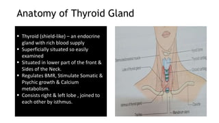
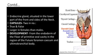
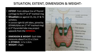
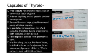
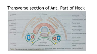
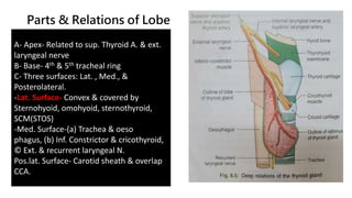
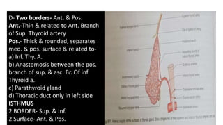
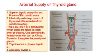
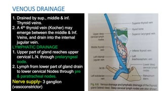
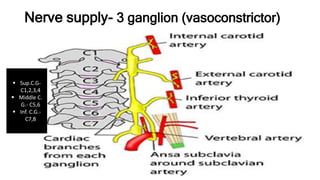
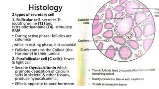
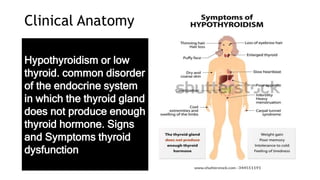
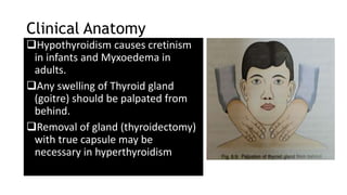
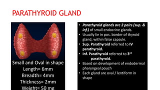
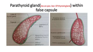
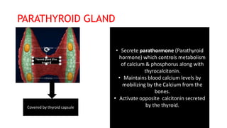
4. Vertical Section of 3 Cervical Fascia
1. Investing
Fascia
2. Pretracheal
Fascia
3. Prevertebral
Fascia
5. Anatomy of Thyroid Gland
Thyroid (shield-like) – an endocrine
gland with rich blood supply
Superficially situated so easily
examined
Situated in lower part of the front &
Sides of the Neck.
Regulates BMR, Stimulate Somatic &
Psychic growth & Calcium
metabolism.
Consists right & left lobe , joined to
each other by isthmus.
6. Contd…
• Endocrine gland, situated in the lower
part of the front and sides of the Neck.
• CAPSULES- Two in no.:
• True & False
• Larger in females than males.
• DEVELOPMENT- From the endoderm of
the Floor of primitive oral cavity in the
region of the Future foramen caecum and
ultimobranchial body.
7. SITUATION, EXTENT, DIMENSION & WEIGHT-
• EXTENT- from oblique line of Thyroid
cartilage to the 5th or 6th tracheal ring.
• Situation-Lie against C5, C6, C7 & T1
vertebra.
• It consist right & Left lobes, joined by
ISTHMUS(lies on 2nd-4th tracheal ring).
• A 3rd Pyramidal lobe may project
upwards from the ISTHMUS.
• DIMENSION & WEIGHT- Each lobe
measures about 5 x 2.5 x 2.5cm
• & ISTHMUS 1.2 x 1.2cm.
• WEIGHT= 25gm
8. Capsules of Thyroid-
True capsule-Peripheral condensation of
connective tissue of gland.
A dense capillary plexus, present deep to
true capsule.
To avoid haemorrhage, gland is removed
along with true capsule.
In prostate, venous plexus lies b/w 2
capsules, therefore during prostectomy,
both capsules are left behind.
False capsule- derived from pretracheal
layer.
It is thin along the pos. border of lobes,
but thick in inner surface (where forms
suspensory ligament- of Berry), Which
connects lobe to Cricoid cartilage.
Thyroid
Prostate
11. D- Two borders- Ant. & Pos.
Ant.-Thin & related to Ant. Branch
of Sup. Thyroid artery
Pos.- Thick & rounded, separates
med. & pos. surface & related to-
a) Inf. Thy. A.
b) Anastomosis between the pos.
branch of sup. & asc. Br. Of inf.
Thyroid a.
c) Parathyroid gland
d) Thoracic duct only in left side
ISTHMUS
2 BORDER- Sup. & Inf.
2 Surface- Ant. & Pos.
12. Arterial Supply of Thyroid gland
1. Superior thyroid artery- first ant.
Branch of Ext. carotid artery
2. Inferior thyroid artery- branch of
thyrocervical trunk (arises from
subclavian artery-
-This a. div. into 4 or 5 glandular br.
Which pierce the fascia to reach
lower pt of gland. One ascending br.
Anastomoses with pos. br. Of sup.
Thyroid a. & supplies the parathyroid
glands.
3. Thyroidea ima a. (lowest thyroid
a.)
4. Accessory thyroid a.
13. VENOUS DRAINAGE
1. Drained by sup., middle & inf.
Thyroid veins.
2. A 4th thyroid vein (Kocher) may
emerge between the middle & Inf.
Veins, and drain into the internal
jugular vein.
LYMPHATIC DRAINAGE
1. Upper part of gland reaches upper
cervical L.N. through prelaryngeal
node.
2. Lymph from lower part of gland drain
to lower cervical Nodes through pre
& paratracheal nodes.
Nerve supply- 3 ganglion
(vasoconstrictor)
15. Development of Thyroid, parathyroid & Thymus Gland
a) Thyroid gland dev.
From median
endodermal thyroid
diverticulum which
grown down in front
of neck from floor of
F. caecum
b) Foramen caecum
c) Thyroglossal duct
d) Developing Thyroid
gland from
thyroglossal duct
16. Histology
2 types of secretary cell
1. Follicular cell- secretes Tr-
iodothyronine (T3) and
tetraiodothyronine (T4)- stimulate
BMR
• During active phase- follicles are
columnar
, while in resting phase, it is cuboidal
• Follicles contains the Colloid (the
Hormone) in their lumina
2. Parafollicular cell (C cells)- fewer
& light cell
• Secrete thyrocalcitonin which
promote deposition of calcium
salts in skeletal & other tissues,
produce hypocalcaemia.
• Effects opposite to parathormone.
17. Clinical Anatomy
Hypothyroidism or low
thyroid. common disorder
of the endocrine system
in which the thyroid gland
does not produce enough
thyroid hormone. Signs
and Symptoms thyroid
dysfunction
18. Clinical Anatomy
Hypothyroidism causes cretinism
in infants and Myxoedema in
adults.
Any swelling of Thyroid gland
(goitre) should be palpated from
behind.
Removal of gland (thyroidectomy)
with true capsule may be
necessary in hyperthyroidism
20. PARATHYROID GLAND
Small and Oval in shape
Length= 6mm
Breadth= 4mm
Thickness= 2mm
Weight= 50 mg
• Parathyroid glands are 2 pairs (sup. &
inf.) of small endocrine glands.
• Usually lie in pos. border of thyroid
gland, within false capsule.
• Sup. Parathyroid referred to IV
parathyroid.
• Inf. Parathyroid referred to 3rd
parathyroid.
• Based on development of endodermal
pharyngeal pouch
• Each gland are oval / lentiform in
shape
22. PARATHYROID GLAND
• Secrete parathormone (Parathyroid
hormone) which controls metabolism
of calcium & phosphorus along with
thyrocalcitonin.
• Maintains blood calcium levels by
mobilizing by the Calcium from the
bones.
• Activate opposite calcitonin secreted
by the thyroid.
Covered by thyroid capsule
23. Location of Parathyroid Gland
(development)
• Sup. Parathyroid
develops from 4th
pharyngeal pouch
• Inf. Parathyroid
develops from 3rd
pharyngeal pouch
27. Histology of Parathyroid gland
Parenchyma consist of 2
types of cell
1. Principal/chief cell-
arranged in sheets
with sinusoids &
capillaries.
2. Oxyphilic cell- few in
no. , occur singly or in
small groups.
Often seen to increase
the age.
-Chief cell secrete
parathormone.
29. Clinical Anatomy
1. Tumours of P.T. glands leads to excessive secretion of
Parathormone (Hyperparathyroidism).
• This leads to increased removal of calcium from bone, making
weak & liable to #.
• Calcium level in blood increase (hypercalcaemia) & increased
urinary excretion of calcium lead to form stones in urinary tract.
2. Hypoparathyroidism- etiology
Spontaneously
Removal of Parathyroid glands during thyroidectomy
SYMPTOMS-
• Tetany (Due to decrease blood calcium level).
Leads hypocalcaemia, increased neuromuscular excitability-
Convulsions & muscular spasm.