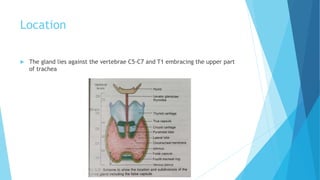
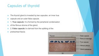
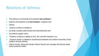
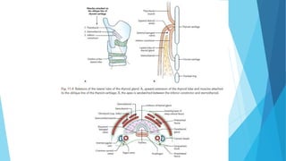
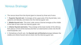
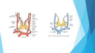
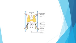
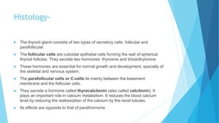
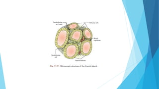
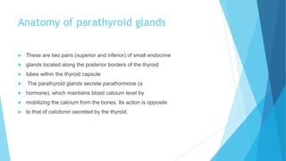
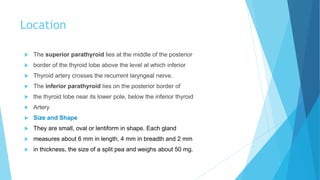
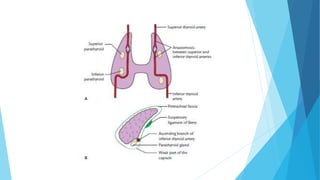
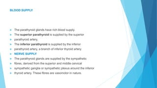
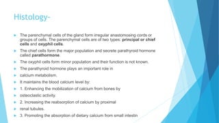
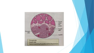
By:- Dr.Divya kumar
Anatomy of Thyroid gland
 The thyroid gland is the largest endocrine gland of the body.
 The hormones secreted by the thyroid gland include triiodothyronine
 (T3), tetraiodothyronine (T4; commonly called
 thyroxine), and calcitonin, which subserve the following
 functions:
 1. Regulate the basal metabolic rate.
 2. Stimulate the psychosomatic growth of the body.
 3. Play an important role in calcium metabolism.
Location
 The gland lies against the vertebrae C5-C7 and T1 embracing the upper part
of trachea
Capsules of thyroid
 The thyroid gland is invested by two capsules: an inner true
 capsule and an outer false capsule.
 1. True capsule: It is formed by the peripheral condensation
 of the fibrous stroma of the gland.
 2. False capsule:t is derived from the splitting of the
 pretracheal fascia.
Weight and Dimensions-
 The thyroid gland weighs about 25 g. Each lobe of the
 thyroid gland is 5 cm long, 3 cm wide, and 2 cm thick
 (5 × 3 × 2 cm). The isthmus measures about 1.25 cm in both,
vertical and transverse diameter
 The thyroid gland is usually larger in females and
 further increases in size during menstruation and pregnancy.
Relations-
Relations of the Thyroid Lobe
 Each lobe of the thyroid gland is roughly pyramidal
 (conical) and presents apex, base, three surfaces (lateral,
 medial, and posterolateral), and two borders (anterior and
 posterior):
 Apex: The apex is directed upwards and slightly laterally.
 It extends up to the oblique line of thyroid cartilage whereit is limited above by
the attachment of sternothyroid
 muscle. The apex is sandwiched between the inferior
 constrictor medially and sternothyroid laterally
Contd.
 Base: The base extends up to the 5th or 6th tracheal ring.
 It is related to inferior thyroid artery and recurrent laryngeal nerve.
 Lateral (superficial) surfaces: It is convex and is covered(a) three strap
muscles (sternothyroid, sternohyoid, and superior belly of omohyoid), and (b)
anterior border of sternocleidomastoid overlapping it inferiorly.
 Medial surface is related to (Fig. 11.6): (a) two tubes: trachea and esophagus,
(b) two muscles: inferior constrictor and cricothyroid, and (c) two cartilages:
cricoid and thyroid.
Contd.
 Posterolateral surface is related to carotid sheath and its contents (common
carotid artery, internal jugular vein, and vagus nerve).
 The ansa-cervicalis is embedded in the anterior wall of the sheath while cervical
sympathetic chain lies posterior to sheath in front of prevertebral fascia.
 Anterior border is thin and separates superficial and medial surfaces. It is
related to anterior branch of the superior thyroid artery.
 Posterior border is thick and rounded. It separates the medial and the
posterior surfaces. It is related to (a) longitudinal arterial anastomosis between
superior and inferior thyroid arteries, and (b) parathyroid glands.
Relations of isthmus-
 The isthmus is horizontal and presents two surfaces—
 anterior and posterior and two borders—superior and
 inferior.
 Anterior surface is related to:
 (a) strap muscles (sternohyoid and sternothyroid) and
 (b) anterior jugular veins.
 Posterior surface is related to 2nd, 3rd, and 4th tracheal rings.
 Superior border is related to anastomosis between the anterior branches of two
superior thyroid arteries.
 Inferior border. Along this border inferior thyroid vein emerge and thyroid artery
(when present) enters.
Arterial Supply-
 The gland is highly vascular and is supplied by the following Arteries
 Superior thyroid artery: It is a branch of the external carotid artery. It runs downwards and
forwards in company with the external laryngeal nerve, which it leaves near the upper pole of the
thyroid lobe Superior thyroid artery supplies the upper one-third of the lobe and upper half of
the isthmus
 Inferior thyroid artery: It is a branch of thyrocervical trunk from the first part of the subclavian
artery
 Inferior thyroid artery: It is a branch of thyrocervical runk from the first part of the subclavian
artery
 Thyroidea ima artery (in 30% cases): It is a branch of the brachiocephalic trunk or may arise
directly from the arch of aorta. It enters the isthmus from below.
 Accessory thyroid arteries: They arise from tracheal and esophageal arteries.
Venous Drainage
 The venous blood from the thyroid gland is drained by three set of veins
 1. Superior thyroid vein: It emerges at the upper pole of the thyroid lobe, runs
upwards and laterally, and drains into the internal jugular vein.
 2. Middle thyroid vein: This short, wide venous channel emerges at the middle
of the lobe to soon enter the internal jugular vein.
 3. Inferior thyroid vein/veins: They emerge at the lower border of the isthmus,
form plexus in front of the trachea and then run downwards to drain into the left
brachiocephalic vein.
 4. Sometimes a fourth vein, the thyroid vein (of Kocher) emerges between the
middle and inferior thyroid veins to drain into the internal jugular vein.
Lymphatic Drainage
 The lymph vessels draining the thyroid gland are arranged into
 two groups, upper and lower, and they follow the arteries:
 1. The upper group drains into the prelaryngeal (lying in
 front of the larynx) and upper deep cervical (jugulodigastric)
 lymph nodes.
 2. The lower group drains into pretracheal and lower deep
 cervical lymph nodes and group of lymph nodes along
 the recurrent laryngeal nerves. Those from lower part of
 isthmus drain into retrosternal or brachiocephalic nodes
 lying in the superior mediastinum.
 The upper lymphatics follow superior thyroid artery and
 lower lymphatics follow the inferior thyroid arteries.
Nerve supply-
 The thyroid gland is supplied by both sympathetic and
 parasympathetic nerve fibres:
 1. The parasympathetic supply is derived from the vagus
 and recurrent laryngeal nerves.
 2. The sympathetic supply is derived from the superior,
 middle, and inferior cervical sympathetic ganglia, but
 mainly from the middle one.
Histology-
 The thyroid gland consists of two types of secretory cells: follicular and
parafollicular.
 The follicular cells are cuboidal epithelial cells forming the wall of spherical
thyroid follicles. They secrete two hormones: thyroxine and triiodothyronine.
 These hormones are essential for normal growth and development, specially of
the skeletal and nervous system.
 The parafollicular cells or C-cells lie mainly between the basement
membrane and the follicular cells.
 They secrete a hormone called thyrocalcitonin (also called calcitonin). It
plays an important role in calcium metabolism. It reduces the blood calcium
level by reducing the reabsorption of the calcium by the renal tubules.
 Its effects are opposite to that of parathormone.
Anatomy of parathyroid glands
 These are two pairs (superior and inferior) of small endocrine
 glands located along the posterior borders of the thyroid
 lobes within the thyroid capsule
 The parathyroid glands secrete parathormone (a
 hormone), which maintains blood calcium level by
 mobilizing the calcium from the bones. Its action is opposite
 to that of calcitonin secreted by the thyroid.
Location
 The superior parathyroid lies at the middle of the posterior
 border of the thyroid lobe above the level at which inferior
 Thyroid artery crosses the recurrent laryngeal nerve.
 The inferior parathyroid lies on the posterior border of
 the thyroid lobe near its lower pole, below the inferior thyroid
 Artery
 Size and Shape
 They are small, oval or lentiform in shape. Each gland
 measures about 6 mm in length, 4 mm in breadth and 2 mm
 in thickness, the size of a split pea and weighs about 50 mg.
BLOOD SUPPLY
 The parathyroid glands have rich blood supply.
 The superior parathyroid is supplied by the superior
 parathyroid artery,
 The inferior parathyroid is supplied by the inferior
 parathyroid artery, a branch of inferior thyroid artery
 NERVE SUPPLY
 The parathyroid glands are supplied by the sympathetic
 fibres, derived from the superior and middle cervical
 sympathetic ganglia or sympathetic plexus around the inferior
 thyroid artery. These fibres are vasomotor in nature.
Histology-
 The parenchymal cells of the gland form irregular anastomosing cords or
groups of cells. The parenchymal cells are of two types: principal or chief
cells and oxyphil cells.
 The chief cells form the major population and secrete parathyroid hormone
called parathormone.
 The oxyphil cells form minor population and their function is not known.
 The parathyroid hormone plays an important role in
 calcium metabolism.
 It maintains the blood calcium level by:
 1. Enhancing the mobilization of calcium from bones by
 osteoclastic activity.
 2. Increasing the reabsorption of calcium by proximal
 renal tubules.
 3. Promoting the absorption of dietary calcium from small intestin
Thyroid and Parathyroid Gland.

Thyroid and Parathyroid Gland.

  • 1.
  • 2.
    Anatomy of Thyroidgland  The thyroid gland is the largest endocrine gland of the body.  The hormones secreted by the thyroid gland include triiodothyronine  (T3), tetraiodothyronine (T4; commonly called  thyroxine), and calcitonin, which subserve the following  functions:  1. Regulate the basal metabolic rate.  2. Stimulate the psychosomatic growth of the body.  3. Play an important role in calcium metabolism.
  • 3.
    Location  The glandlies against the vertebrae C5-C7 and T1 embracing the upper part of trachea
  • 4.
    Capsules of thyroid The thyroid gland is invested by two capsules: an inner true  capsule and an outer false capsule.  1. True capsule: It is formed by the peripheral condensation  of the fibrous stroma of the gland.  2. False capsule:t is derived from the splitting of the  pretracheal fascia.
  • 5.
    Weight and Dimensions- The thyroid gland weighs about 25 g. Each lobe of the  thyroid gland is 5 cm long, 3 cm wide, and 2 cm thick  (5 × 3 × 2 cm). The isthmus measures about 1.25 cm in both, vertical and transverse diameter  The thyroid gland is usually larger in females and  further increases in size during menstruation and pregnancy.
  • 6.
    Relations- Relations of theThyroid Lobe  Each lobe of the thyroid gland is roughly pyramidal  (conical) and presents apex, base, three surfaces (lateral,  medial, and posterolateral), and two borders (anterior and  posterior):  Apex: The apex is directed upwards and slightly laterally.  It extends up to the oblique line of thyroid cartilage whereit is limited above by the attachment of sternothyroid  muscle. The apex is sandwiched between the inferior  constrictor medially and sternothyroid laterally
  • 7.
    Contd.  Base: Thebase extends up to the 5th or 6th tracheal ring.  It is related to inferior thyroid artery and recurrent laryngeal nerve.  Lateral (superficial) surfaces: It is convex and is covered(a) three strap muscles (sternothyroid, sternohyoid, and superior belly of omohyoid), and (b) anterior border of sternocleidomastoid overlapping it inferiorly.  Medial surface is related to (Fig. 11.6): (a) two tubes: trachea and esophagus, (b) two muscles: inferior constrictor and cricothyroid, and (c) two cartilages: cricoid and thyroid.
  • 8.
    Contd.  Posterolateral surfaceis related to carotid sheath and its contents (common carotid artery, internal jugular vein, and vagus nerve).  The ansa-cervicalis is embedded in the anterior wall of the sheath while cervical sympathetic chain lies posterior to sheath in front of prevertebral fascia.  Anterior border is thin and separates superficial and medial surfaces. It is related to anterior branch of the superior thyroid artery.  Posterior border is thick and rounded. It separates the medial and the posterior surfaces. It is related to (a) longitudinal arterial anastomosis between superior and inferior thyroid arteries, and (b) parathyroid glands.
  • 9.
    Relations of isthmus- The isthmus is horizontal and presents two surfaces—  anterior and posterior and two borders—superior and  inferior.  Anterior surface is related to:  (a) strap muscles (sternohyoid and sternothyroid) and  (b) anterior jugular veins.  Posterior surface is related to 2nd, 3rd, and 4th tracheal rings.  Superior border is related to anastomosis between the anterior branches of two superior thyroid arteries.  Inferior border. Along this border inferior thyroid vein emerge and thyroid artery (when present) enters.
  • 11.
    Arterial Supply-  Thegland is highly vascular and is supplied by the following Arteries  Superior thyroid artery: It is a branch of the external carotid artery. It runs downwards and forwards in company with the external laryngeal nerve, which it leaves near the upper pole of the thyroid lobe Superior thyroid artery supplies the upper one-third of the lobe and upper half of the isthmus  Inferior thyroid artery: It is a branch of thyrocervical trunk from the first part of the subclavian artery  Inferior thyroid artery: It is a branch of thyrocervical runk from the first part of the subclavian artery  Thyroidea ima artery (in 30% cases): It is a branch of the brachiocephalic trunk or may arise directly from the arch of aorta. It enters the isthmus from below.  Accessory thyroid arteries: They arise from tracheal and esophageal arteries.
  • 12.
    Venous Drainage  Thevenous blood from the thyroid gland is drained by three set of veins  1. Superior thyroid vein: It emerges at the upper pole of the thyroid lobe, runs upwards and laterally, and drains into the internal jugular vein.  2. Middle thyroid vein: This short, wide venous channel emerges at the middle of the lobe to soon enter the internal jugular vein.  3. Inferior thyroid vein/veins: They emerge at the lower border of the isthmus, form plexus in front of the trachea and then run downwards to drain into the left brachiocephalic vein.  4. Sometimes a fourth vein, the thyroid vein (of Kocher) emerges between the middle and inferior thyroid veins to drain into the internal jugular vein.
  • 14.
    Lymphatic Drainage  Thelymph vessels draining the thyroid gland are arranged into  two groups, upper and lower, and they follow the arteries:  1. The upper group drains into the prelaryngeal (lying in  front of the larynx) and upper deep cervical (jugulodigastric)  lymph nodes.  2. The lower group drains into pretracheal and lower deep  cervical lymph nodes and group of lymph nodes along  the recurrent laryngeal nerves. Those from lower part of  isthmus drain into retrosternal or brachiocephalic nodes  lying in the superior mediastinum.  The upper lymphatics follow superior thyroid artery and  lower lymphatics follow the inferior thyroid arteries.
  • 15.
    Nerve supply-  Thethyroid gland is supplied by both sympathetic and  parasympathetic nerve fibres:  1. The parasympathetic supply is derived from the vagus  and recurrent laryngeal nerves.  2. The sympathetic supply is derived from the superior,  middle, and inferior cervical sympathetic ganglia, but  mainly from the middle one.
  • 17.
    Histology-  The thyroidgland consists of two types of secretory cells: follicular and parafollicular.  The follicular cells are cuboidal epithelial cells forming the wall of spherical thyroid follicles. They secrete two hormones: thyroxine and triiodothyronine.  These hormones are essential for normal growth and development, specially of the skeletal and nervous system.  The parafollicular cells or C-cells lie mainly between the basement membrane and the follicular cells.  They secrete a hormone called thyrocalcitonin (also called calcitonin). It plays an important role in calcium metabolism. It reduces the blood calcium level by reducing the reabsorption of the calcium by the renal tubules.  Its effects are opposite to that of parathormone.
  • 19.
    Anatomy of parathyroidglands  These are two pairs (superior and inferior) of small endocrine  glands located along the posterior borders of the thyroid  lobes within the thyroid capsule  The parathyroid glands secrete parathormone (a  hormone), which maintains blood calcium level by  mobilizing the calcium from the bones. Its action is opposite  to that of calcitonin secreted by the thyroid.
  • 20.
    Location  The superiorparathyroid lies at the middle of the posterior  border of the thyroid lobe above the level at which inferior  Thyroid artery crosses the recurrent laryngeal nerve.  The inferior parathyroid lies on the posterior border of  the thyroid lobe near its lower pole, below the inferior thyroid  Artery  Size and Shape  They are small, oval or lentiform in shape. Each gland  measures about 6 mm in length, 4 mm in breadth and 2 mm  in thickness, the size of a split pea and weighs about 50 mg.
  • 22.
    BLOOD SUPPLY  Theparathyroid glands have rich blood supply.  The superior parathyroid is supplied by the superior  parathyroid artery,  The inferior parathyroid is supplied by the inferior  parathyroid artery, a branch of inferior thyroid artery  NERVE SUPPLY  The parathyroid glands are supplied by the sympathetic  fibres, derived from the superior and middle cervical  sympathetic ganglia or sympathetic plexus around the inferior  thyroid artery. These fibres are vasomotor in nature.
  • 23.
    Histology-  The parenchymalcells of the gland form irregular anastomosing cords or groups of cells. The parenchymal cells are of two types: principal or chief cells and oxyphil cells.  The chief cells form the major population and secrete parathyroid hormone called parathormone.  The oxyphil cells form minor population and their function is not known.  The parathyroid hormone plays an important role in  calcium metabolism.  It maintains the blood calcium level by:  1. Enhancing the mobilization of calcium from bones by  osteoclastic activity.  2. Increasing the reabsorption of calcium by proximal  renal tubules.  3. Promoting the absorption of dietary calcium from small intestin