This document provides information about HELLP syndrome, including:
1. HELLP syndrome is characterized by hemolysis, elevated liver enzymes, and low platelet count. It occurs in 0.2-0.6% of pregnancies and is caused by abnormal vascular tone and coagulation defects.
2. Clinical presentation is often unclear but includes right upper quadrant pain, nausea, and headache. Diagnosis is based on low platelets (<100,000/μl), elevated liver enzymes, and signs of hemolysis on blood smear.
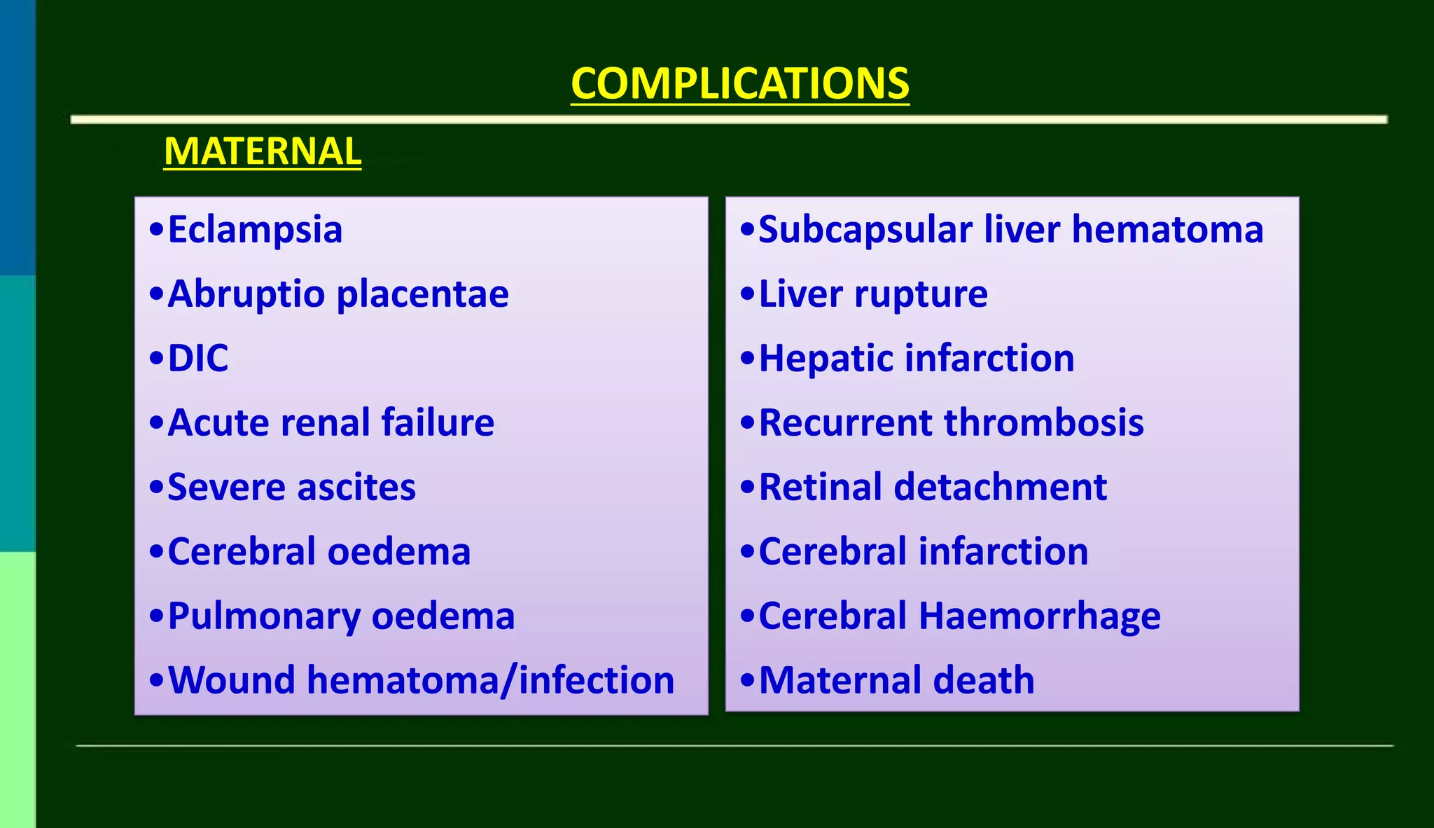
3. Complications include eclampsia, abruption, renal failure, and liver hematoma rupture. Management involves delivery after 34 weeks if possible, with
HELLP Syndrome ischaracterized by
Hepatic endothelial disruption followed by platelet
activation, aggregation and consumption,
ultimately resulting in ischemia and hepatocyte
death
3.
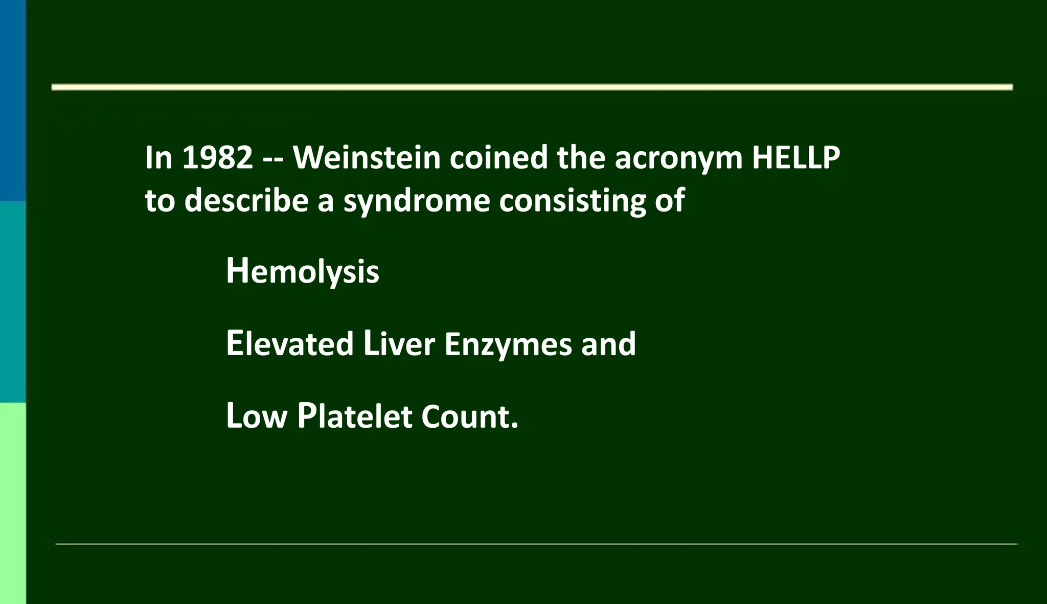
In 1982 --Weinstein coined the acronym HELLP
to describe a syndrome consisting of
Hemolysis
Elevated Liver Enzymes and
Low Platelet Count.
4.
INCIDENCE
• HELLP Syndromeoccurs in 0.2 to 0.6% of all pregnancies
• Incidence of HELLP in women with pre eclampsia is
20 %
70% cases are diagnosed in antenatal period
30% after delivery.
5.
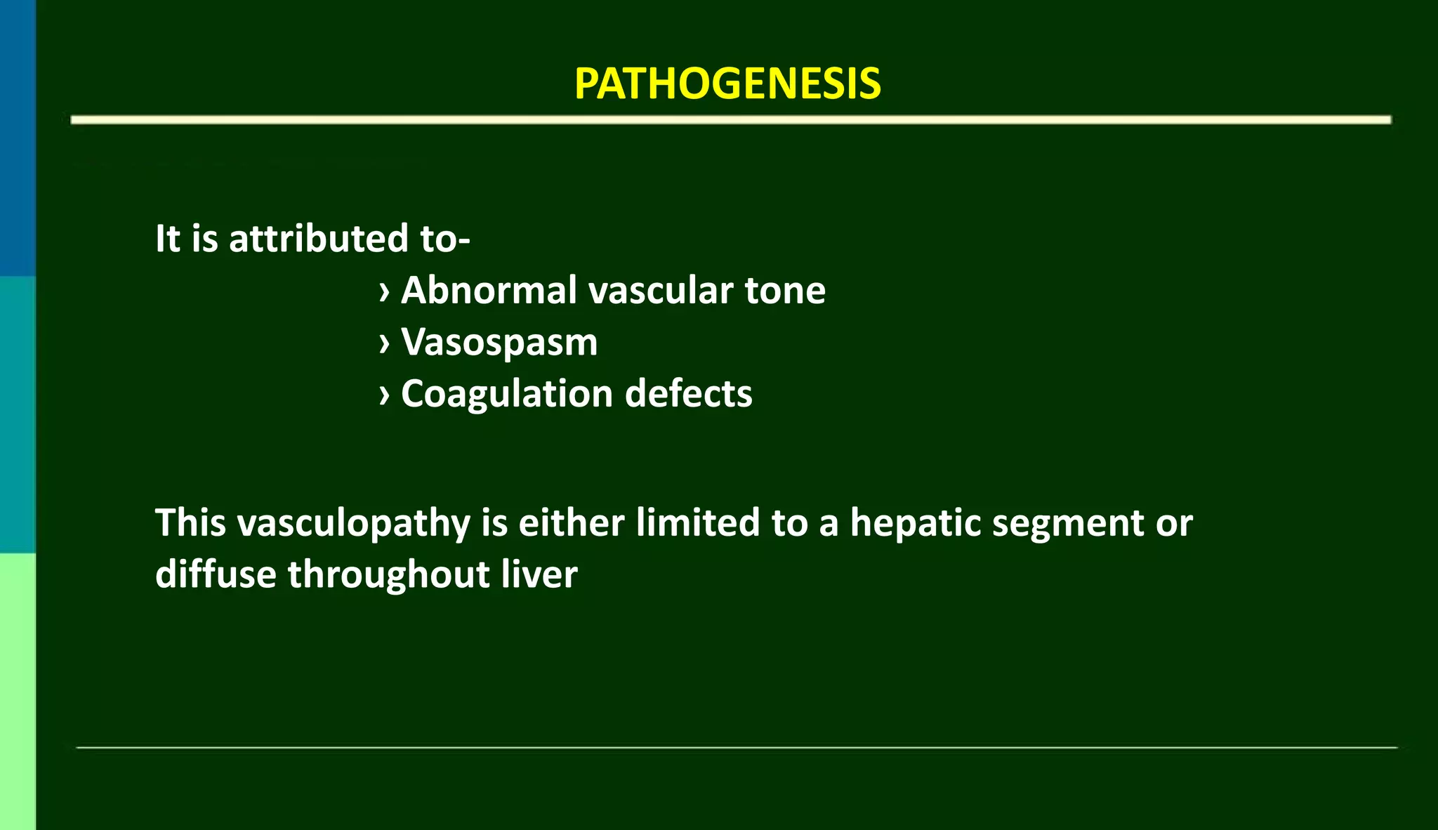
PATHOGENESIS
It is attributedto-
› Abnormal vascular tone
› Vasospasm
› Coagulation defects
This vasculopathy is either limited to a hepatic segment or
diffuse throughout liver
6.
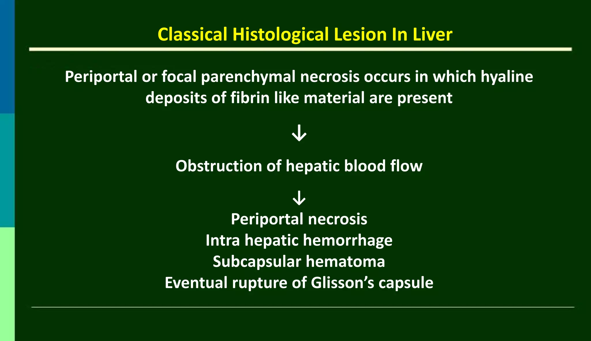
Classical Histological LesionIn Liver
Periportal or focal parenchymal necrosis occurs in which hyaline
deposits of fibrin like material are present
↓
Obstruction of hepatic blood flow
↓
Periportal necrosis
Intra hepatic hemorrhage
Subcapsular hematoma
Eventual rupture of Glisson’s capsule
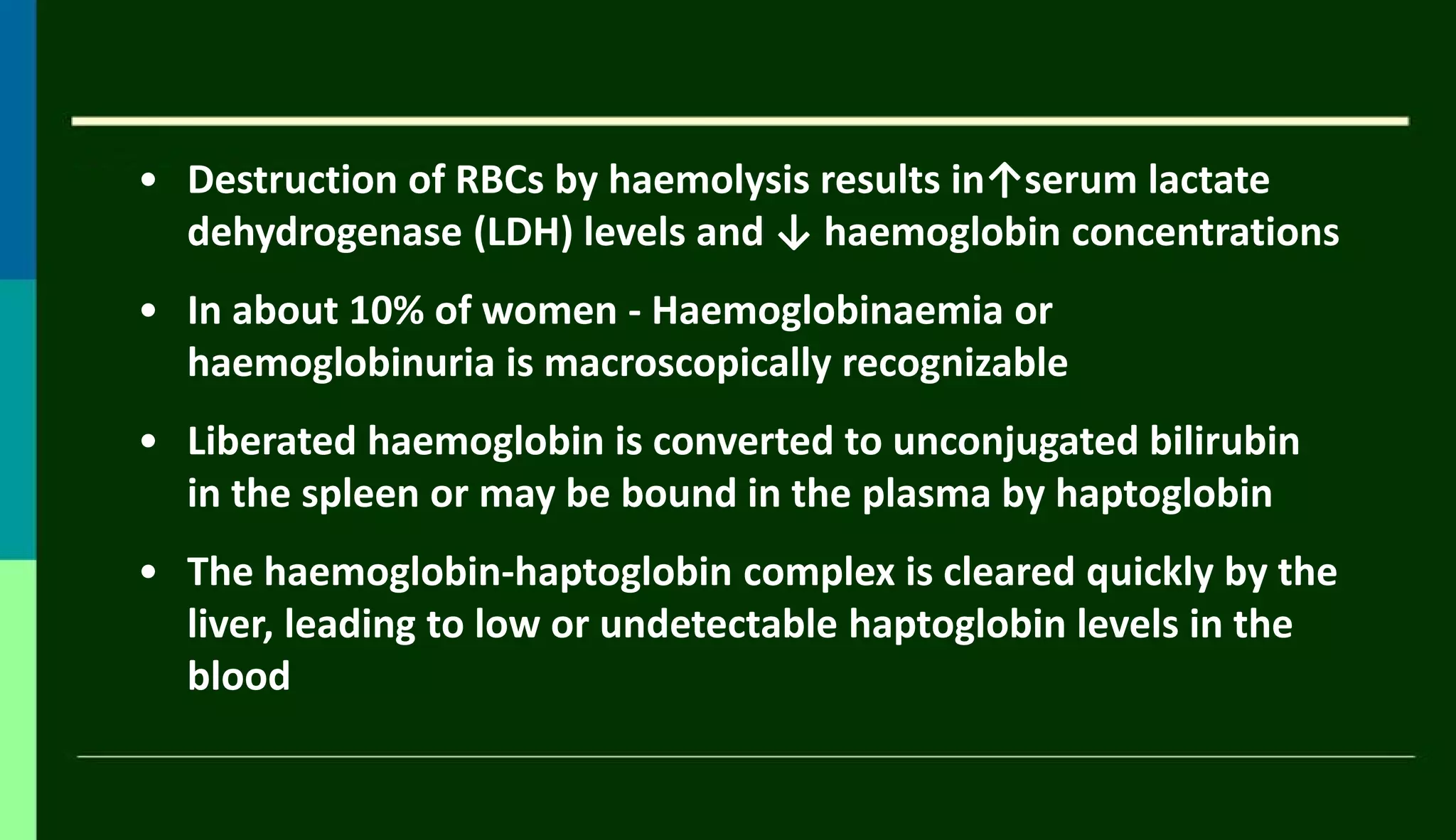
• Destruction ofRBCs by haemolysis results in↑serum lactate
dehydrogenase (LDH) levels and ↓ haemoglobin concentrations
• In about 10% of women - Haemoglobinaemia or
haemoglobinuria is macroscopically recognizable
• Liberated haemoglobin is converted to unconjugated bilirubin
in the spleen or may be bound in the plasma by haptoglobin
• The haemoglobin-haptoglobin complex is cleared quickly by the
liver, leading to low or undetectable haptoglobin levels in the
blood
10.
But
The demonstration oflow or undetectable haptoglobin
concentration is a more specific indicator.
The diagnosis of haemolysis is supported by -
- high LDH concentration and
- presence of unconjugated bilirubin
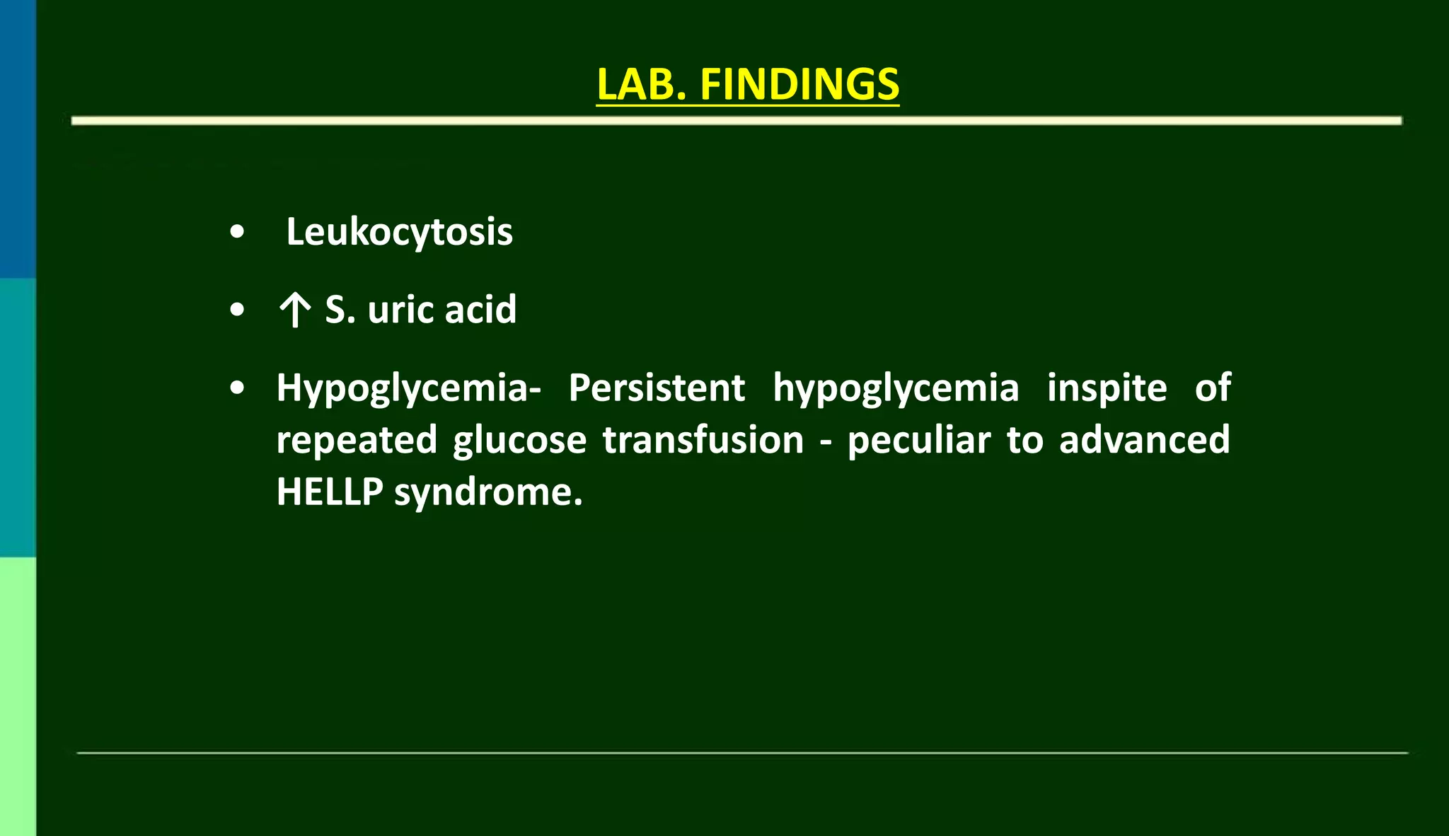
THROMBOCYTOPENIA
• ↓Platelet countin HELLP syndrome is due to their
↑consumption
• Platelets are activated, and adhere to damaged vascular
endothelial cells, resulting in ↑ platelet turnover with shorter
lifespan
Patient with well developed HELLP syndrome may develop
DIC
13.
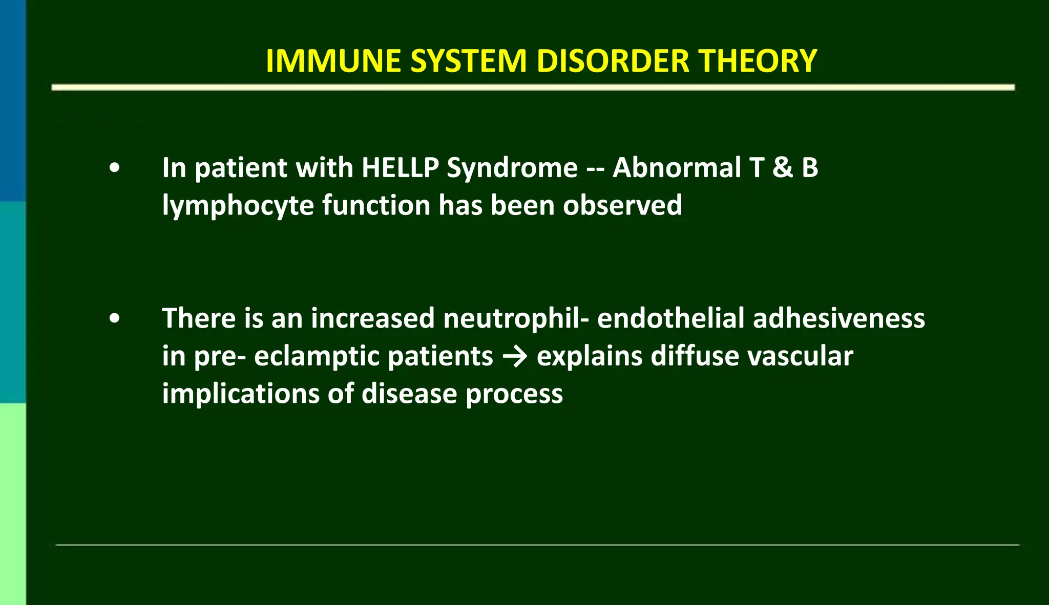
IMMUNE SYSTEM DISORDERTHEORY
• In patient with HELLP Syndrome -- Abnormal T & B
lymphocyte function has been observed
• There is an increased neutrophil- endothelial adhesiveness
in pre- eclamptic patients → explains diffuse vascular
implications of disease process
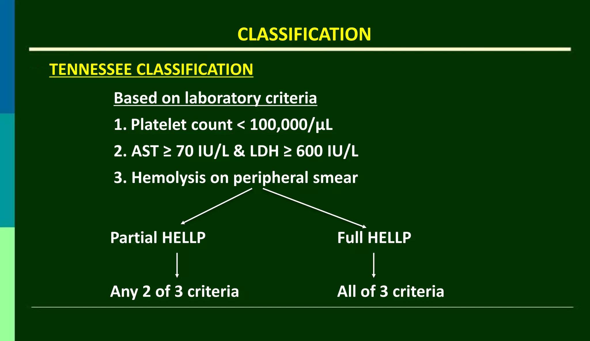
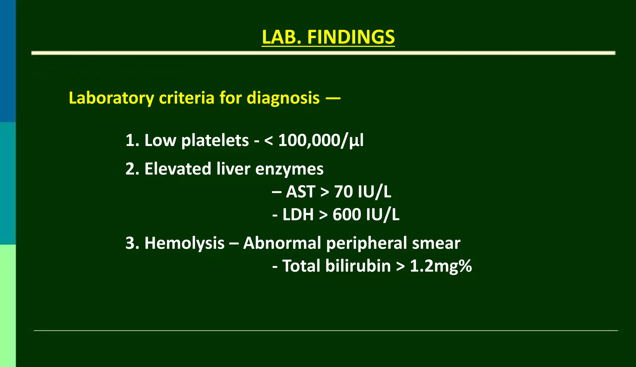
CLASSIFICATION
TENNESSEE CLASSIFICATION
Based onlaboratory criteria
1. Platelet count < 100,000/µL
2. AST ≥ 70 IU/L & LDH ≥ 600 IU/L
3. Hemolysis on peripheral smear
Partial HELLP Full HELLP
Any 2 of 3 criteria All of 3 criteria
18.
CLASSIFICATION
MISSISIPI CLASSIFICATION
CLASS I
›Platelet ≤ 50,000/µL(severe
thrombocytopenia)
› AST ≥ 70 IU/L› LDH ≥ 600 IU/L
› Hemolysis on smear
CLASS II
› Platelet 50,000/µL to
100,000/µL (moderate
thrombocytopenia)
› AST ≥ 70 IU/L› LDH ≥ 600 IU/L
› Hemolysis on smear
CLASS III
› Platelet 100,000/µL to150,000/µL
(mild thrombocytopenia)
› AST ≥ 40 IU/L › LDH ≥ 600 IU/L
› Hemolysis on smear
19.
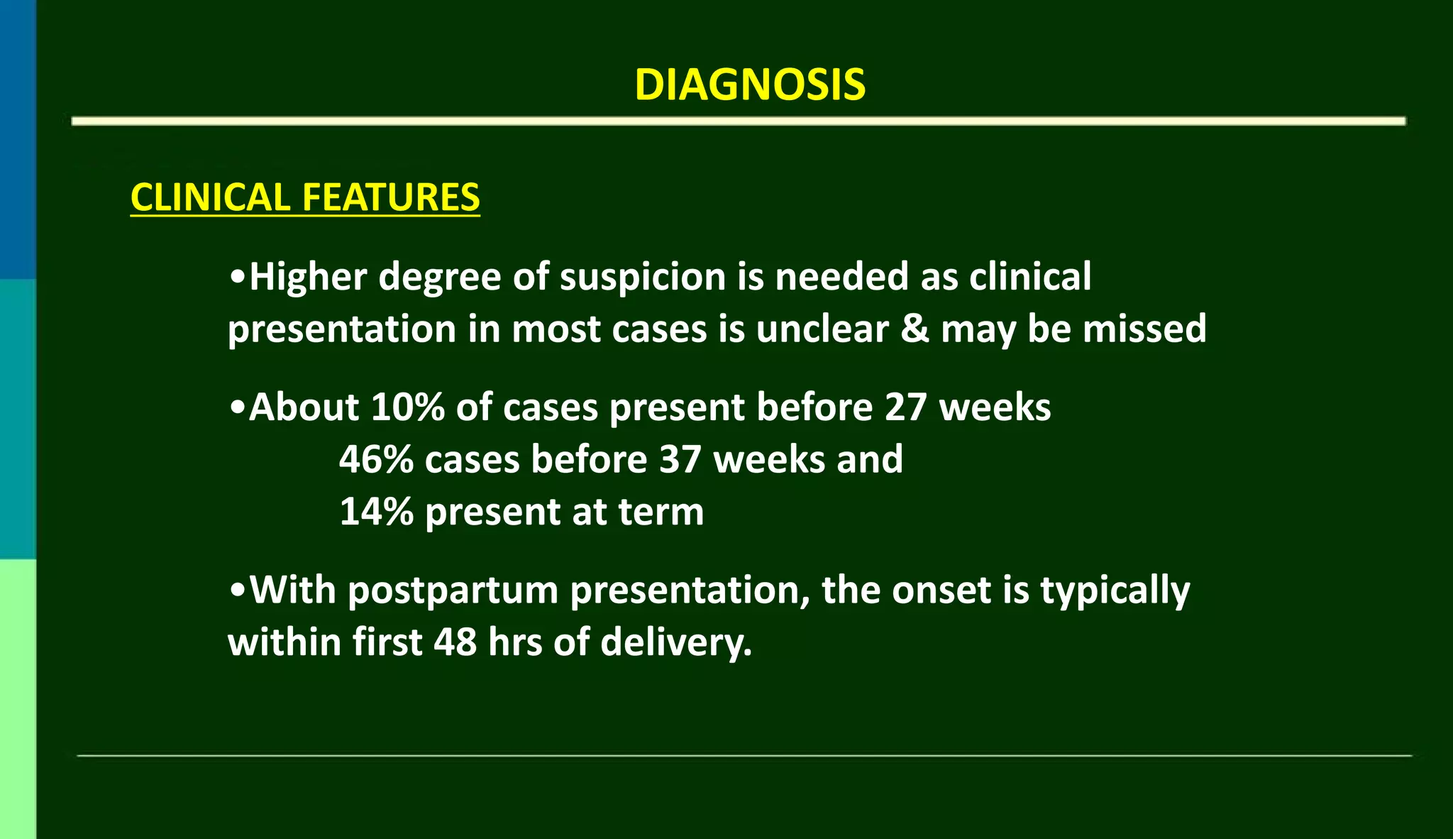
DIAGNOSIS
CLINICAL FEATURES
•Higher degreeof suspicion is needed as clinical
presentation in most cases is unclear & may be missed
•About 10% of cases present before 27 weeks
46% cases before 37 weeks and
14% present at term
•With postpartum presentation, the onset is typically
within first 48 hrs of delivery.
20.
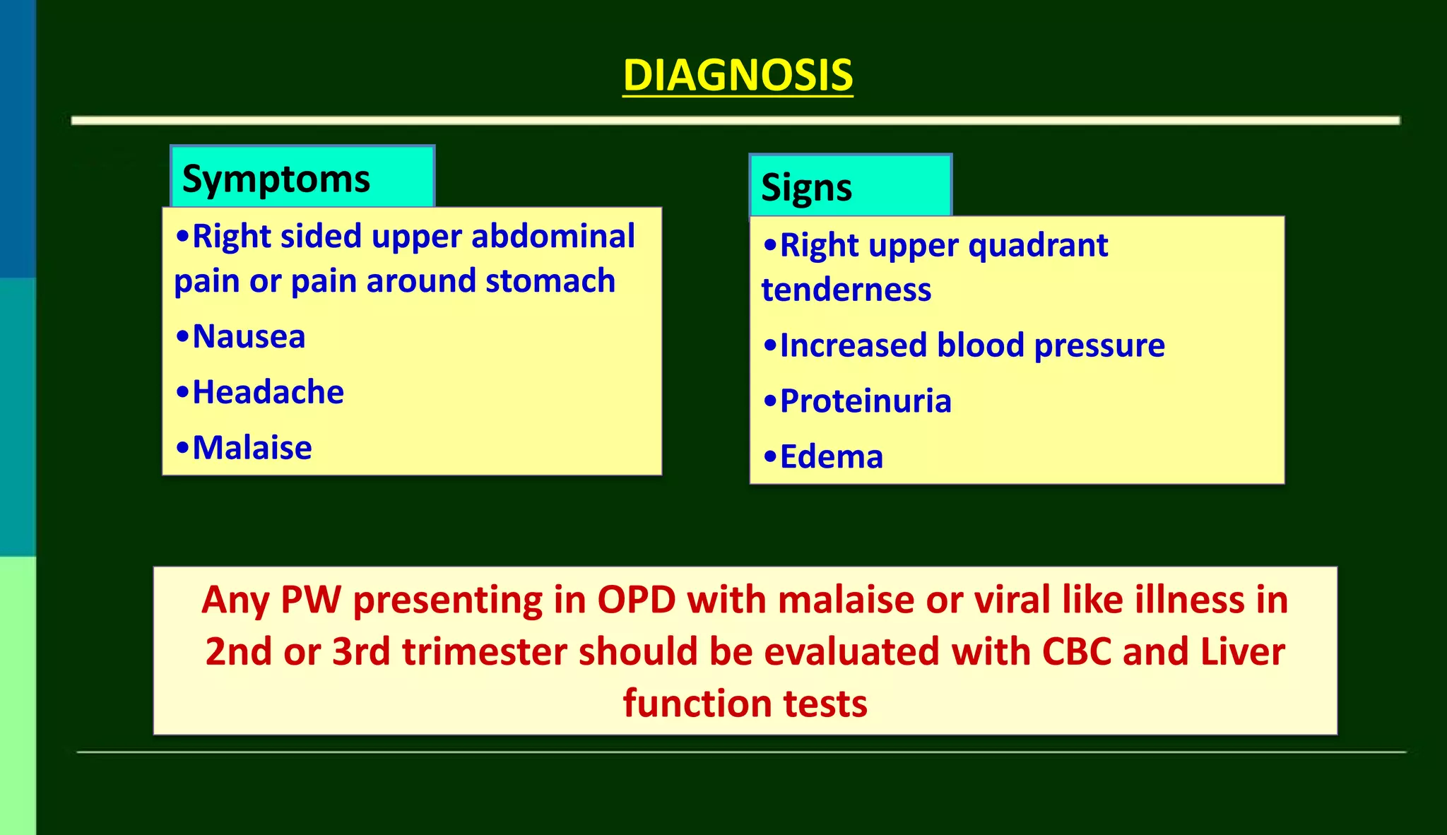
DIAGNOSIS
Symptoms
•Right sided upperabdominal
pain or pain around stomach
•Nausea
•Headache
•Malaise
Signs
•Right upper quadrant
tenderness
•Increased blood pressure
•Proteinuria
•Edema
Any PW presenting in OPD with malaise or viral like illness in
2nd or 3rd trimester should be evaluated with CBC and Liver
function tests
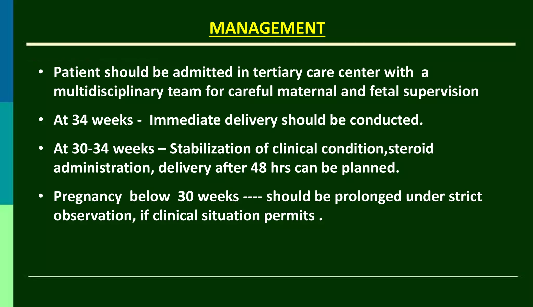
• Patient shouldbe admitted in tertiary care center with a
multidisciplinary team for careful maternal and fetal supervision
• At 34 weeks - Immediate delivery should be conducted.
• At 30-34 weeks – Stabilization of clinical condition,steroid
administration, delivery after 48 hrs can be planned.
• Pregnancy below 30 weeks ---- should be prolonged under strict
observation, if clinical situation permits .
MANAGEMENT
27.
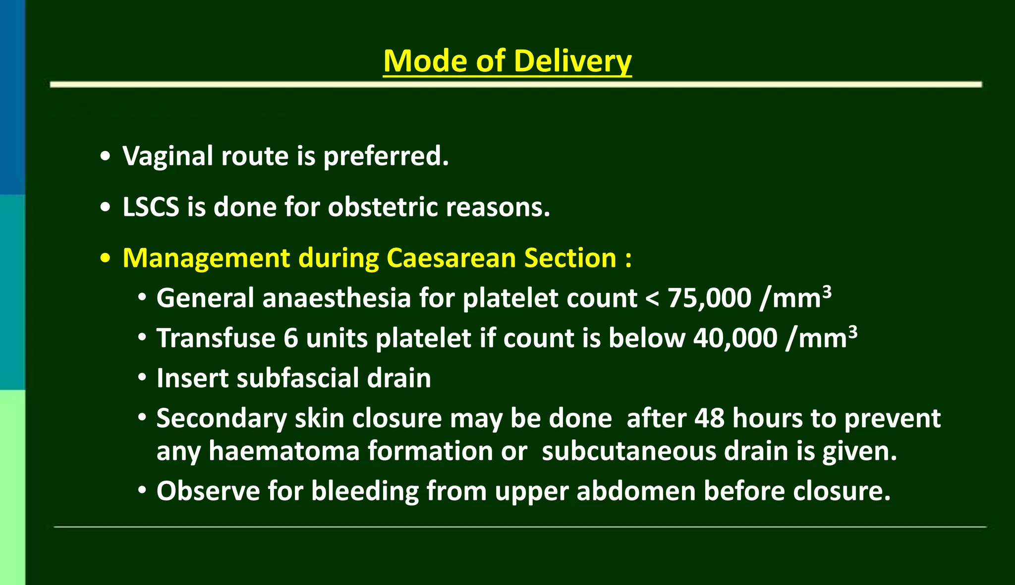
Mode of Delivery
•Vaginal route is preferred.
• LSCS is done for obstetric reasons.
• Management during Caesarean Section :
• General anaesthesia for platelet count < 75,000 /mm3
• Transfuse 6 units platelet if count is below 40,000 /mm3
• Insert subfascial drain
• Secondary skin closure may be done after 48 hours to prevent
any haematoma formation or subcutaneous drain is given.
• Observe for bleeding from upper abdomen before closure.
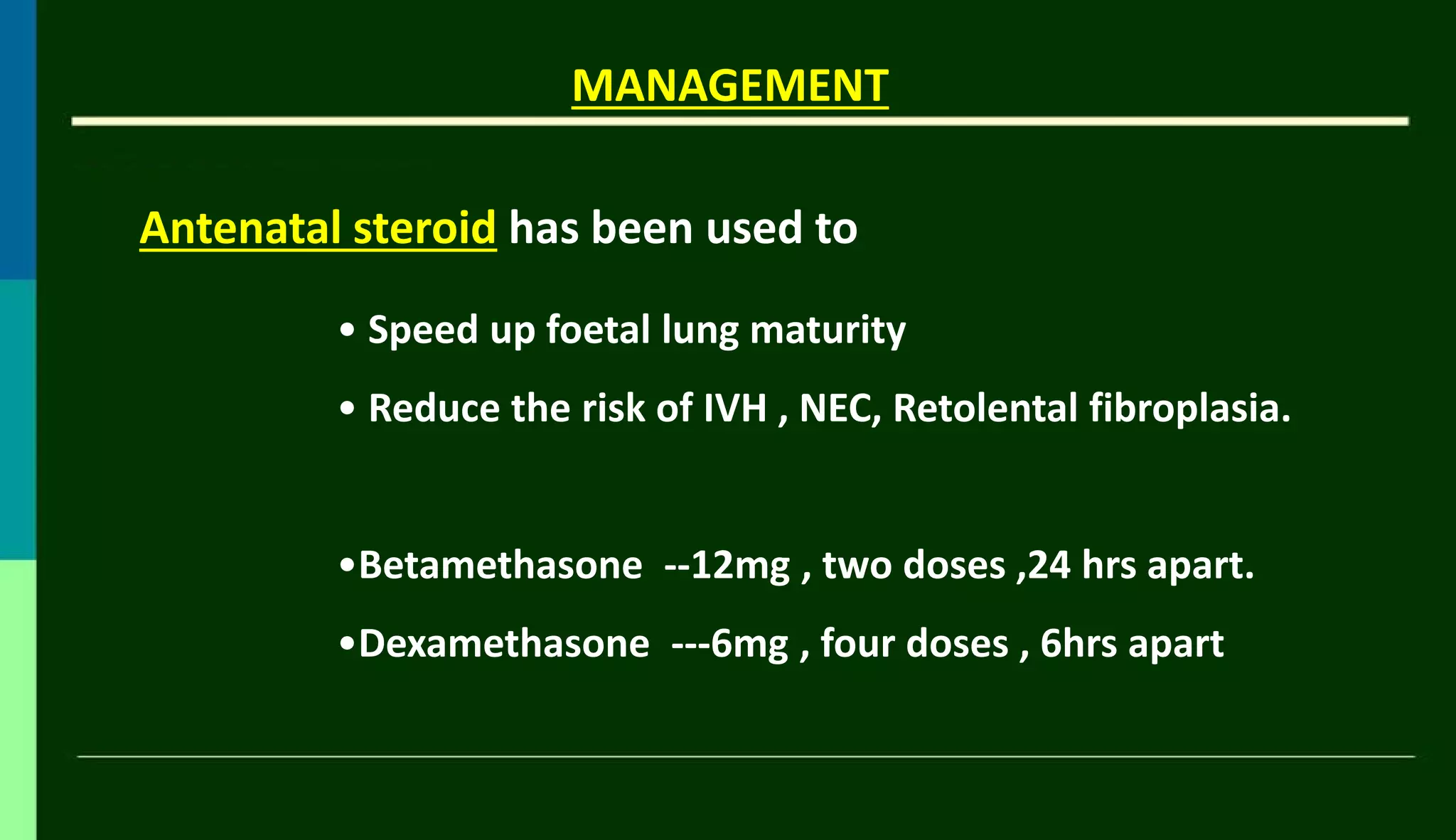
Antenatal steroid hasbeen used to
MANAGEMENT
• Speed up foetal lung maturity
• Reduce the risk of IVH , NEC, Retolental fibroplasia.
•Betamethasone --12mg , two doses ,24 hrs apart.
•Dexamethasone ---6mg , four doses , 6hrs apart
30.
Steroid Treatment
Benefit ofsteroid treatment for HELLP syndrome was first reported in
1984
Mech. of Action- Unknown
Proposed mech. - Diminishes oedema, inhibits endothelial
activation and reduces endothelial dysfunction
↓
Prevention of thrombotic microangiopathic anaemia, Inhibition
of cytokine production
↓
Induces anti-inflammatory effects in HELLP syndrome
MANAGEMENT
31.
MANAGEMENT
Available evidence doesnot support high and repeated dose of
corticosteroid treatment
Can improve the outcome of pregnancy affected by HELLP
syndrome
For selected high risk cases with profound thrombocytopenia
with CNS dysfunction
- 20mg IV dexamethasone every 6hrs up to 4 doses is
given.
32.
MANAGEMENT
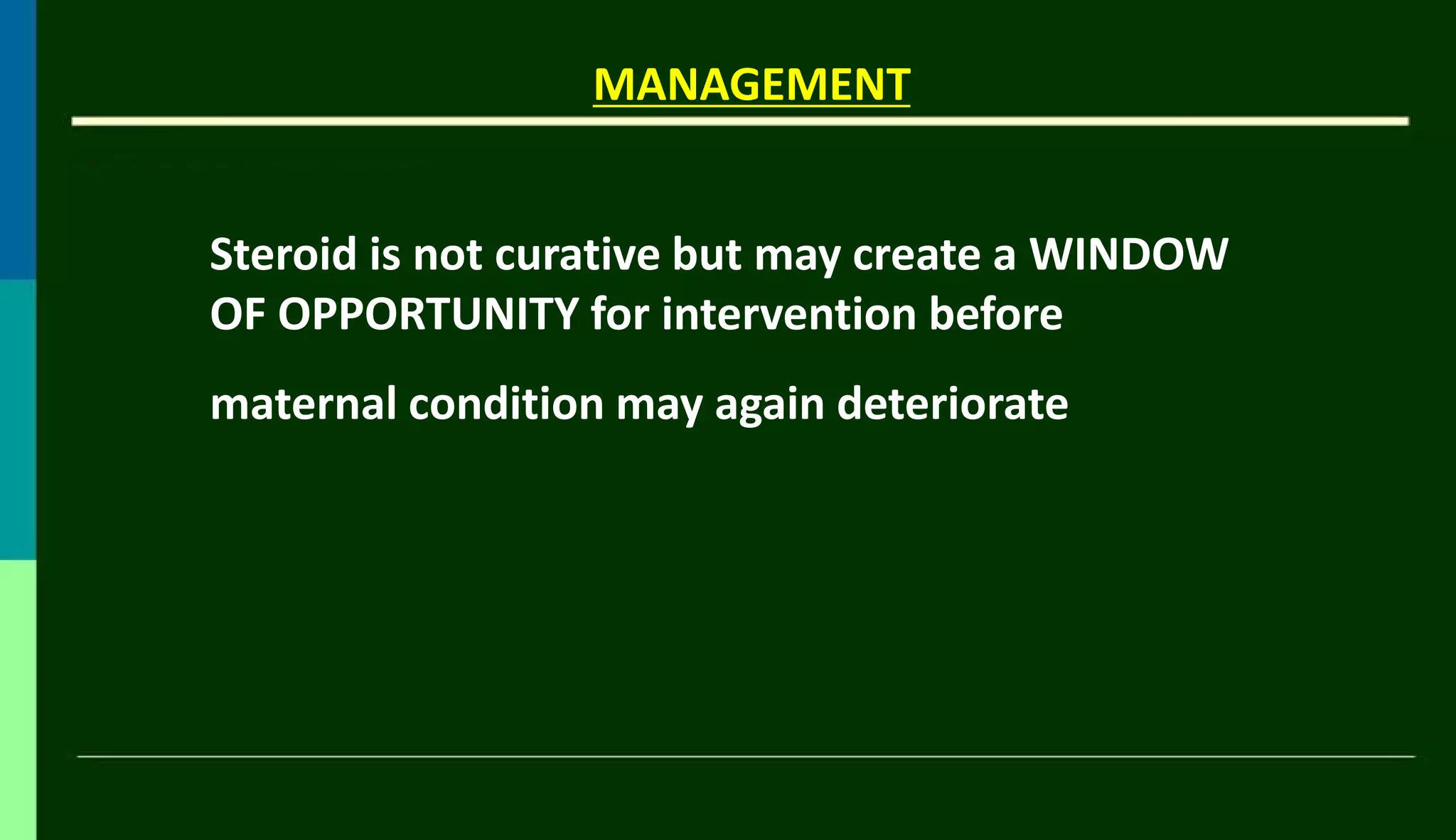
Steroid is notcurative but may create a WINDOW
OF OPPORTUNITY for intervention before
maternal condition may again deteriorate
33.
MANAGEMENT
• If plateletcount <40,000/µL, 6 – 10 U of platelet
transfusion is required.
• Platelet transfusion is required if there is bleeding from
wound or intra peritoneal bleeding.
• PRBC and FFP is required if coagulopathy is present.
34.
MANAGEMENT
Antithrombin III transfusion: -
- Corrects hypercoagulability,
- Stimulates prostacyclin production,
- Regulates thrombin-induced vasoconstriction,
- Improves foetal status
35.
Management of postpartum HELLP Syndrome
• About 30% of HELLP syndromes develop after birth
• The time of onset ranges from few hrs to 7 days but the
majority within the first 48 hours after delivery
• In post-partum HELLP syndrome, risk of renal failure and
pulmonary oedema is ↑ hence intensive monitoring of the
mother is required.
In most women, the maternal platelet count starts decreasing
immediately post-partum with an increasing trend on the third
day
36.
Management of postpartum HELLP Syndrome
• Women with HELLP syndrome who show progressive ↑
of bilirubin or creatinine for > 72 hours after delivery
may benefit from plasma exchange with fresh frozen
plasma
• In case of continuing haemolysis, persistent
thrombocytopenia and hypoproteinaemia -- PRBC,
Platelets as well as Albumin supplementation is
required
37.
Management of postpartum HELLP Syndrome
Recurrence rate - 20% in subsequent pregnancies.
Women with a history of HELLP syndrome at or before 28
weeks gestation during the index pregnancy are at ↑ risk for
several obstetric complications like:
- Preterm birth
- Pregnancy- induced hypertension
- Increased neonatal mortality in a subsequent pregnancy.
38.
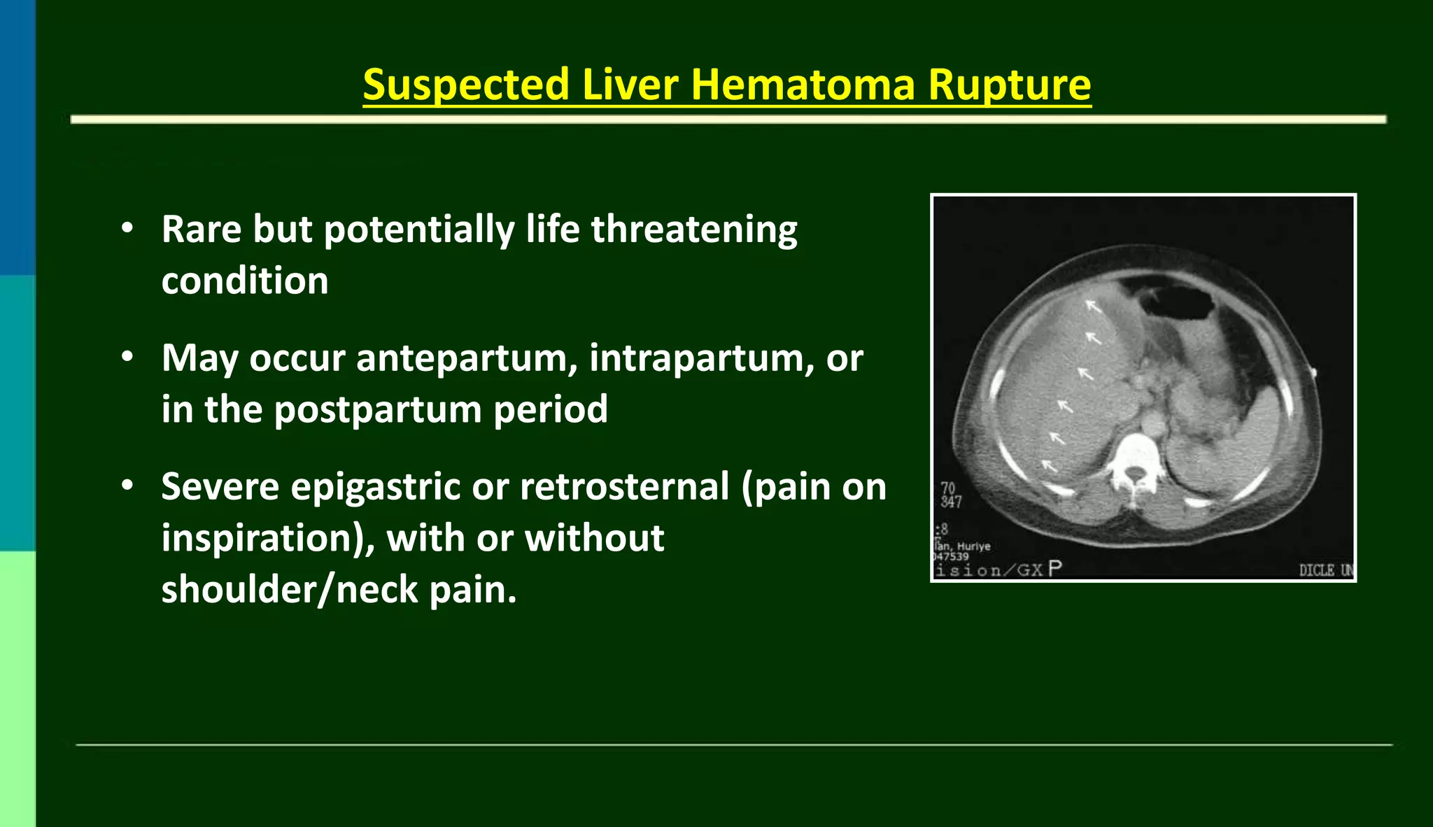
Suspected Liver HematomaRupture
• Rare but potentially life threatening
condition
• May occur antepartum, intrapartum, or
in the postpartum period
• Severe epigastric or retrosternal (pain on
inspiration), with or without
shoulder/neck pain.
39.
• Rupture Occurs1 in 40,000 to 1 in 250,000 deliveries and about
1% to < 2% of the cases with HELLP syndrome
• Should be suspected when profound hypovolemic shock occurs in
a previously hypertensive patient
• Diagnosis can be made by ultrasound or CT of the liver which can
diagnose intraperitoneal bleeding.
• In most cases, rupture involves the right lobe of the liver and is
preceded by a parenchymal liver hematoma.
Suspected Liver Hematoma Rupture
40.
• Maternal andfetal mortality increases substantially when a
subcapsular liver hematoma is present.
• Mortality may exceed 50% when frank rupture of the capsule
involves liver tissue.
• Management depends on maternal hemodynamic status,
integrity of the capsule (ruptured or intact), and the fetal
condition.
Liver Hematoma Rupture
41.
Conservative management shouldbe done in hemodynamically stable
women with an unruptured hematoma.
Liver Hematoma Rupture
– Close monitoring of the patient’s hemodynamic and
coagulation status
-- Serial assessment of the hematoma with ultrasound or
CT scan
• Exogenous trauma to the liver should be avoided, such as frequent
abdominal palpation, emesis, or convulsions.
• Any sudden increase in intra-abdominal pressure can lead to rupture
of hematoma
42.
When rupture occurs
LiverHematoma Rupture
-- It is a surgical emergency
-- Maternal resuscitation with fluids and PRBC to
maintain blood pressure and tissue perfusion should
be done.
-- Correction of coagulopathy with fresh frozen
plasma and platelets is required.
43.
– Packing anddrainage (preferred)
– Ligation of the hepatic lacerations
– Embolization of the hepatic artery to the affected liver
segment, and loosely suturing omentum or surgical
mesh to the liver surface
– Recombinant factor VII A
Liver Hematoma Rupture
Options at laparotomy include : –
44.
PROGNOSIS OF HELLPSYNDROME
• Maternal mortality -- 0 to 15%.
• Maternal morbidity - Acute renal failure, Hepatic infarct,
Hepatic hematoma, Hepatic rupture,
• Disseminated intravascular coagulation, Post-partum
hemorrhage
• Pulmonary edema may occur
• There are usually no long term maternal complications.


![Understanding Parkinson’s Disease: Causes, Symptoms, and Treatment [2025]](https://cdn.slidesharecdn.com/ss_thumbnails/understandingparkinson-251208102525-80ba3223-thumbnail.jpg?width=640&height=640&fit=bounds)


