The document provides a comprehensive overview of the pelvis, detailing its structure, parts, types, and functions, as well as common conditions affecting it. Various types of pelvis are described, including gynecoid, android, anthropoid, and platypelloid, each characterized by distinct shapes. Additionally, the text discusses pelvic ligaments and common disorders such as osteitis pubis and fractures.
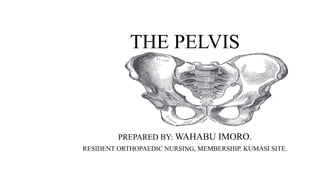


![STRUCTURE OF THE PELVIS
• Anterior view of the pelvis
Anterior view of the pelvic girdle. Adapted from [Van de Graaff, 2001, Ch. 7]](https://image.slidesharecdn.com/thepelvis-220506140656-5fa6dc2a/85/The-Pelvis-pptx-4-320.jpg)


