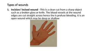
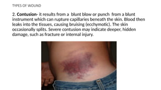
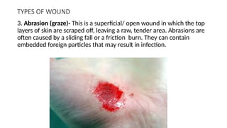
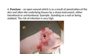
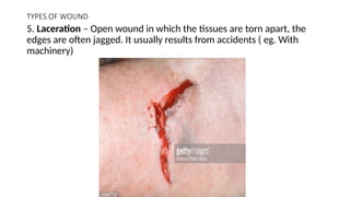
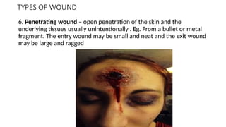
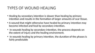
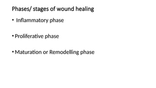
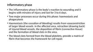
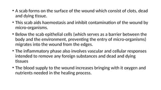
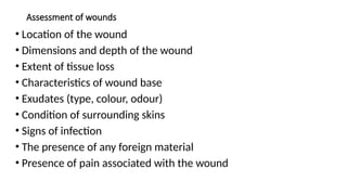
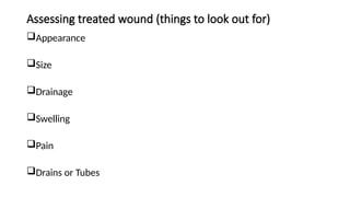
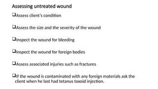
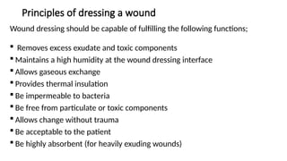
SKIN INTEGRITY AND WOUND CARE
Introduction
• The skin is the largest organ in the body
• It serves a variety of important functions in maintaining
health and protecting the body from injury
What is skin integrity?
• It refers to the presence of normal skin and skin layers
uninterrupted by wounds
• The appearance of the skin and skin integrity are influenced
by intrinsic factors and extrinsic factors.
Introduction
• Intrinsic factors – eg. Genetics/heredity (influences skin
colour, sensitivity to sunlight and allergies), age and the
underlying health of the individual (eg. Chronic illness such
as people with impaired arterial circulation may have skin on
the legs that appears shiny and damages easily)
• Extrinsic factors – eg. Nutrition, smoking and activity (eg.
Some medications such as certain antibiotics, chemotherapy
drugs, psychotherapeutic drugs and some corticosteroids
causes thinning of the skin)
Types of wounds
1. Incision/ incised wound - This is a clean cut from a sharp object
such as a broken glass or knife. The blood vessels at the wound
edges are cut straight across hence the is profuse bleeding. It is an
open wound which may be deep or shallow
TYPES OF WOUND
2. Contusion- it results from a blunt blow or punch from a blunt
instrument which can rupture capillaries beneath the skin. Blood then
leaks into the tissues, causing bruising (ecchymotic). The skin
occasionally splits. Severe contusion may indicate deeper, hidden
damage, such as fracture or internal injury.
TYPES OF WOUND
3. Abrasion (graze)- This is a superficial/ open wound in which the top
layers of skin are scraped off, leaving a raw, tender area. Abrasions are
often caused by a sliding fall or a friction burn. They can contain
embedded foreign particles that may result in infection.
4. Puncture – an open wound which is as a result of penetration of the
skin and often the underlying tissues by a sharp instrument, either
intentional or unintentional. Example. Standing on a nail or being
stabbed. The risk of infection is very high.
TYPES OF WOUND
5. Laceration – Open wound in which the tissues are torn apart, the
edges are often jagged. It usually results from accidents ( eg. With
machinery)
TYPES OF WOUND
6. Penetrating wound – open penetration of the skin and the
underlying tissues usually unintentionally . Eg. From a bullet or metal
fragment. The entry wound may be small and neat and the exit wound
may be large and ragged
TISSUE REPAIR AND WOUND HEALING
• Body organs and structures contain two types of tissues:
• parenchymal and stromal.
• The parenchymal tissues contain the functioning cells of an organ or
body part (e.g., hepatocytes, renal tubular cells).
• The stromal tissues consist of the supporting connective tissues,
blood vessels, and nerve fibers.
• Injured tissues are repaired by regeneration of parenchymal cells or
by connective tissue repair in which scar tissue is substituted for the
parenchymal cells of the injured tissue
TISSUE REPAIR AND WOUND HEALING
• The primary objective of the healing process is to fill the gap created
by tissue destruction and to restore the structural continuity of the
injured part.
• When regeneration cannot occur, healing by replacement with
connective scar tissue provides the means for maintaining this
continuity.
• Although scar tissue fills the gap created by tissue death, it does not
repair the structure with functioning parenchymal cells.
• Because the regenerative capabilities of most tissues are limited,
wound healing usually involves some connective tissue repair.
Regeneration
• Regeneration involves replacement of the injured tissue with cells of
the same parenchymal type, leaving little or no evidence of the
previous injury.
• The ability to regenerate varies with the tissue and cell type.
• Body cells are divided into three types according to their ability to
undergo regeneration:
• labile,
• stable
• permanent cells.
Labile
• Labile cells are those that continue to divide and replicate throughout
life, replacing cells that are continually being destroyed.
• Labile cells can be found in tissues that have a daily turnover of cells.
• They include the surface epithelial cells of the skin, the oral cavity,
vagina, and cervix;
• the columnar epithelium of the gastrointestinal tract, uterus, and
fallopian tubes;
• the transitional epithelium of the urinary tract; and bone marrow cells
Stable cells
• Stable cells are those that normally stop dividing when growth ceases.
• However, these cells are capable of undergoing regeneration when
confronted with an appropriate stimulus.
• For stable cells to regenerate and restore tissues to their original state,
the supporting stromal framework must be present.
• When this framework has been destroyed, the replacement of tissues is
haphazard.
• The hepatocytes of the liver are one form of stable cell, and the
importance of the supporting framework to regeneration is evidenced
by two forms of liver disease.
Permanent
• Permanent or fixed cells cannot undergo mitotic division.
• The fixed cells include nerve cells, skeletal muscle cells, and cardiac
muscle cells.
• These cells cannot regenerate; once destroyed, they are replaced
with fibrous scar tissue that lacks the functional characteristics of the
destroyed tissue.
• For example, the scar tis sue that develops in the heart after a heart
attack can not conduct impulses or contract to pump blood.
TYPES OF WOUND HEALING
• Depending on the extent of tissue loss, wound closure and healing
occur by primary or secondary intention.
• A sutured surgical incision is an example of healing by primary
intention.
• Larger wounds (e.g., burns and large surface wounds) that have a
greater loss of tissue and contamination, heal by secondary intention.
TYPES OF WOUND HEALING
• Healing by secondary intention is slower than healing by primary
intention and results in the formation of larger amounts of scar tissue.
• A wound that might otherwise have healed by primary intention may
become infected and heal by secondary intention.
• In wounds healing by secondary intention, the process depends on
the extent of injury and the healing environment.
• In wounds healing by primary intention, the duration of the phases is
fairly predictable
Phases/ stages of wound healing
• Inflammatory phase
•Proliferative phase
•Maturation or Remodelling phase
Inflammatory phase
• The inflammatory phase is the body’s reaction to wounding and it
begins with minutes of injury and last for 3 to 6 days.
• Two major processes occur during this phase: haemostasis and
phagocytosis
• Haemostasis (the cessation of bleeding) results from vasoconstriction
of larger blood vessels in the affected area, retraction (drawing back)
of injured blood vessels, the deposition of fibrin (connective tissue)
and the formation of blood clots in the area.
• The blood clots formed from the blood platelets, provide a matrix of
fibrin that becomes the framework for cell repair.
• A scab forms on the surface of the wound which consist of clots, dead
and dying tissue.
• This scab aids haemostasis and inhibit contamination of the wound by
micro-organisms.
• Below the scab epithelial cells (which serves as a barrier between the
body and the environment, preventing the entry of micro-organisms)
migrates into the wound from the edges.
• The inflammatory phase also involves vascular and cellular responses
intended to remove any foreign substances and dead and dying
tissues
• The blood supply to the wound increases bringing with it oxygen and
nutrients needed in the healing process.
• The area appears reddened and oedematous as a result
• During cell migration, leukocytes (specifically, neutrophils) move into
the interstitial space.
• These are replaced about 24 hours after injury by macrophages,
which arise from the blood monocytes.
• These macrophages engulf micro-organisms and cellular debris by a
process known as phagocytosis
• The macrophages also excrete an angiogenesis factor (AGF), which
stimulates the formation of epithelial buds at the end of injured blood
vessels.
• The microcirculatory network that results sustains the healing process
and the wound during its life.
Proliferative phase
• The phase in healing, extents from the 3rd
or 4th
day to about the 21st
post-injury.
• Fibroblast(connective tissue cell) which migrate into the wound
starting about 24 hours after injury begin to synthesize collage.
• Collagen- which is a whitish protein substance that adds tensile
strength to the wound.
• As the amount of collagen increases, so does the strength of the
wound, thus the chance that the wound will open decreases.
• If the wound is sutured, a raised “healing ridge” appears under the
intact suture line. In a wound that is not sutured the new collagen is
often visible.
• Capillaries grow across the wound increasing the blood supply.
Fibroblast move from the bloodstream into the wound depositing
fibrin.
• As the capillary network develops the tissue becomes a translucent
red colour, this tissue known as granulation tissue is fragile and
bleeds easily.
• When the skin edges are not sutured, the area must be filled in with
granulation tissue. When the granulation tissue matures, marginal
epithelial cells migrate to it, proliferating over its connective tissue
base to fill the wound
• If the wound does not close by epithelializaion, the area becomes
covered with dried plasma proteins and dead cells. This is known as
eschar
• Initally wounds healing by secondary intention seep blood-tinge
(serosanguineous) drainage.
• Later if they are not covered with thick, grey, fibrinous tissue that is
converted into dense scar tissue.
Maturation phase
• This begins around the 21st
day and can extend one or two years after
the injury.
• Fibroblast continue to synthesis collagen. The collagen fibres which
where initially laid in a haphazard fashion, reorganize into a more
orderly structure
• During maturation, the wound is remodeled and contracted.
• The scar becomes stronger but the repaired area is never as strong as
the original tissue.
• In some individuals eg. Dark skinned persons, an abnormal amount of
collagen is laid down. This can result in a hypertrophic scar or keloid
Kinds of wound drain
• Exudate is material such as fluid and cells, which has escaped from
blood vessels during the inflammatory process and its deposited in
tissue or on tissue surfaces.
• The nature and amount of exudate vary according to the tissue
involved, the intensity and duration of the inflammation and the
presence of micro-organisms.
• There are three major types of exudate;
 Serous
Purulent
Sanguineous (haemorrhagic)
SEROUS EXUDATE- consist of serum (the clear portion of the blood)
derived from blood and the serous membranes of the body. It looks watery
and has few cells. Example – the fluid in a blister from a burn
PURULENT EXUDATE – this is thicker than serous exudate because of the
presence of pus, which consist of leukocytes, liquefied dead tissue debris,
dead and living bacteria. Purulent exudates vary in colour, some acquiring
tinges of blue, green or yellow. The colour depends on the causative
organism.
HAEMORRHAGIC EXUDATE- consist of large amounts of red blood cells,
indicating damage to capillaries that is severe enough to allow the escape
of red blood cells from plasma. This type of exudate is frequently seen in
open wounds.
Complications of wound healing
• Heamorrhage
• Infection
Pain
Change in the colour of the wound bed
Malodorous (offensive smelling) exudate
Heat
Swelling
• Dehiscence (wound ruptures along a surgical incision)
Assessment of wounds
• Location of the wound
• Dimensions and depth of the wound
• Extent of tissue loss
• Characteristics of wound base
• Exudates (type, colour, odour)
• Condition of surrounding skins
• Signs of infection
• The presence of any foreign material
• Presence of pain associated with the wound
Assessing treated wound (things to look out for)
Appearance
Size
Drainage
Swelling
Pain
Drains or Tubes
Assessing untreated wound
Assess client’s condition
Assess the size and the severity of the wound
Inspect the wound for bleeding
Inspect the wound for foreign bodies
Assess associated injuries such as fractures
If the wound is contaminated with any foreign materials ask the
client when he last had tetanus toxoid injection.
Principles of dressing a wound
Wound dressing should be capable of fulfilling the following functions;
 Removes excess exudate and toxic components
 Maintains a high humidity at the wound dressing interface
 Allows gaseous exchange
 Provides thermal insulation
 Be impermeable to bacteria
 Be free from particulate or toxic components
 Allows change without trauma
 Be acceptable to the patient
 Be highly absorbent (for heavily exuding wounds)
Cont’d
 Be cost effective
 Provide mechanical protection
 Be conformable and mouldable
 Be able to be sterilized
Observations to make during wound dressing
1. The surface area or volume of the wound should be measured – this can
be done using a planimetry measurements using a ruler, tracing the wound
unto grid paper or Vistrak.
o photograph of the wound can be taken and used as a useful record of
wound progression
2. The amount and type of drainage
3. Odour and colour
4. Condition of surrounding skin - edges of the wound
5. Signs of Inflamation and infection
6. Pain
7. Episodes of bleeding
Setting trolley for wound dressing
Top shelf
A sterile pack containing;
• 3 gallipots for lotions
• 2 pairs of dissecting forceps
• Dressing forceps
• Probe
• Stitches scissors
• Covered bowl containing sterile cotton wool and gauze swabs
• Covered bowl containing dressing towel and clip remover if necessary
Setting trolley for wound dressing cont’d
Bottom shelf
• Cleaning solution
• Dressing solution
• Sterile cotton pack
• Sterile gauze pack
• Reciever for used cotton
• Draw sheet
• Draw mackintosh
Cont’d
• adhesive plaster
• bandage
• Scissors
• Gloves
• Covered receiver containing parazone (1:10) for soiled instruments
• Receptacle for soiled dressings.
Factors inhibiting wound healing in older adults
• Many local and systemic factors influence wound healing.
• Although there are many factors that impair healing, science has found a few
ways to hasten the normal process of wound repair.
• Among the causes of impaired wound healing are
• Malnutrition
• impaired blood flow and oxygen delivery
• impaired inflammatory and immune responses
• infection,
• wound separation
• foreign bodies
• age effects
Malnutrition
• Successful wound healing depends in part on adequate stores of
proteins, carbohydrates, fats, vitamins, and minerals.
• It is well recognized that malnutrition slows the healing process,
causing wounds to heal inadequately or incompletely.
• Protein deficiencies prolong the inflammatory phase of healing and
impair fibroblast proliferation, collagen and protein matrix synthesis,
angiogenesis, and wound remodeling.
• Carbohydrates are needed as an energy source for white blood cells.
Malnutrition
• Carbohydrates also have a protein-sparing effect and help to prevent the
use of amino acids for fuel when they are needed for the healing process.
• Fats are essential constituents of cell membranes and are needed for the
synthesis of new cells.
• Although most vitamins are essential cofactors for the daily functions of
the body, only vitamins A and C have been shown to play an essential role
in the healing process.
• Vitamin C is needed for collagen synthesis.
• Vitamin A functions in stimulating and supporting epithelialization,
capillary formation, and collagen synthesis.
Blood Flow and Oxygen Delivery
• For healing to occur, wounds must have adequate blood flow to
supply the necessary nutrients and to remove the resulting waste,
local toxins, bacteria, and other debris.
• Impaired wound healing caused by poor blood flow may occur as a
result of wound conditions (e.g., swelling) or pre-existing health
problems.
• Arterial disease and venous pathology are well-documented causes of
impaired wound healing.
Blood Flow and Oxygen Delivery
• In situations of trauma, a decrease in blood volume may cause a
reduction in blood flow to injured tissues.
• Molecular oxygen is required for collagen synthesis.
• It has been shown that even a temporary lack of oxygen can result in
the formation of less stable collagen.
• Wounds in ischemic tissue become infected more frequently than do
wounds in well-vascularized tissue.
Impaired Inflammatory and Immune Responses
• Inflammatory and immune mechanisms function in wound healing.
• Inflammation is essential to the first phase of wound healing, and
immune mechanisms prevent infections that impair wound healing.
• Among the conditions that impair inflammation and immune function
are disorders of phagocytic function, diabetes mellitus, and
therapeutic administration of corticosteroid drugs
Infection, Wound Separation, and Foreign Bodies
• Wound contamination, wound separation, and foreign bodies delay wound
healing.
• Infection impairs all dimensions of wound healing.
• It prolongs the inflammatory phase, impairs the formation of granulation tissue,
and inhibits proliferation of fibroblasts and deposition of collagen fibers.
• All wounds are contaminated at the time of injury.
• Although body defenses can handle the invasion of microorganisms at the time of
wounding, badly contaminated wounds can overwhelm host defenses.
• Trauma and existing impairment of host defenses also can contribute to the
development of wound infections

SKIN INTEGRITY AND WOUND CAREd RNAC2pptx

  • 1.
  • 2.
    Introduction • The skinis the largest organ in the body • It serves a variety of important functions in maintaining health and protecting the body from injury What is skin integrity? • It refers to the presence of normal skin and skin layers uninterrupted by wounds • The appearance of the skin and skin integrity are influenced by intrinsic factors and extrinsic factors.
  • 3.
    Introduction • Intrinsic factors– eg. Genetics/heredity (influences skin colour, sensitivity to sunlight and allergies), age and the underlying health of the individual (eg. Chronic illness such as people with impaired arterial circulation may have skin on the legs that appears shiny and damages easily) • Extrinsic factors – eg. Nutrition, smoking and activity (eg. Some medications such as certain antibiotics, chemotherapy drugs, psychotherapeutic drugs and some corticosteroids causes thinning of the skin)
  • 4.
    Types of wounds 1.Incision/ incised wound - This is a clean cut from a sharp object such as a broken glass or knife. The blood vessels at the wound edges are cut straight across hence the is profuse bleeding. It is an open wound which may be deep or shallow
  • 5.
    TYPES OF WOUND 2.Contusion- it results from a blunt blow or punch from a blunt instrument which can rupture capillaries beneath the skin. Blood then leaks into the tissues, causing bruising (ecchymotic). The skin occasionally splits. Severe contusion may indicate deeper, hidden damage, such as fracture or internal injury.
  • 6.
    TYPES OF WOUND 3.Abrasion (graze)- This is a superficial/ open wound in which the top layers of skin are scraped off, leaving a raw, tender area. Abrasions are often caused by a sliding fall or a friction burn. They can contain embedded foreign particles that may result in infection.
  • 7.
    4. Puncture –an open wound which is as a result of penetration of the skin and often the underlying tissues by a sharp instrument, either intentional or unintentional. Example. Standing on a nail or being stabbed. The risk of infection is very high.
  • 8.
    TYPES OF WOUND 5.Laceration – Open wound in which the tissues are torn apart, the edges are often jagged. It usually results from accidents ( eg. With machinery)
  • 9.
    TYPES OF WOUND 6.Penetrating wound – open penetration of the skin and the underlying tissues usually unintentionally . Eg. From a bullet or metal fragment. The entry wound may be small and neat and the exit wound may be large and ragged
  • 10.
    TISSUE REPAIR ANDWOUND HEALING • Body organs and structures contain two types of tissues: • parenchymal and stromal. • The parenchymal tissues contain the functioning cells of an organ or body part (e.g., hepatocytes, renal tubular cells). • The stromal tissues consist of the supporting connective tissues, blood vessels, and nerve fibers. • Injured tissues are repaired by regeneration of parenchymal cells or by connective tissue repair in which scar tissue is substituted for the parenchymal cells of the injured tissue
  • 11.
    TISSUE REPAIR ANDWOUND HEALING • The primary objective of the healing process is to fill the gap created by tissue destruction and to restore the structural continuity of the injured part. • When regeneration cannot occur, healing by replacement with connective scar tissue provides the means for maintaining this continuity. • Although scar tissue fills the gap created by tissue death, it does not repair the structure with functioning parenchymal cells. • Because the regenerative capabilities of most tissues are limited, wound healing usually involves some connective tissue repair.
  • 12.
    Regeneration • Regeneration involvesreplacement of the injured tissue with cells of the same parenchymal type, leaving little or no evidence of the previous injury. • The ability to regenerate varies with the tissue and cell type. • Body cells are divided into three types according to their ability to undergo regeneration: • labile, • stable • permanent cells.
  • 13.
    Labile • Labile cellsare those that continue to divide and replicate throughout life, replacing cells that are continually being destroyed. • Labile cells can be found in tissues that have a daily turnover of cells. • They include the surface epithelial cells of the skin, the oral cavity, vagina, and cervix; • the columnar epithelium of the gastrointestinal tract, uterus, and fallopian tubes; • the transitional epithelium of the urinary tract; and bone marrow cells
  • 14.
    Stable cells • Stablecells are those that normally stop dividing when growth ceases. • However, these cells are capable of undergoing regeneration when confronted with an appropriate stimulus. • For stable cells to regenerate and restore tissues to their original state, the supporting stromal framework must be present. • When this framework has been destroyed, the replacement of tissues is haphazard. • The hepatocytes of the liver are one form of stable cell, and the importance of the supporting framework to regeneration is evidenced by two forms of liver disease.
  • 15.
    Permanent • Permanent orfixed cells cannot undergo mitotic division. • The fixed cells include nerve cells, skeletal muscle cells, and cardiac muscle cells. • These cells cannot regenerate; once destroyed, they are replaced with fibrous scar tissue that lacks the functional characteristics of the destroyed tissue. • For example, the scar tis sue that develops in the heart after a heart attack can not conduct impulses or contract to pump blood.
  • 16.
    TYPES OF WOUNDHEALING • Depending on the extent of tissue loss, wound closure and healing occur by primary or secondary intention. • A sutured surgical incision is an example of healing by primary intention. • Larger wounds (e.g., burns and large surface wounds) that have a greater loss of tissue and contamination, heal by secondary intention.
  • 17.
    TYPES OF WOUNDHEALING • Healing by secondary intention is slower than healing by primary intention and results in the formation of larger amounts of scar tissue. • A wound that might otherwise have healed by primary intention may become infected and heal by secondary intention. • In wounds healing by secondary intention, the process depends on the extent of injury and the healing environment. • In wounds healing by primary intention, the duration of the phases is fairly predictable
  • 20.
    Phases/ stages ofwound healing • Inflammatory phase •Proliferative phase •Maturation or Remodelling phase
  • 21.
    Inflammatory phase • Theinflammatory phase is the body’s reaction to wounding and it begins with minutes of injury and last for 3 to 6 days. • Two major processes occur during this phase: haemostasis and phagocytosis • Haemostasis (the cessation of bleeding) results from vasoconstriction of larger blood vessels in the affected area, retraction (drawing back) of injured blood vessels, the deposition of fibrin (connective tissue) and the formation of blood clots in the area. • The blood clots formed from the blood platelets, provide a matrix of fibrin that becomes the framework for cell repair.
  • 22.
    • A scabforms on the surface of the wound which consist of clots, dead and dying tissue. • This scab aids haemostasis and inhibit contamination of the wound by micro-organisms. • Below the scab epithelial cells (which serves as a barrier between the body and the environment, preventing the entry of micro-organisms) migrates into the wound from the edges. • The inflammatory phase also involves vascular and cellular responses intended to remove any foreign substances and dead and dying tissues • The blood supply to the wound increases bringing with it oxygen and nutrients needed in the healing process.
  • 23.
    • The areaappears reddened and oedematous as a result • During cell migration, leukocytes (specifically, neutrophils) move into the interstitial space. • These are replaced about 24 hours after injury by macrophages, which arise from the blood monocytes. • These macrophages engulf micro-organisms and cellular debris by a process known as phagocytosis • The macrophages also excrete an angiogenesis factor (AGF), which stimulates the formation of epithelial buds at the end of injured blood vessels. • The microcirculatory network that results sustains the healing process and the wound during its life.
  • 24.
    Proliferative phase • Thephase in healing, extents from the 3rd or 4th day to about the 21st post-injury. • Fibroblast(connective tissue cell) which migrate into the wound starting about 24 hours after injury begin to synthesize collage. • Collagen- which is a whitish protein substance that adds tensile strength to the wound. • As the amount of collagen increases, so does the strength of the wound, thus the chance that the wound will open decreases. • If the wound is sutured, a raised “healing ridge” appears under the intact suture line. In a wound that is not sutured the new collagen is often visible.
  • 25.
    • Capillaries growacross the wound increasing the blood supply. Fibroblast move from the bloodstream into the wound depositing fibrin. • As the capillary network develops the tissue becomes a translucent red colour, this tissue known as granulation tissue is fragile and bleeds easily. • When the skin edges are not sutured, the area must be filled in with granulation tissue. When the granulation tissue matures, marginal epithelial cells migrate to it, proliferating over its connective tissue base to fill the wound • If the wound does not close by epithelializaion, the area becomes covered with dried plasma proteins and dead cells. This is known as eschar
  • 26.
    • Initally woundshealing by secondary intention seep blood-tinge (serosanguineous) drainage. • Later if they are not covered with thick, grey, fibrinous tissue that is converted into dense scar tissue.
  • 27.
    Maturation phase • Thisbegins around the 21st day and can extend one or two years after the injury. • Fibroblast continue to synthesis collagen. The collagen fibres which where initially laid in a haphazard fashion, reorganize into a more orderly structure • During maturation, the wound is remodeled and contracted. • The scar becomes stronger but the repaired area is never as strong as the original tissue. • In some individuals eg. Dark skinned persons, an abnormal amount of collagen is laid down. This can result in a hypertrophic scar or keloid
  • 28.
    Kinds of wounddrain • Exudate is material such as fluid and cells, which has escaped from blood vessels during the inflammatory process and its deposited in tissue or on tissue surfaces. • The nature and amount of exudate vary according to the tissue involved, the intensity and duration of the inflammation and the presence of micro-organisms. • There are three major types of exudate;  Serous Purulent Sanguineous (haemorrhagic)
  • 29.
    SEROUS EXUDATE- consistof serum (the clear portion of the blood) derived from blood and the serous membranes of the body. It looks watery and has few cells. Example – the fluid in a blister from a burn PURULENT EXUDATE – this is thicker than serous exudate because of the presence of pus, which consist of leukocytes, liquefied dead tissue debris, dead and living bacteria. Purulent exudates vary in colour, some acquiring tinges of blue, green or yellow. The colour depends on the causative organism. HAEMORRHAGIC EXUDATE- consist of large amounts of red blood cells, indicating damage to capillaries that is severe enough to allow the escape of red blood cells from plasma. This type of exudate is frequently seen in open wounds.
  • 30.
    Complications of woundhealing • Heamorrhage • Infection Pain Change in the colour of the wound bed Malodorous (offensive smelling) exudate Heat Swelling • Dehiscence (wound ruptures along a surgical incision)
  • 31.
    Assessment of wounds •Location of the wound • Dimensions and depth of the wound • Extent of tissue loss • Characteristics of wound base • Exudates (type, colour, odour) • Condition of surrounding skins • Signs of infection • The presence of any foreign material • Presence of pain associated with the wound
  • 32.
    Assessing treated wound(things to look out for) Appearance Size Drainage Swelling Pain Drains or Tubes
  • 33.
    Assessing untreated wound Assessclient’s condition Assess the size and the severity of the wound Inspect the wound for bleeding Inspect the wound for foreign bodies Assess associated injuries such as fractures If the wound is contaminated with any foreign materials ask the client when he last had tetanus toxoid injection.
  • 34.
    Principles of dressinga wound Wound dressing should be capable of fulfilling the following functions;  Removes excess exudate and toxic components  Maintains a high humidity at the wound dressing interface  Allows gaseous exchange  Provides thermal insulation  Be impermeable to bacteria  Be free from particulate or toxic components  Allows change without trauma  Be acceptable to the patient  Be highly absorbent (for heavily exuding wounds)
  • 35.
    Cont’d  Be costeffective  Provide mechanical protection  Be conformable and mouldable  Be able to be sterilized
  • 36.
    Observations to makeduring wound dressing 1. The surface area or volume of the wound should be measured – this can be done using a planimetry measurements using a ruler, tracing the wound unto grid paper or Vistrak. o photograph of the wound can be taken and used as a useful record of wound progression 2. The amount and type of drainage 3. Odour and colour 4. Condition of surrounding skin - edges of the wound 5. Signs of Inflamation and infection 6. Pain 7. Episodes of bleeding
  • 37.
    Setting trolley forwound dressing Top shelf A sterile pack containing; • 3 gallipots for lotions • 2 pairs of dissecting forceps • Dressing forceps • Probe • Stitches scissors • Covered bowl containing sterile cotton wool and gauze swabs • Covered bowl containing dressing towel and clip remover if necessary
  • 38.
    Setting trolley forwound dressing cont’d Bottom shelf • Cleaning solution • Dressing solution • Sterile cotton pack • Sterile gauze pack • Reciever for used cotton • Draw sheet • Draw mackintosh
  • 39.
    Cont’d • adhesive plaster •bandage • Scissors • Gloves • Covered receiver containing parazone (1:10) for soiled instruments • Receptacle for soiled dressings.
  • 40.
    Factors inhibiting woundhealing in older adults • Many local and systemic factors influence wound healing. • Although there are many factors that impair healing, science has found a few ways to hasten the normal process of wound repair. • Among the causes of impaired wound healing are • Malnutrition • impaired blood flow and oxygen delivery • impaired inflammatory and immune responses • infection, • wound separation • foreign bodies • age effects
  • 41.
    Malnutrition • Successful woundhealing depends in part on adequate stores of proteins, carbohydrates, fats, vitamins, and minerals. • It is well recognized that malnutrition slows the healing process, causing wounds to heal inadequately or incompletely. • Protein deficiencies prolong the inflammatory phase of healing and impair fibroblast proliferation, collagen and protein matrix synthesis, angiogenesis, and wound remodeling. • Carbohydrates are needed as an energy source for white blood cells.
  • 42.
    Malnutrition • Carbohydrates alsohave a protein-sparing effect and help to prevent the use of amino acids for fuel when they are needed for the healing process. • Fats are essential constituents of cell membranes and are needed for the synthesis of new cells. • Although most vitamins are essential cofactors for the daily functions of the body, only vitamins A and C have been shown to play an essential role in the healing process. • Vitamin C is needed for collagen synthesis. • Vitamin A functions in stimulating and supporting epithelialization, capillary formation, and collagen synthesis.
  • 43.
    Blood Flow andOxygen Delivery • For healing to occur, wounds must have adequate blood flow to supply the necessary nutrients and to remove the resulting waste, local toxins, bacteria, and other debris. • Impaired wound healing caused by poor blood flow may occur as a result of wound conditions (e.g., swelling) or pre-existing health problems. • Arterial disease and venous pathology are well-documented causes of impaired wound healing.
  • 44.
    Blood Flow andOxygen Delivery • In situations of trauma, a decrease in blood volume may cause a reduction in blood flow to injured tissues. • Molecular oxygen is required for collagen synthesis. • It has been shown that even a temporary lack of oxygen can result in the formation of less stable collagen. • Wounds in ischemic tissue become infected more frequently than do wounds in well-vascularized tissue.
  • 45.
    Impaired Inflammatory andImmune Responses • Inflammatory and immune mechanisms function in wound healing. • Inflammation is essential to the first phase of wound healing, and immune mechanisms prevent infections that impair wound healing. • Among the conditions that impair inflammation and immune function are disorders of phagocytic function, diabetes mellitus, and therapeutic administration of corticosteroid drugs
  • 46.
    Infection, Wound Separation,and Foreign Bodies • Wound contamination, wound separation, and foreign bodies delay wound healing. • Infection impairs all dimensions of wound healing. • It prolongs the inflammatory phase, impairs the formation of granulation tissue, and inhibits proliferation of fibroblasts and deposition of collagen fibers. • All wounds are contaminated at the time of injury. • Although body defenses can handle the invasion of microorganisms at the time of wounding, badly contaminated wounds can overwhelm host defenses. • Trauma and existing impairment of host defenses also can contribute to the development of wound infections