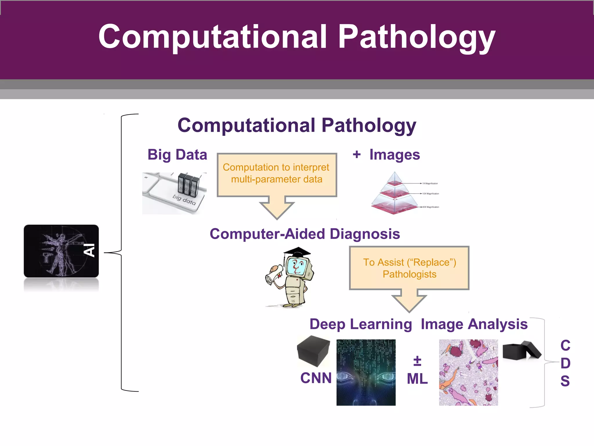
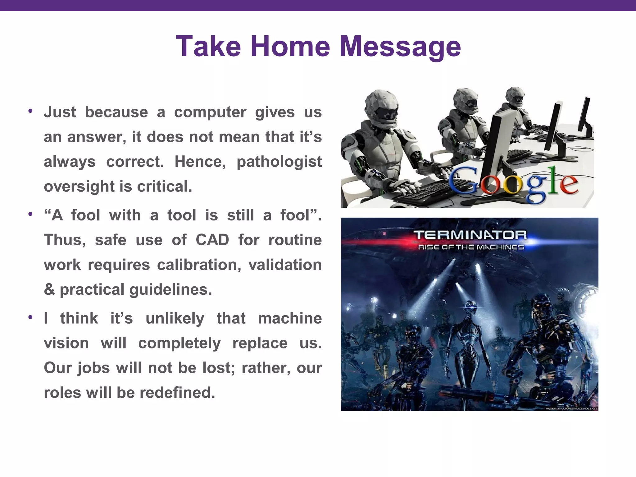
Computer Aided Diagnosis in
Pathology: Pros & Cons
Liron Pantanowitz MD
Director of Pathology Informatics & Cytology
Professor of Pathology & Biomedical Informatics
University of Pittsburgh Medical Center (UPMC)
pantanowitzl@upmc.edu
Disclosure
Consultant for Hamamatsu
Objectives
Overview of CAD in Pathology
 Pros & cons of image analysis
Recommendations for the field
AI General Facts
• AI is no longer just a science-fiction Hollywood story.
• 61% of people see AI as making the world a better place.
• 55% of people would trust using a self-driving car.
• 57% would prefer an AI doctor to perform an eye exam.
• Major investments in AI predicted to grow 300% in 2017.
• Workers do fear they could be replaced by machines!
AI in Healthcare
• Use of algorithms & software to
approximate human cognition to
analyze complex medical data.
• Early phases used fuzzy logic,
Bayesian networks & artificial
neural networks.
• Improved computing, EHRs,
growth of health-related data +
computer vision.
• Recent projects include Google
DeepMind & IBM Watson.
Google uses AI to detect
lymph node metastatic
breast carcinoma
Image Analysis Trends
• Business analyses predict software is high-growth market
• New players entering the market post FDA-approval of WSI
• Image analysis is indeed the “holy grail” of digital pathology
• Transition from qualitative (descriptive) to quantitative science
• Precision medicine currently demands precision diagnostics
• Current shift from research to more useful clinical applications
• Much hype surrounding automated Computer Aided Diagnosis
Computational Pathology
Big Data + Images
Computer-Aided Diagnosis
Deep Learning Image Analysis
Computation to interpret
multi-parameter data
To Assist (“Replace”)
Pathologists
C
D
S
±
MLCNN
AI
Why use image analysis?
Edward Adelson checkershadow illusion
Perception of color (human limitation)
Deep Learning for HER2
• Diagnostic discordance was caused by perception differences in
assessing HER2 due to stain heterogeneity.
Vandenberghe ME et al. Sci Reports 2017.
Image Analysis Benefits
• Better accuracy (more precise quantitative measurements).
• Standardization (more reproducible results, especially for
intermediate categories & complex scoring systems).
• Efficiency (reduce time consumption for pathologists, especially for
performing mundane tasks like counting, & triage cases – such as
weed out negative cases).
• CAD will soon help pathologists find, diagnose & grade cancer.
Killer App in Digital Pathology
Does something the microscope can’t
accomplish
Immunoscore
Predictive Medicine (Imaging vs Omics)
- Developed C-Path (Computational Pathologist)
system to measure a rich quantitative feature set
from breast cancer epithelium and stroma (6642
features).
- Included both standard morphometric
descriptors of image objects and higher-level
contextual, relational, and global image features.
- Their findings implicated stromal morphologic
structure as a previously unrecognized
prognostic determinant for breast cancer.
Google Image Search Engine (GISE)
50%
83%
Developing Image Algorithms
Machine Learning Approaches
Image
Analysis
Approach
Traditional Algorithm Deep Learning
App design Expert annotation Training dataset
Application Calibration Re-Train
Drawback Human dependent Data size & quality
Regulations Expected Plausible
Classic Image Analysis Steps
1. Image pre-processing (e.g. color normalization)
2. Classification
3. Detection (identification)
4. Segmentation
5. Feature extraction
6. Quantification
PD-L1 (CD274)
Teixido et al. Cancer Biol Med. 2015; 12(3):259IC = immune cells
Image courtesy of Dr. H. Guo
Variables
• Pre-analytical
– Tissue handling (collection, fixation, processing)
– Slide preparation (section thickness, artifacts like folds)
– Stain variation (IHC platform, color variation)
– Image acquisition (scanner difference, compression, etc.)
• Analytical
– Algorithms limited by file format & magnification
– Measurements vary with different algorithms
– Do you analyze regions of interest (ROI) vs. WSI
– Tumor heterogeneity (e.g. “hotspots”)
– Artifacts (tissue folds, air bubbles, crushed tissue, overlapping cells)
– Counting errors (e.g. cells between frames)
• Post-analytical
– e.g. Human interpretation, IT support
Image Compression (IHC view)
Image Compression (IA view)
Brightness Contrast
Compression Blurring
Her2/neu
Slide & Tissue ArtifactsSlide & Tissue Artifacts
Comparison of Algorithms
Combrinck M, Fine J, Pantanowitz L. J Pathol Inform 2015, 6:S3-S4.
ER Ki67
Within Scanner Variability
Automated prostate gland segmentation
Aperio Phillips Ventana
QIA Guideline from CAP
• Scope:
– Provide recommendations for improving reproducibility, precision,
& accuracy of QIA for HER2 by IHC
• Topics:
– Algorithm selection (e.g. locked down, FDA-approved only?)
– System validation (what is appropriate for clinical use?)
– Calibration (reproducibility of results and controls to be used?)
– Training & operation (which staff to involve, ROI selection?)
– Performance monitoring (QA and change control process?)
• Methods:
– Expert & advisory panels
– Systematic literature review
– Publication expected soon
Platforms
Hazards of AI & Data Mining
• Many failed projects
• Inaccurate predictions
• Inappropriate modeling
• Reliability of input data
• Technological mistrust
• Accountability
GIGO Principle
DATA MODEL RESULT
DATA MODEL RESULT
DATA MODEL RESULT
DATA MODEL RESULT
e.g. Criminal Machine Learning
• Wu & Zhang. Automated
inference on criminality using
face images. arXive. Nov 2016.
• ML to detect human face features
(1,800+ photos).
• Accurately (90%) distinguished
criminals vs. non-criminals.
• Only non-criminals were faintly
smiling!
Pathologists as Information Scientists
• Pathologists have always embraced technology in the lab.
• Some tasks once performed manually have been automated
(e.g. cell counts, Pap tests), leaving pathologists with more
complex tasks.
Jha & Topol. JAMA 2016; 316 (22)
• But can AI perform the more complex tasks of pathologists?
• And, in some instances, with superior accuracy?
Take Home Message
• Just because a computer gives us
an answer, it does not mean that it’s
always correct. Hence, pathologist
oversight is critical.
• “A fool with a tool is still a fool”.
Thus, safe use of CAD for routine
work requires calibration, validation
& practical guidelines.
• I think it’s unlikely that machine
vision will completely replace us.
Our jobs will not be lost; rather, our
roles will be redefined.
Questions?

Computer Aided Diagnosis in Pathology: Pros & Cons by Dr. Liron Pantanowitz

  • 1.
    Computer Aided Diagnosisin Pathology: Pros & Cons Liron Pantanowitz MD Director of Pathology Informatics & Cytology Professor of Pathology & Biomedical Informatics University of Pittsburgh Medical Center (UPMC) pantanowitzl@upmc.edu
  • 2.
  • 3.
    Objectives Overview of CADin Pathology  Pros & cons of image analysis Recommendations for the field
  • 4.
    AI General Facts •AI is no longer just a science-fiction Hollywood story. • 61% of people see AI as making the world a better place. • 55% of people would trust using a self-driving car. • 57% would prefer an AI doctor to perform an eye exam. • Major investments in AI predicted to grow 300% in 2017. • Workers do fear they could be replaced by machines!
  • 5.
    AI in Healthcare •Use of algorithms & software to approximate human cognition to analyze complex medical data. • Early phases used fuzzy logic, Bayesian networks & artificial neural networks. • Improved computing, EHRs, growth of health-related data + computer vision. • Recent projects include Google DeepMind & IBM Watson.
  • 6.
    Google uses AIto detect lymph node metastatic breast carcinoma
  • 7.
    Image Analysis Trends •Business analyses predict software is high-growth market • New players entering the market post FDA-approval of WSI • Image analysis is indeed the “holy grail” of digital pathology • Transition from qualitative (descriptive) to quantitative science • Precision medicine currently demands precision diagnostics • Current shift from research to more useful clinical applications • Much hype surrounding automated Computer Aided Diagnosis
  • 8.
    Computational Pathology Computational Pathology BigData + Images Computer-Aided Diagnosis Deep Learning Image Analysis Computation to interpret multi-parameter data To Assist (“Replace”) Pathologists C D S ± MLCNN AI
  • 9.
    Why use imageanalysis? Edward Adelson checkershadow illusion Perception of color (human limitation)
  • 10.
    Deep Learning forHER2 • Diagnostic discordance was caused by perception differences in assessing HER2 due to stain heterogeneity. Vandenberghe ME et al. Sci Reports 2017.
  • 12.
    Image Analysis Benefits •Better accuracy (more precise quantitative measurements). • Standardization (more reproducible results, especially for intermediate categories & complex scoring systems). • Efficiency (reduce time consumption for pathologists, especially for performing mundane tasks like counting, & triage cases – such as weed out negative cases). • CAD will soon help pathologists find, diagnose & grade cancer.
  • 14.
    Killer App inDigital Pathology Does something the microscope can’t accomplish
  • 15.
  • 16.
  • 17.
    - Developed C-Path(Computational Pathologist) system to measure a rich quantitative feature set from breast cancer epithelium and stroma (6642 features). - Included both standard morphometric descriptors of image objects and higher-level contextual, relational, and global image features. - Their findings implicated stromal morphologic structure as a previously unrecognized prognostic determinant for breast cancer.
  • 18.
    Google Image SearchEngine (GISE) 50% 83%
  • 20.
  • 21.
    Machine Learning Approaches Image Analysis Approach TraditionalAlgorithm Deep Learning App design Expert annotation Training dataset Application Calibration Re-Train Drawback Human dependent Data size & quality Regulations Expected Plausible
  • 22.
    Classic Image AnalysisSteps 1. Image pre-processing (e.g. color normalization) 2. Classification 3. Detection (identification) 4. Segmentation 5. Feature extraction 6. Quantification
  • 23.
    PD-L1 (CD274) Teixido etal. Cancer Biol Med. 2015; 12(3):259IC = immune cells
  • 26.
  • 27.
  • 28.
    Variables • Pre-analytical – Tissuehandling (collection, fixation, processing) – Slide preparation (section thickness, artifacts like folds) – Stain variation (IHC platform, color variation) – Image acquisition (scanner difference, compression, etc.) • Analytical – Algorithms limited by file format & magnification – Measurements vary with different algorithms – Do you analyze regions of interest (ROI) vs. WSI – Tumor heterogeneity (e.g. “hotspots”) – Artifacts (tissue folds, air bubbles, crushed tissue, overlapping cells) – Counting errors (e.g. cells between frames) • Post-analytical – e.g. Human interpretation, IT support
  • 29.
  • 30.
  • 31.
  • 32.
    Her2/neu Slide & TissueArtifactsSlide & Tissue Artifacts
  • 33.
    Comparison of Algorithms CombrinckM, Fine J, Pantanowitz L. J Pathol Inform 2015, 6:S3-S4. ER Ki67
  • 34.
  • 35.
    Automated prostate glandsegmentation Aperio Phillips Ventana
  • 37.
    QIA Guideline fromCAP • Scope: – Provide recommendations for improving reproducibility, precision, & accuracy of QIA for HER2 by IHC • Topics: – Algorithm selection (e.g. locked down, FDA-approved only?) – System validation (what is appropriate for clinical use?) – Calibration (reproducibility of results and controls to be used?) – Training & operation (which staff to involve, ROI selection?) – Performance monitoring (QA and change control process?) • Methods: – Expert & advisory panels – Systematic literature review – Publication expected soon
  • 39.
  • 40.
    Hazards of AI& Data Mining • Many failed projects • Inaccurate predictions • Inappropriate modeling • Reliability of input data • Technological mistrust • Accountability
  • 43.
  • 44.
  • 45.
  • 46.
  • 47.
  • 48.
    e.g. Criminal MachineLearning • Wu & Zhang. Automated inference on criminality using face images. arXive. Nov 2016. • ML to detect human face features (1,800+ photos). • Accurately (90%) distinguished criminals vs. non-criminals. • Only non-criminals were faintly smiling!
  • 49.
    Pathologists as InformationScientists • Pathologists have always embraced technology in the lab. • Some tasks once performed manually have been automated (e.g. cell counts, Pap tests), leaving pathologists with more complex tasks. Jha & Topol. JAMA 2016; 316 (22) • But can AI perform the more complex tasks of pathologists? • And, in some instances, with superior accuracy?
  • 50.
    Take Home Message •Just because a computer gives us an answer, it does not mean that it’s always correct. Hence, pathologist oversight is critical. • “A fool with a tool is still a fool”. Thus, safe use of CAD for routine work requires calibration, validation & practical guidelines. • I think it’s unlikely that machine vision will completely replace us. Our jobs will not be lost; rather, our roles will be redefined.
  • 51.

Editor's Notes

  • #26 … … So, what was going on in the image? … … It may seem easy to lots of people who grow up in this country, but it was not easy for me. I got it wrong. Yes, I know who this guy is. But, I don’t know the other guys. I thought that a group of world leaders are taking turns to measure their body weight. I was wondering what kind of meeting were they having. For some reason, Obama was impatient and can not wait for his turn. And he jumped the gun. But, I did not understand what he and the other guys was laughing about. I was wrong because I grow up in a different country where nobody cares about their body weight. Also, I did not know these people are friends and they work together.
  • #27 Now, let me use this example to walk you through VisioPharm. The purpose is to find jackets, faces, ceiling, floor, and lockers. So, I draw different labels on different objects.
  • #28 Not too bad. Here is the finding by VisioPharm. Let’s compare to the original picture and see how good it is. Suits =Hair = Shoes
  • #35 This graph shows 4 immunostained prostate tissue cores that were scanned on the same WSI scanner every week for 12 weeks and analyzed (IHC quantified) using the same image analysis algorithm. This illustrates the marked variability of outcome data based on variations in image acquisition. The major drop in measurements at week 9 was related to changing the light bulb in the WSI scanner