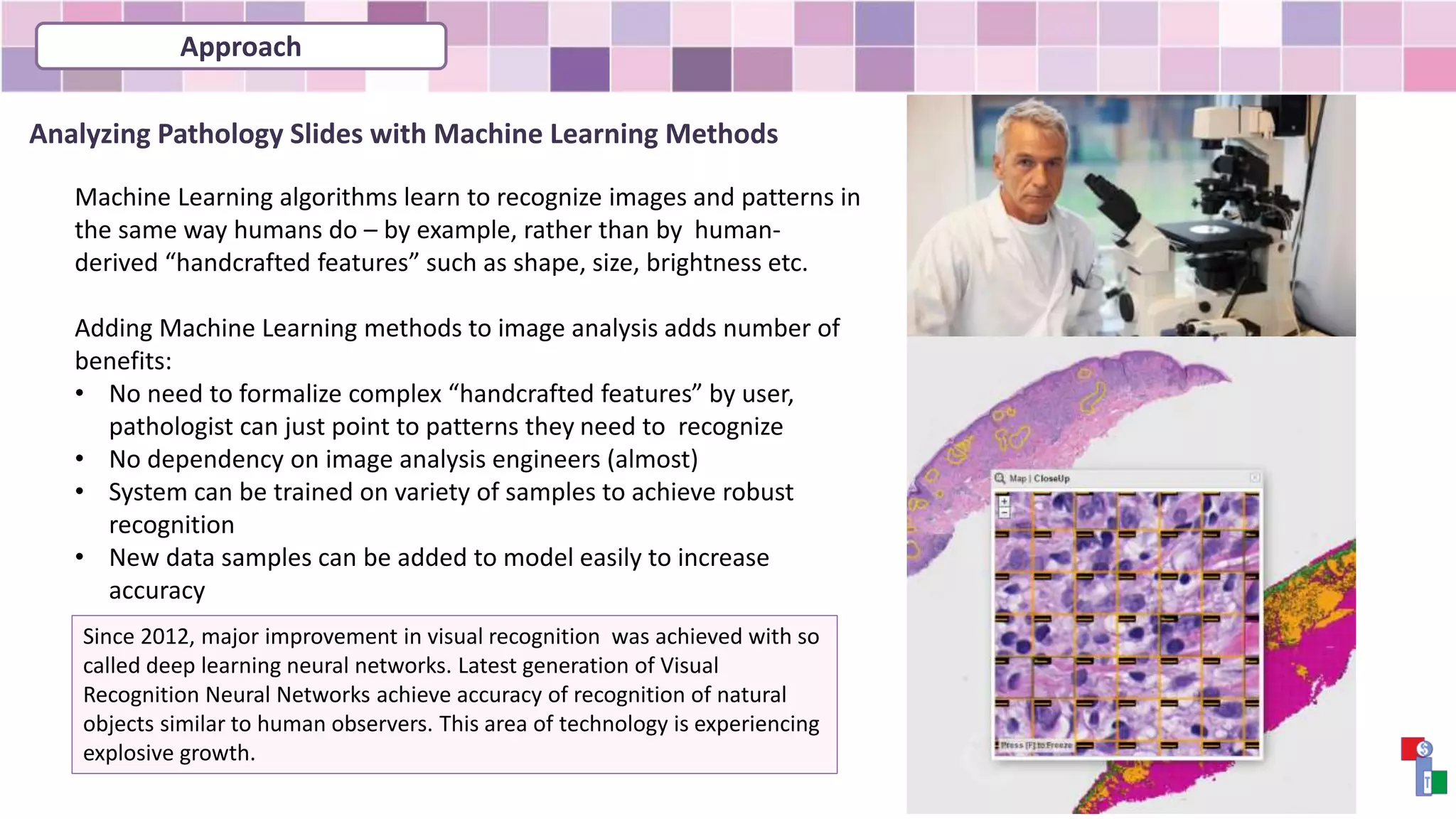
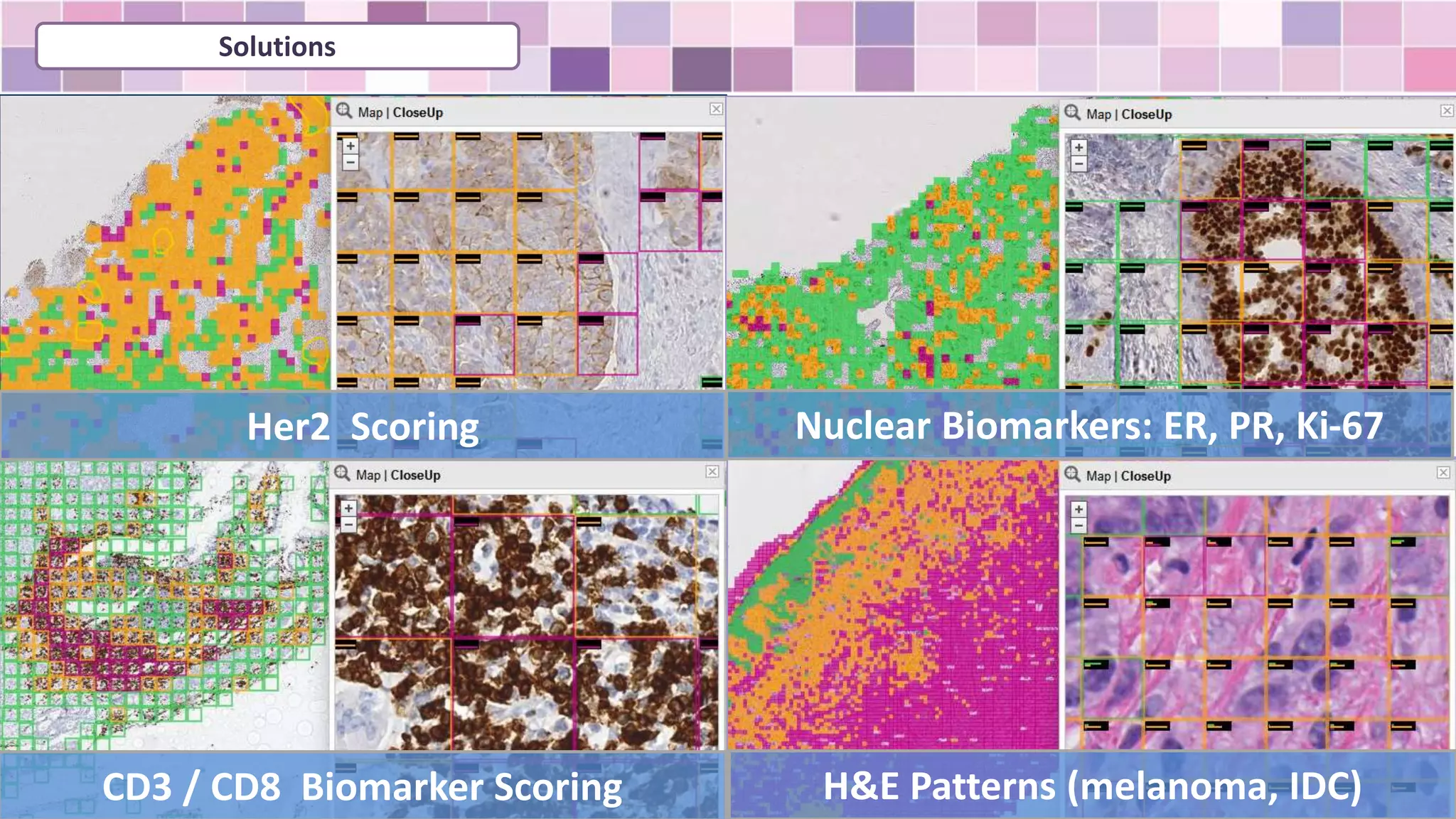
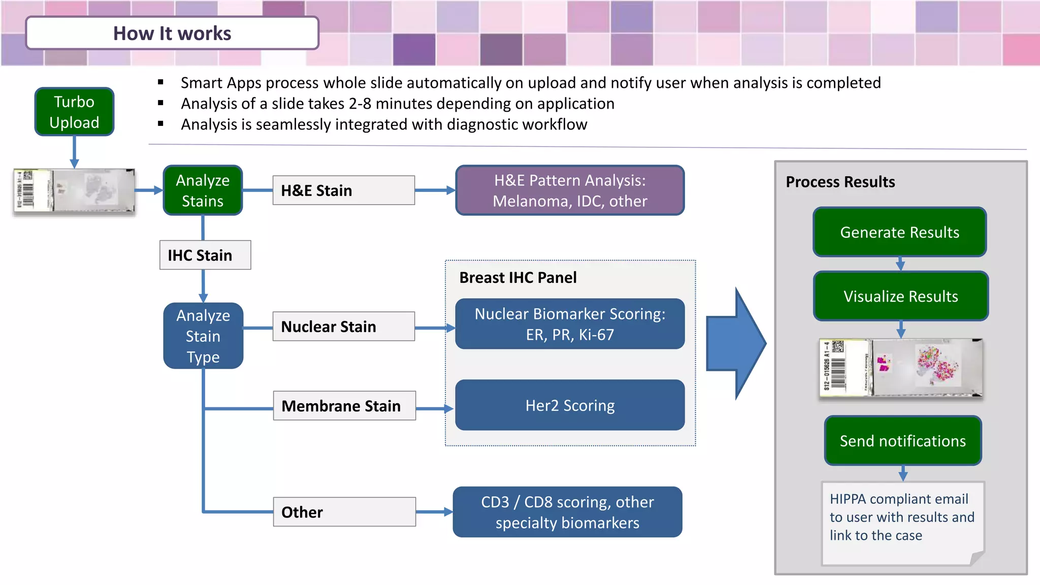
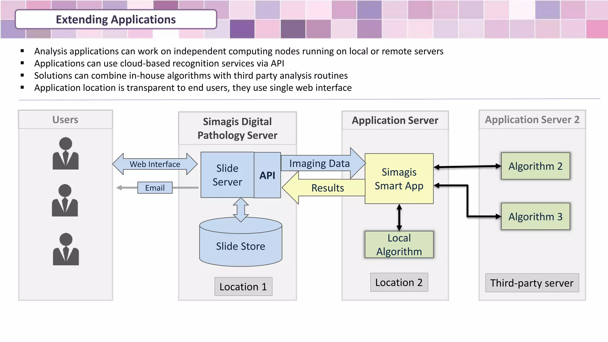
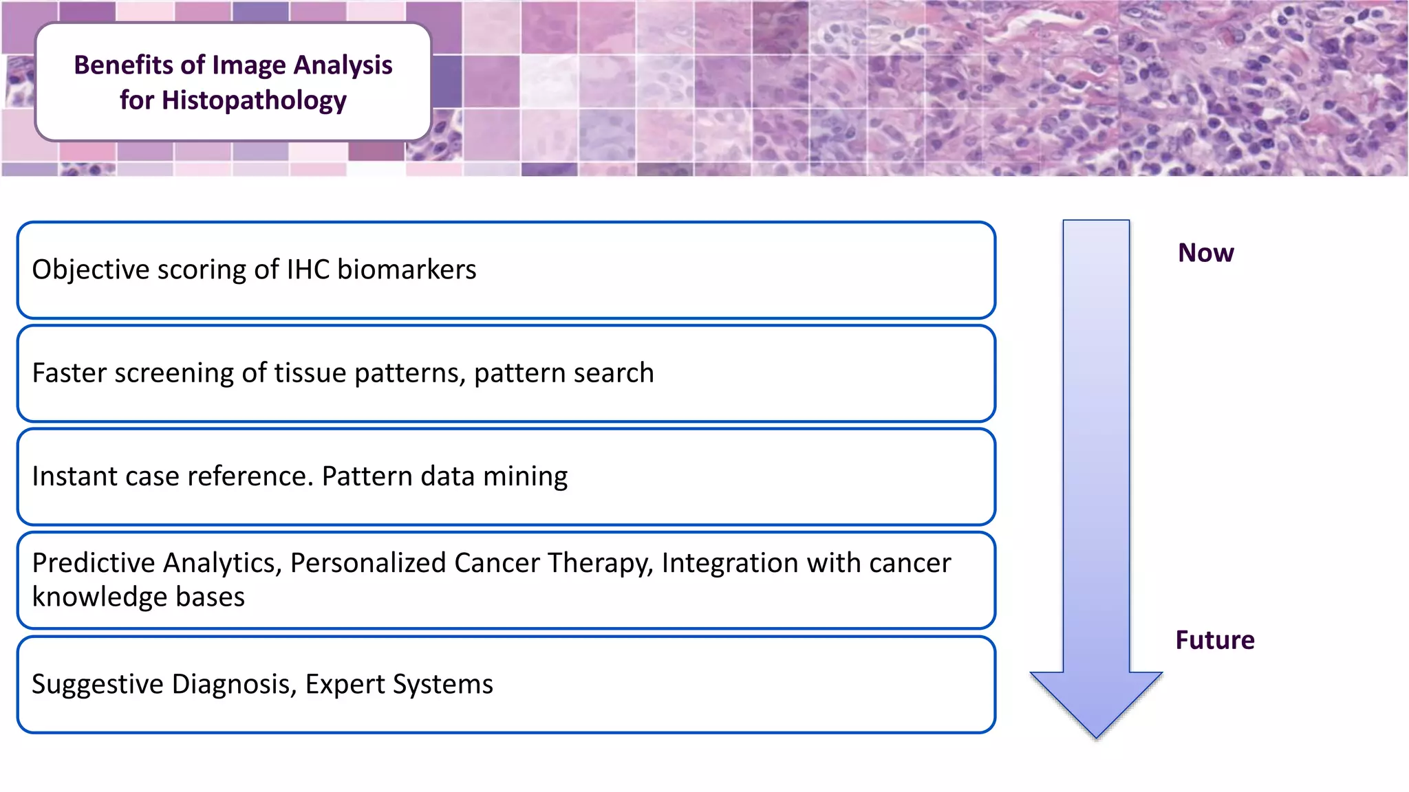
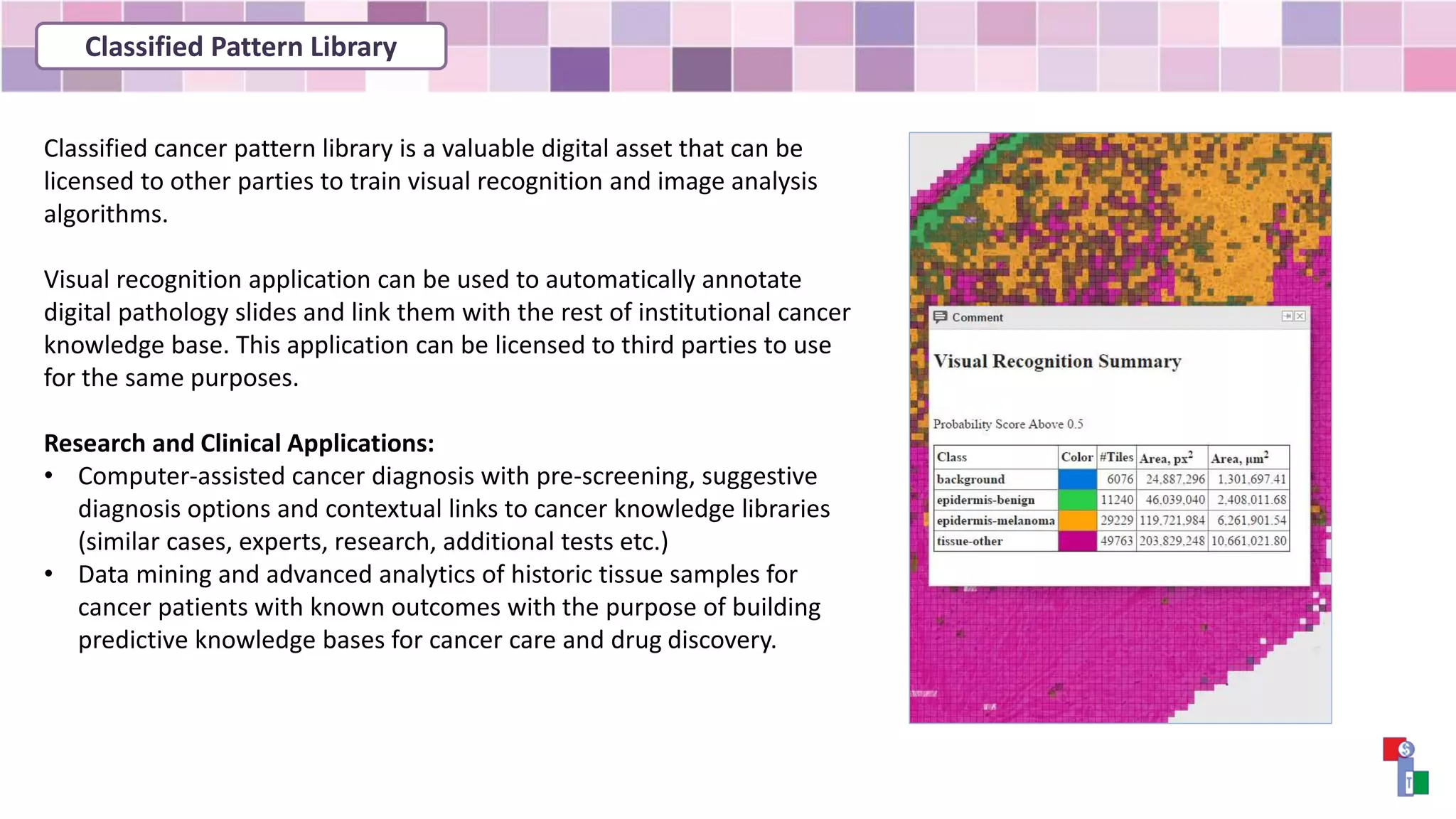
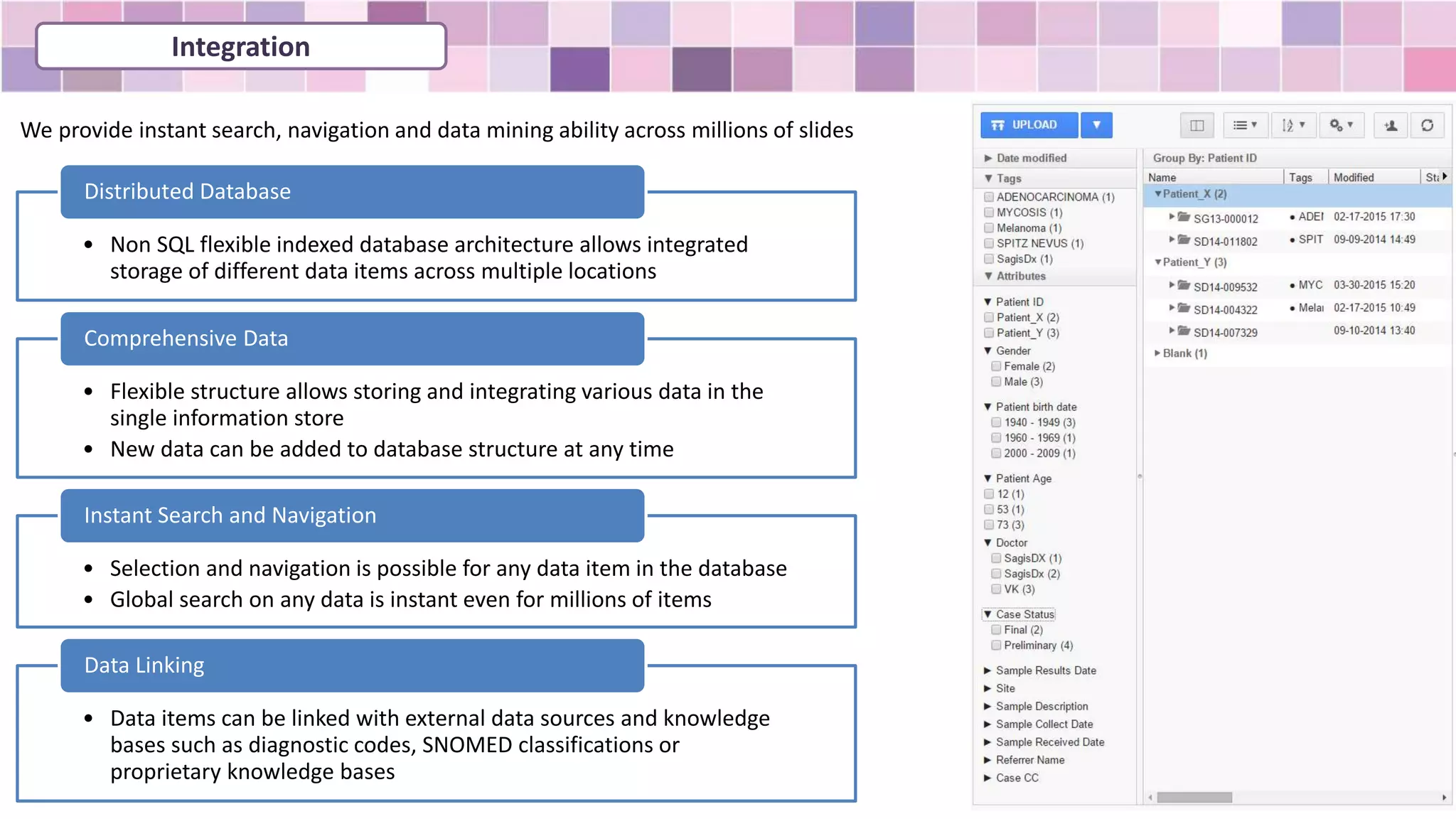
SMART IMAGING TECHNOLOGIES web-pathology.net
Machine Learning
in Digital Pathology
Analyzing Pathology Slides with Machine Learning Methods
Machine Learning algorithms learn to recognize images and patterns in
the same way humans do – by example, rather than by human-
derived “handcrafted features” such as shape, size, brightness etc.
Adding Machine Learning methods to image analysis adds number of
benefits:
• No need to formalize complex “handcrafted features” by user,
pathologist can just point to patterns they need to recognize
• No dependency on image analysis engineers (almost)
• System can be trained on variety of samples to achieve robust
recognition
• New data samples can be added to model easily to increase
accuracy
Since 2012, major improvement in visual recognition was achieved with so
called deep learning neural networks. Latest generation of Visual
Recognition Neural Networks achieve accuracy of recognition of natural
objects similar to human observers. This area of technology is experiencing
explosive growth.
Approach
Solutions
Her2 Scoring Nuclear Biomarkers: ER, PR, Ki-67
CD3 / CD8 Biomarker Scoring H&E Patterns (melanoma, IDC)
How It works
Turbo
Upload
Analyze
Stains
H&E Pattern Analysis:
Melanoma, IDC, other
H&E Stain
Analyze
Stain
Type
Nuclear Biomarker Scoring:
ER, PR, Ki-67
Her2 Scoring
Breast IHC Panel
CD3 / CD8 scoring, other
specialty biomarkers
Nuclear Stain
Other
Membrane Stain
IHC Stain
Generate Results
Visualize Results
Send notifications
HIPPA compliant email
to user with results and
link to the case
 Smart Apps process whole slide automatically on upload and notify user when analysis is completed
 Analysis of a slide takes 2-8 minutes depending on application
 Analysis is seamlessly integrated with diagnostic workflow
Process Results
Extending Applications
 Analysis applications can work on independent computing nodes running on local or remote servers
 Applications can use cloud-based recognition services via API
 Solutions can combine in-house algorithms with third party analysis routines
 Application location is transparent to end users, they use single web interface
Slide Store
Slide
Server
Simagis
Smart App
Simagis Digital
Pathology Server
Local
Algorithm
Algorithm 2
Third-party server
Algorithm 3
API
Imaging Data
Application Server
Results
Application Server 2
Web Interface
Users
Location 2Location 1
Email
Objective scoring of IHC biomarkers
Faster screening of tissue patterns, pattern search
Instant case reference. Pattern data mining
Predictive Analytics, Personalized Cancer Therapy, Integration with cancer
knowledge bases
Suggestive Diagnosis, Expert Systems
Now
Future
Benefits of Image Analysis
for Histopathology
Classified cancer pattern library is a valuable digital asset that can be
licensed to other parties to train visual recognition and image analysis
algorithms.
Visual recognition application can be used to automatically annotate
digital pathology slides and link them with the rest of institutional cancer
knowledge base. This application can be licensed to third parties to use
for the same purposes.
Research and Clinical Applications:
• Computer-assisted cancer diagnosis with pre-screening, suggestive
diagnosis options and contextual links to cancer knowledge libraries
(similar cases, experts, research, additional tests etc.)
• Data mining and advanced analytics of historic tissue samples for
cancer patients with known outcomes with the purpose of building
predictive knowledge bases for cancer care and drug discovery.
Classified Pattern Library
• Non SQL flexible indexed database architecture allows integrated
storage of different data items across multiple locations
Distributed Database
• Flexible structure allows storing and integrating various data in the
single information store
• New data can be added to database structure at any time
Comprehensive Data
• Selection and navigation is possible for any data item in the database
• Global search on any data is instant even for millions of items
Instant Search and Navigation
• Data items can be linked with external data sources and knowledge
bases such as diagnostic codes, SNOMED classifications or
proprietary knowledge bases
Data Linking
We provide instant search, navigation and data mining ability across millions of slides
Integration
Integration: Information Systems
• RESTful API with live examples and templates provide easy
integration with third-party applications
Easy API
• Integration with other medical information systems is available
via HL7 Integration Engine (Rhapsody by Orion Health)
LIS / EMR Systems
• Third party image analysis application can access images and
metadata
Algorithms
• Information in the database can be integrated with other web
based knowledge system via standard integration protocols
Knowledge Bases
Our product includes standard industry data exchange protocols and
APIs for integration with any third party application
Integration
Over 2000 registered product users on all
continents
US Consultation Network of over 200 Pathologists
with experts in every specialty
Clients and Partners

Machine Learning in Pathology Diagnostics with Simagis Live

  • 1.
    SMART IMAGING TECHNOLOGIESweb-pathology.net Machine Learning in Digital Pathology
  • 2.
    Analyzing Pathology Slideswith Machine Learning Methods Machine Learning algorithms learn to recognize images and patterns in the same way humans do – by example, rather than by human- derived “handcrafted features” such as shape, size, brightness etc. Adding Machine Learning methods to image analysis adds number of benefits: • No need to formalize complex “handcrafted features” by user, pathologist can just point to patterns they need to recognize • No dependency on image analysis engineers (almost) • System can be trained on variety of samples to achieve robust recognition • New data samples can be added to model easily to increase accuracy Since 2012, major improvement in visual recognition was achieved with so called deep learning neural networks. Latest generation of Visual Recognition Neural Networks achieve accuracy of recognition of natural objects similar to human observers. This area of technology is experiencing explosive growth. Approach
  • 3.
    Solutions Her2 Scoring NuclearBiomarkers: ER, PR, Ki-67 CD3 / CD8 Biomarker Scoring H&E Patterns (melanoma, IDC)
  • 4.
    How It works Turbo Upload Analyze Stains H&EPattern Analysis: Melanoma, IDC, other H&E Stain Analyze Stain Type Nuclear Biomarker Scoring: ER, PR, Ki-67 Her2 Scoring Breast IHC Panel CD3 / CD8 scoring, other specialty biomarkers Nuclear Stain Other Membrane Stain IHC Stain Generate Results Visualize Results Send notifications HIPPA compliant email to user with results and link to the case  Smart Apps process whole slide automatically on upload and notify user when analysis is completed  Analysis of a slide takes 2-8 minutes depending on application  Analysis is seamlessly integrated with diagnostic workflow Process Results
  • 5.
    Extending Applications  Analysisapplications can work on independent computing nodes running on local or remote servers  Applications can use cloud-based recognition services via API  Solutions can combine in-house algorithms with third party analysis routines  Application location is transparent to end users, they use single web interface Slide Store Slide Server Simagis Smart App Simagis Digital Pathology Server Local Algorithm Algorithm 2 Third-party server Algorithm 3 API Imaging Data Application Server Results Application Server 2 Web Interface Users Location 2Location 1 Email
  • 6.
    Objective scoring ofIHC biomarkers Faster screening of tissue patterns, pattern search Instant case reference. Pattern data mining Predictive Analytics, Personalized Cancer Therapy, Integration with cancer knowledge bases Suggestive Diagnosis, Expert Systems Now Future Benefits of Image Analysis for Histopathology
  • 7.
    Classified cancer patternlibrary is a valuable digital asset that can be licensed to other parties to train visual recognition and image analysis algorithms. Visual recognition application can be used to automatically annotate digital pathology slides and link them with the rest of institutional cancer knowledge base. This application can be licensed to third parties to use for the same purposes. Research and Clinical Applications: • Computer-assisted cancer diagnosis with pre-screening, suggestive diagnosis options and contextual links to cancer knowledge libraries (similar cases, experts, research, additional tests etc.) • Data mining and advanced analytics of historic tissue samples for cancer patients with known outcomes with the purpose of building predictive knowledge bases for cancer care and drug discovery. Classified Pattern Library
  • 8.
    • Non SQLflexible indexed database architecture allows integrated storage of different data items across multiple locations Distributed Database • Flexible structure allows storing and integrating various data in the single information store • New data can be added to database structure at any time Comprehensive Data • Selection and navigation is possible for any data item in the database • Global search on any data is instant even for millions of items Instant Search and Navigation • Data items can be linked with external data sources and knowledge bases such as diagnostic codes, SNOMED classifications or proprietary knowledge bases Data Linking We provide instant search, navigation and data mining ability across millions of slides Integration
  • 9.
    Integration: Information Systems •RESTful API with live examples and templates provide easy integration with third-party applications Easy API • Integration with other medical information systems is available via HL7 Integration Engine (Rhapsody by Orion Health) LIS / EMR Systems • Third party image analysis application can access images and metadata Algorithms • Information in the database can be integrated with other web based knowledge system via standard integration protocols Knowledge Bases Our product includes standard industry data exchange protocols and APIs for integration with any third party application Integration
  • 10.
    Over 2000 registeredproduct users on all continents US Consultation Network of over 200 Pathologists with experts in every specialty Clients and Partners