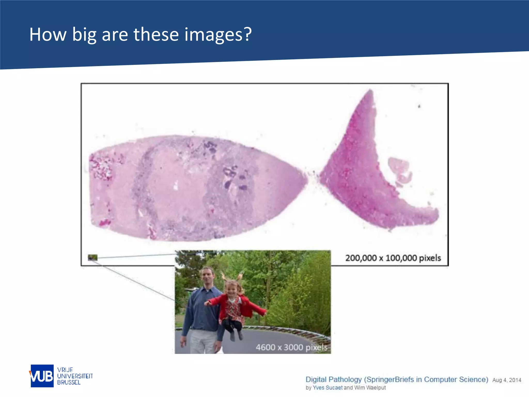
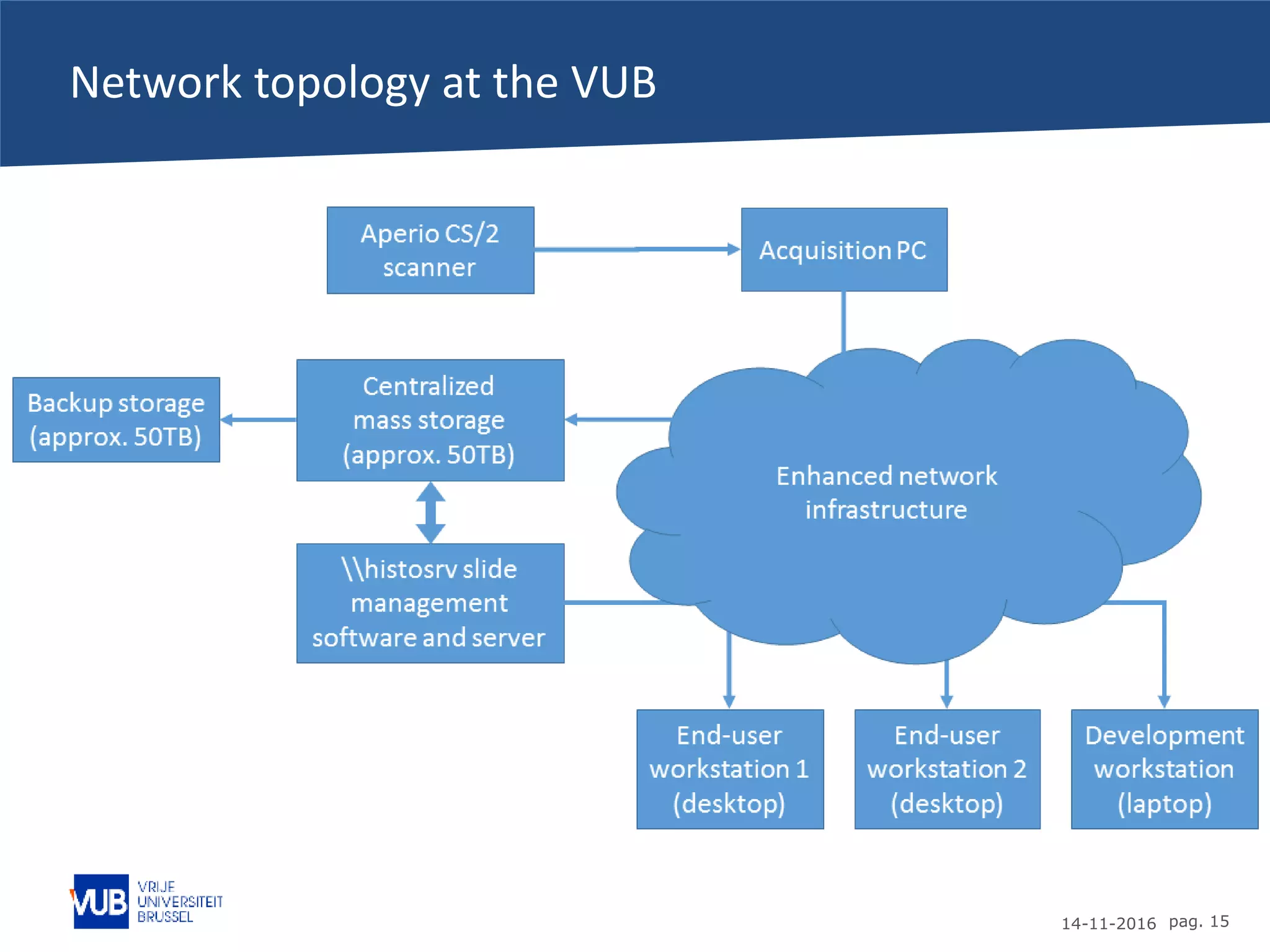
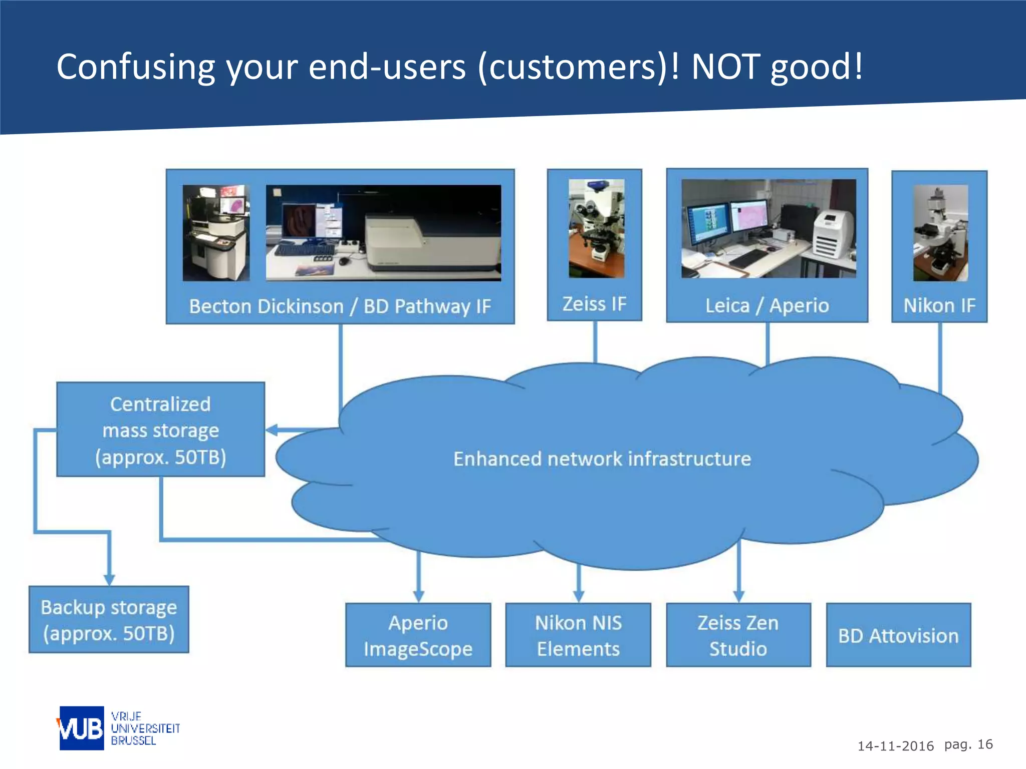
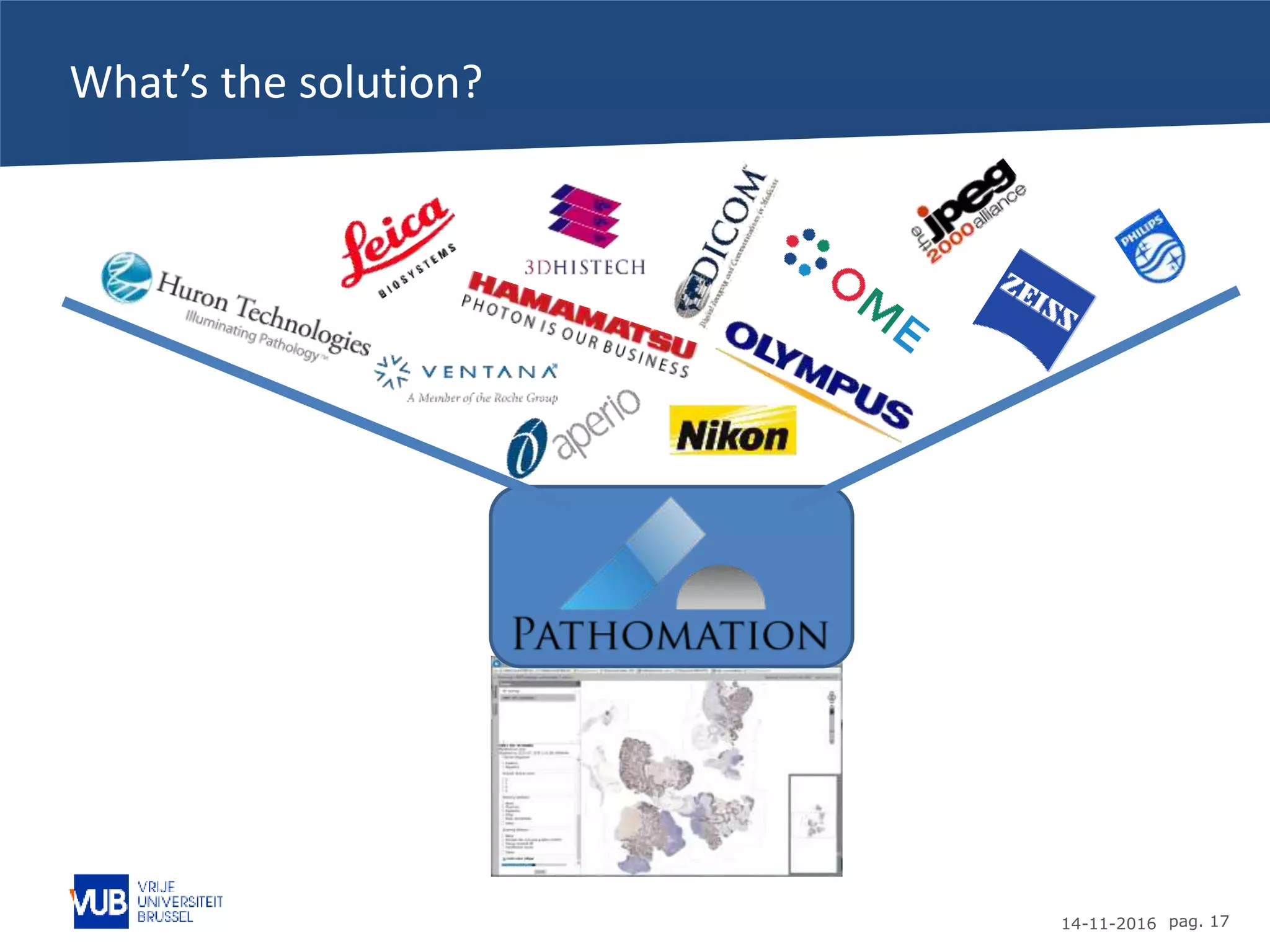
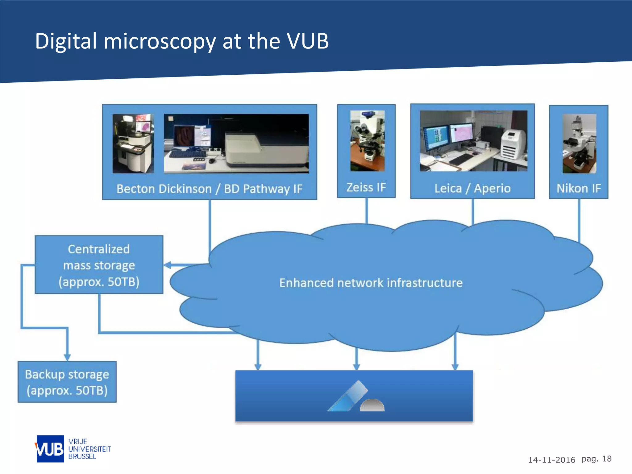
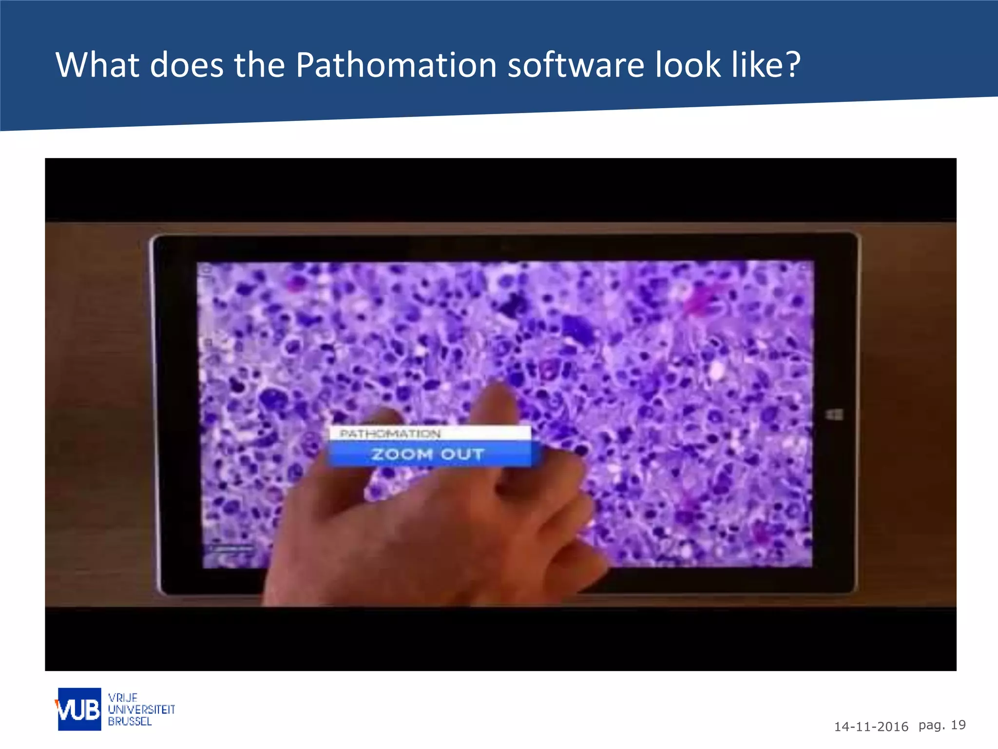
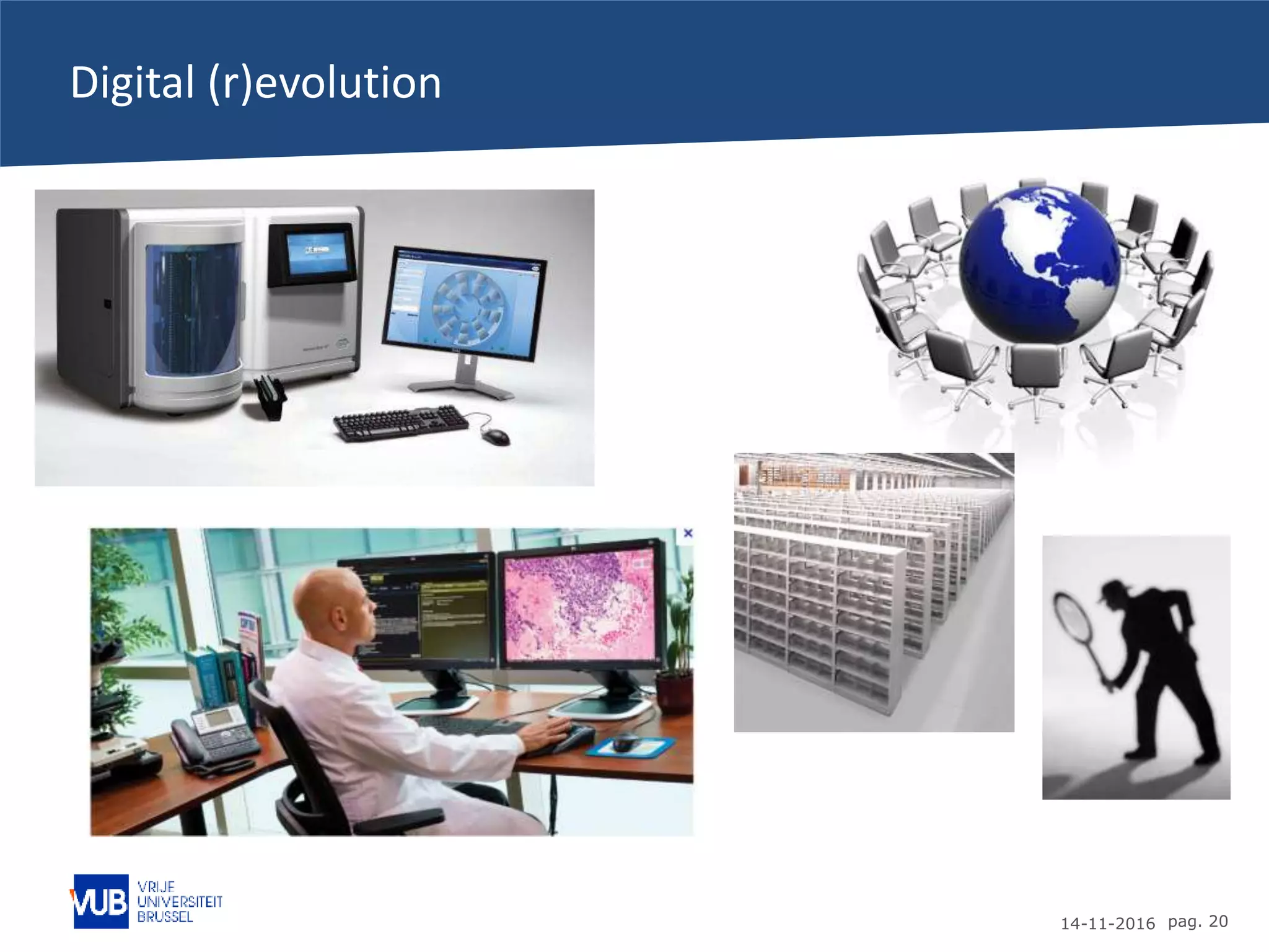
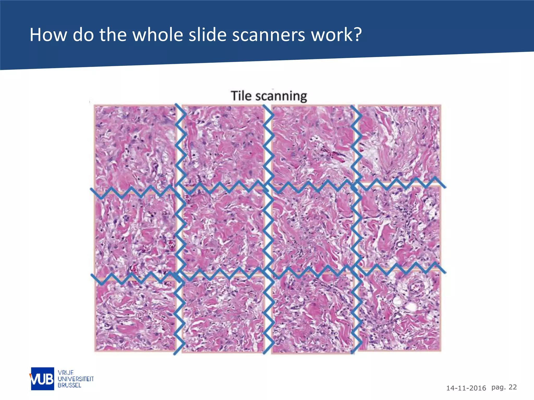
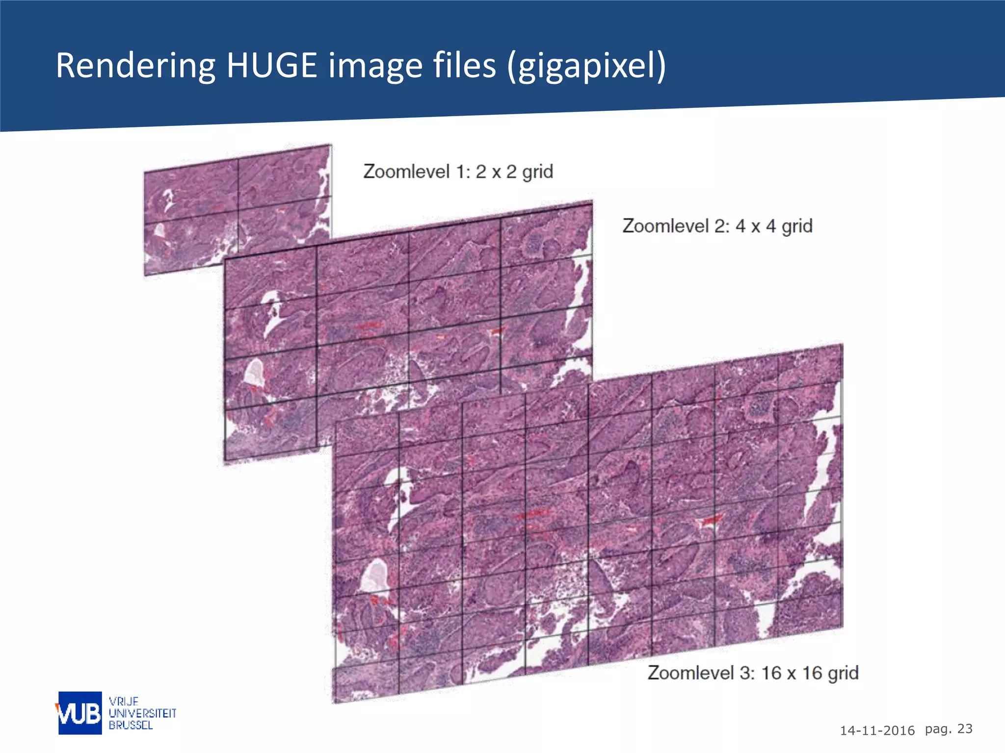
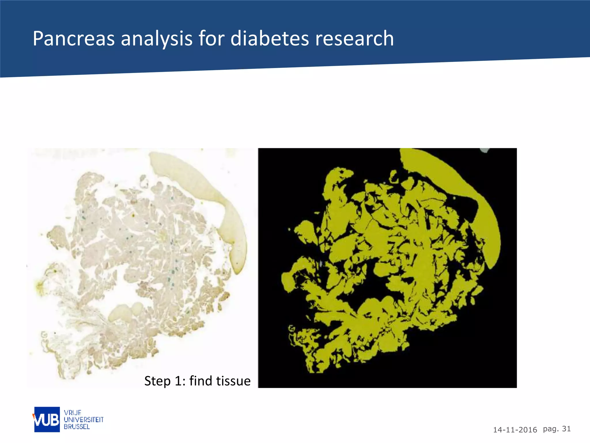
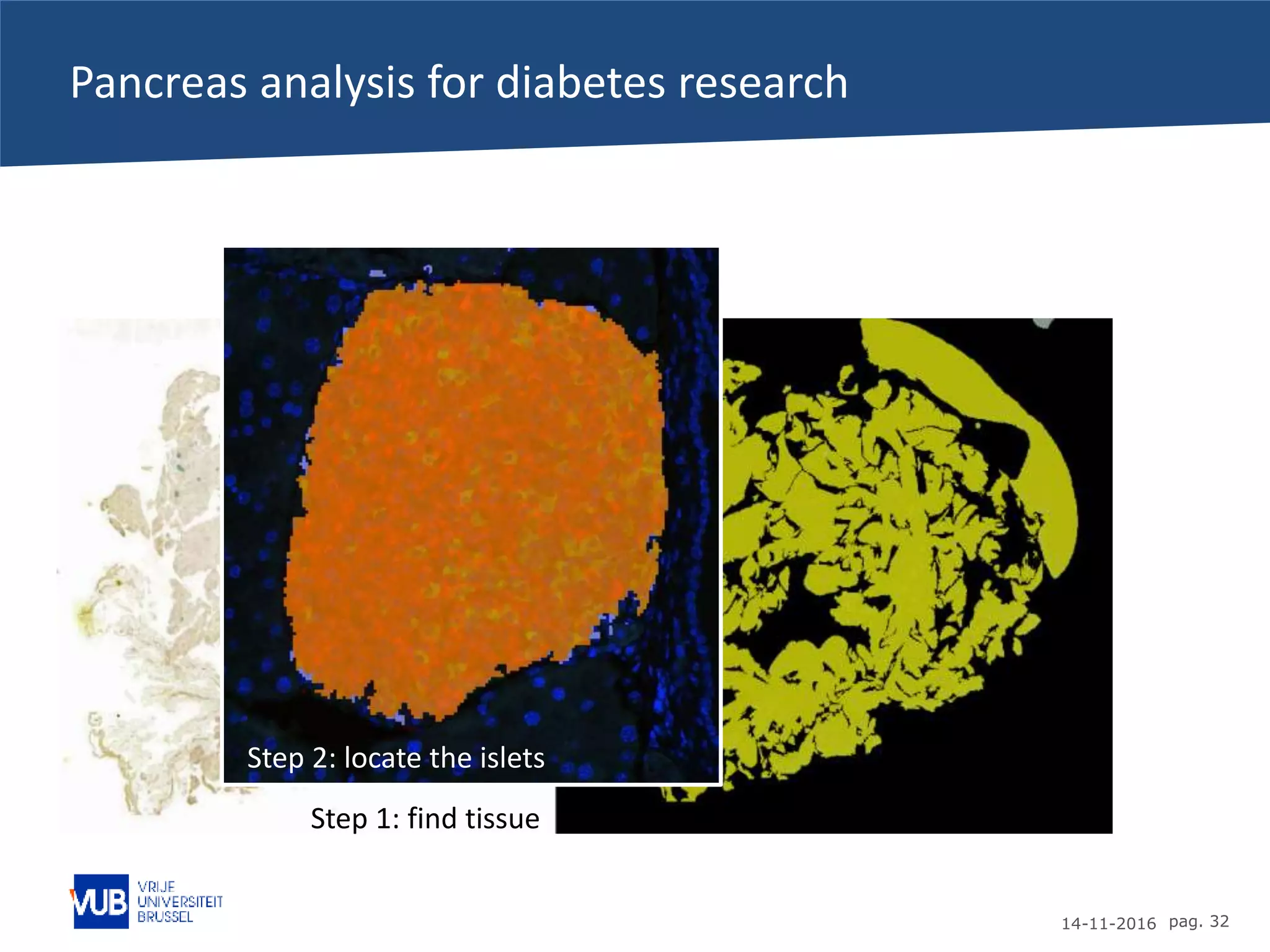
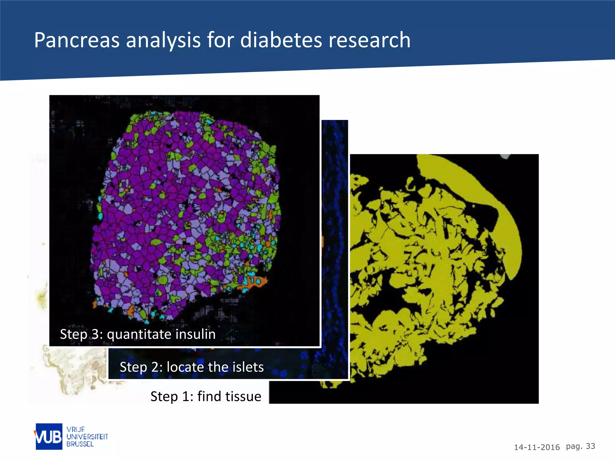
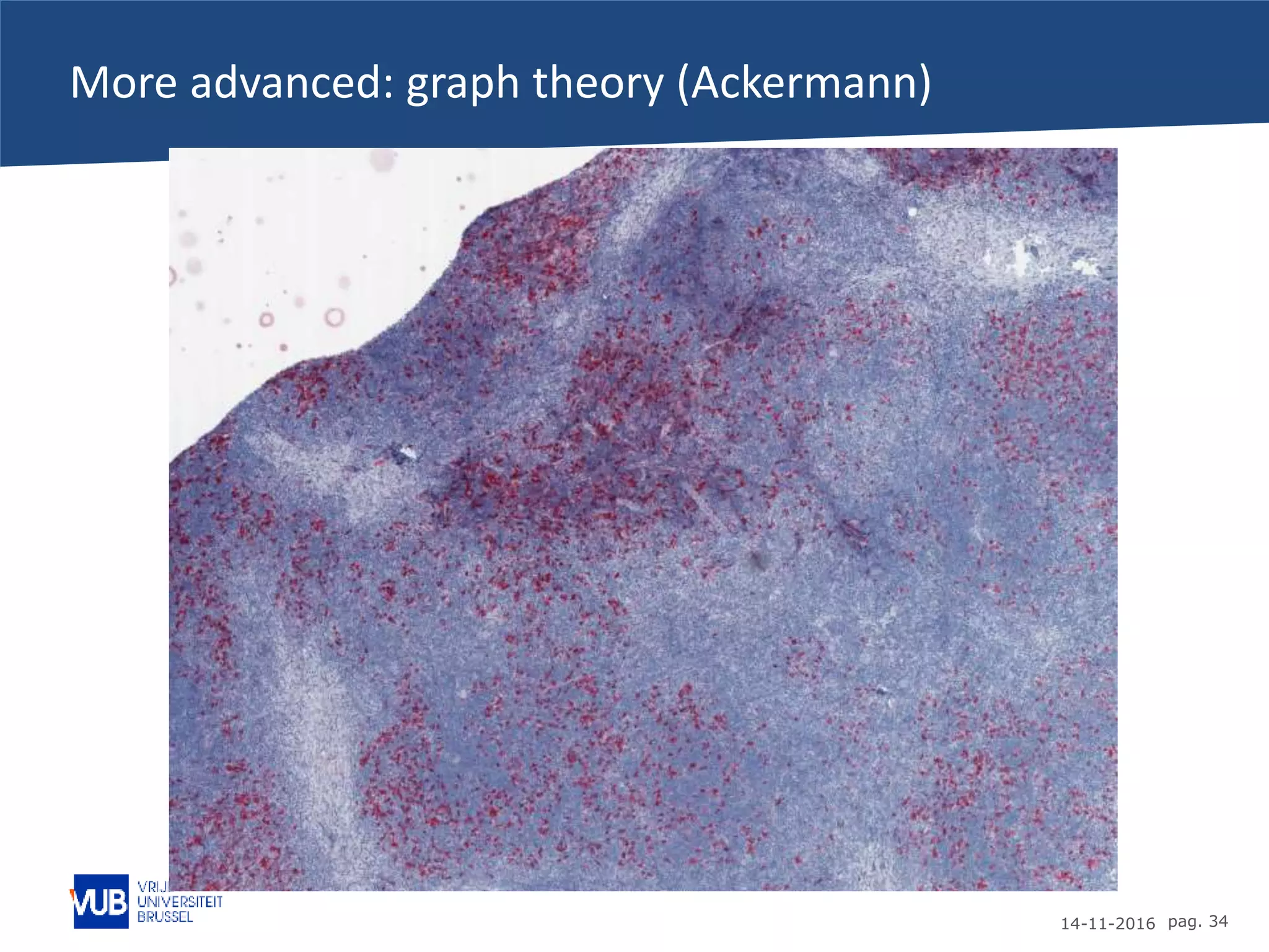
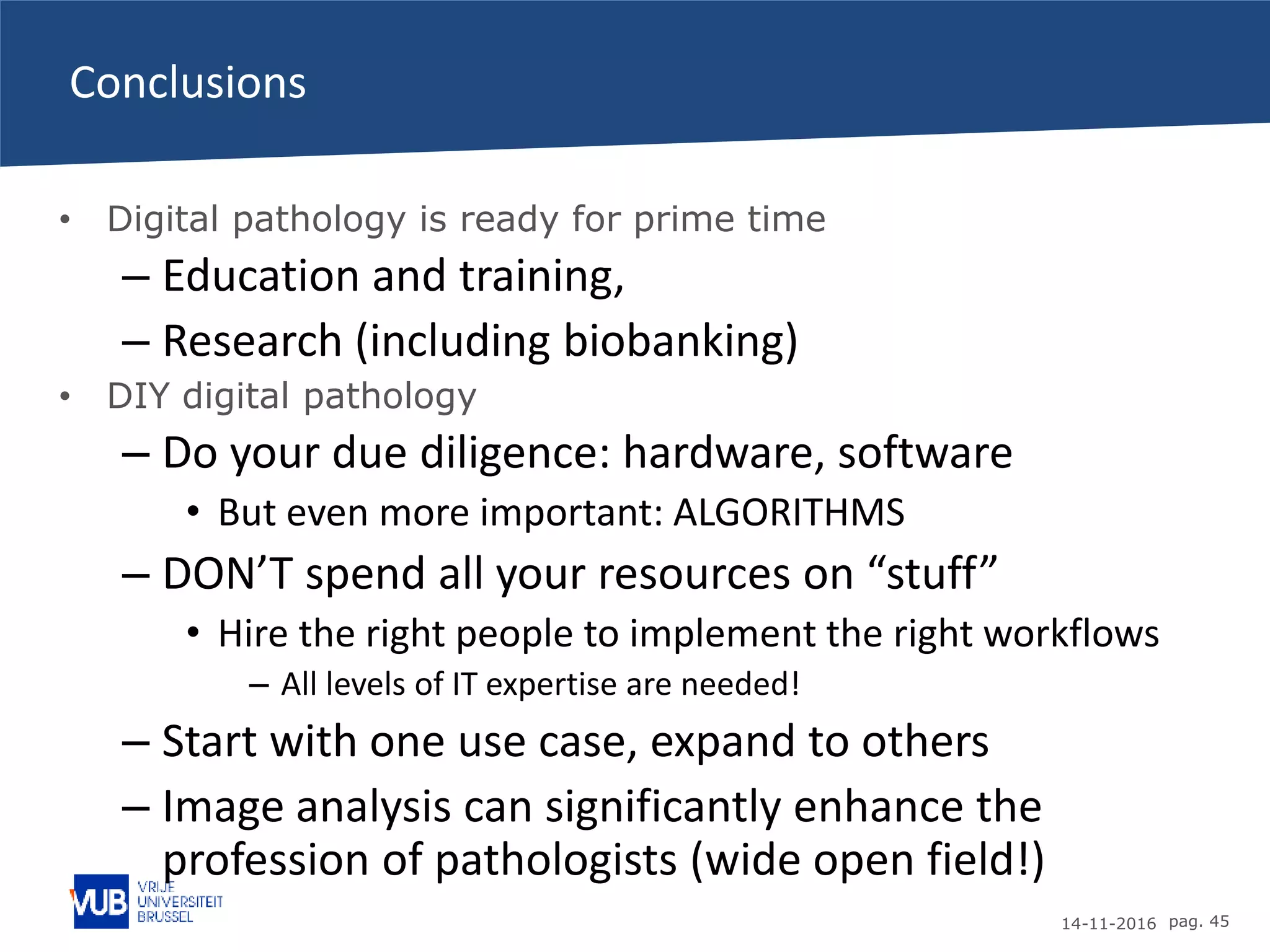
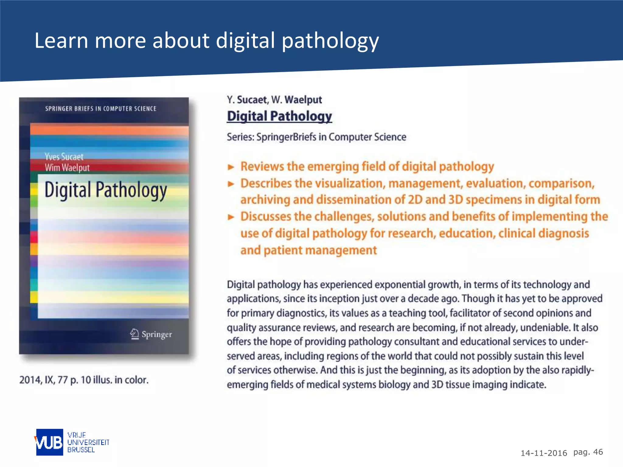
The advent of
digital
microscopy
Yves Sucaet,
Wim Waelput,
Peter In’t Veld
IT / ComS edition 
14-11-2016 pag. 2
Financial disclosure
• Yves Sucaet and Wim Waelput are co-founders and
shareholders in Pathomation, an innovative company founded
in 2012. The company strives to offer the most
comprehensive software platform for digital pathology
possible. The focus is on integration, scalability, and user-
friendliness. Pathomation implements digital pathology in a
variety of use cases and scenarios.
14-11-2016 pag. 3
Prelude
• In October 2016, I was honored at Troy University as one of
its 2016 “alumni of the year” during the annual homecoming
activities.
• In the following week, I gave several guest lectures in various
departments across campus.
• This is the lecture as presented for the Information Systems
(IT/ComS) department on Thursday, October 20, 2016.
14-11-2016 pag. 4
Topics for today
• How did I get here?
• Digital microscopy/pathology
• How does it work (technology)
• Big images, Big data, and deep learning
PERSONAL BACKGROUND
14-11-2016 pag. 6
Who am I (education)?
• 1998-2000: Hogeschool Gent (BE)
– BS Computer Sciences
• 2001-2005: Troy State University (US)
– Exchange program
• Developed an interest in using ComS to help (molecular) biologists
– MS Biological Sciences
• Research in yeast genetics with Dr. Christi Magrath (NSF fellowship)
• 2005-2010: Iowa State University
– PhD Bioinformatics & Computational Biology
Education
14-11-2016 pag. 7
Who am I (professional)
Professional
• 2000-2001: Becton Dickinson
• 2010-2013: HistoGeneX
• Section head Data Management & Bioinformatics
• 2012-now: Pathomation
• Chief Technology Officer
• 2014-Q1 2017: VUB
• Digital Pathology Manager
• 2016-now: HistoGeneX
• Data scientist
WHAT IS DIGITAL
MICROSCOPY/PATHOLOGY?
14-11-2016 pag. 9
This is not a digital microscope
14-11-2016 pag. 10
Getting started with digital microscopy
14-11-2016 pag. 11
Whole slide imaging (single slide)
14-11-2016 pag. 12
Hardware
14-11-2016 pag. 13
Software stack
Very
large
image!
14-11-2016 pag. 14
How big are these images?
14-11-2016 pag. 15
Network topology at the VUB
14-11-2016 pag. 16
Confusing your end-users (customers)! NOT good!
14-11-2016 pag. 17
What’s the solution?
14-11-2016 pag. 18
Digital microscopy at the VUB
14-11-2016 pag. 19
What does the Pathomation software look like?
14-11-2016 pag. 20
Digital (r)evolution
HOW DOES IT WORK?
14-11-2016 pag. 22
How do the whole slide scanners work?
14-11-2016 pag. 23
Rendering HUGE image files (gigapixel)
14-11-2016 pag. 24
How much data are you transferring?
• It depends
• The original file is about 1GB
– But you only transfer data in packages of 512x512 px
– Optimize the speed of transfer by toggling the
compression ratio
• No impact on diagnostic accuracy!
– Tiles are downloaded in parallel
• Browser initiates 6 parallel downloads
– Tiles 7, 8, 9… are queued
• Optimize tile size for screen size
– Mobile devices vs. 4K screens
14-11-2016 pag. 25
So… how much data ARE you transferring?
• We wrote a profiler application
14-11-2016 pag. 26
Time taken to serve tiles
Time is milliseconds
Percentofcontentserved
91.86% of the tiles were
served below 200 ms,
including network time.
14-11-2016 pag. 27
And does it scale?
Numberoftilesservedwithin10minutetimeframe
10 minute intervals
14-11-2016 pag. 28
Facilitating the European Society of Pathology
BIG IMAGES, BIG DATA, AND DEEP
LEARNING
14-11-2016 pag. 30
Once you have image data…
• Commercially
available
• Free-of-charge, open
source
14-11-2016 pag. 31
Pancreas analysis for diabetes research
Step 1: find tissue
14-11-2016 pag. 32
Pancreas analysis for diabetes research
Step 1: find tissue
Step 2: locate the islets
14-11-2016 pag. 33
Pancreas analysis for diabetes research
Step 1: find tissue
Step 2: locate the islets
Step 3: quantitate insulin
14-11-2016 pag. 34
More advanced: graph theory (Ackermann)
14-11-2016 pag. 35
More advanced: graph theory (Ackermann)
14-11-2016 pag. 36
More advanced: graph theory (Ackermann)
14-11-2016 pag. 37
More advanced: graph theory (Ackermann)
14-11-2016 pag. 38
But what does the graph mean (Ackermann)?
• Measured degree distributions show that:
– Cell positions are not random in the tissue
– CD30+ cells cluster in the tissue
– the cell graphs are not scale-free
• NextGen Sequencing, proteomics, microarrays etc…
– Are NOT the “answer to everything”
– Tissue is STILL the issue
• Topology matters!
14-11-2016 pag. 39
Deep learning as the new frontier (Van der Laak)
14-11-2016 pag. 40
Another way of looking at deep learning (Van der Laak)
14-11-2016 pag. 41
Do this for histology (Van der Laak)
14-11-2016 pag. 42
Does it work (Van der Laak)?
Pathologist Exb METURadboudumcHMS & MIT
14-11-2016 pag. 43
Computational pathology as a decision support tool
Tumour
IN CLOSING
14-11-2016 pag. 45
Conclusions
• Digital pathology is ready for prime time
– Education and training,
– Research (including biobanking)
• DIY digital pathology
– Do your due diligence: hardware, software
• But even more important: ALGORITHMS
– DON’T spend all your resources on “stuff”
• Hire the right people to implement the right workflows
– All levels of IT expertise are needed!
– Start with one use case, expand to others
– Image analysis can significantly enhance the
profession of pathologists (wide open field!)
14-11-2016 pag. 46
Learn more about digital pathology
14-11-2016 pag. 47
Continue the conversation
• Email: yves.sucaet@gmail.com
Thank you for inviting me!

The Troy lectures: The advent of digital microscopy (IT/ComS edition)