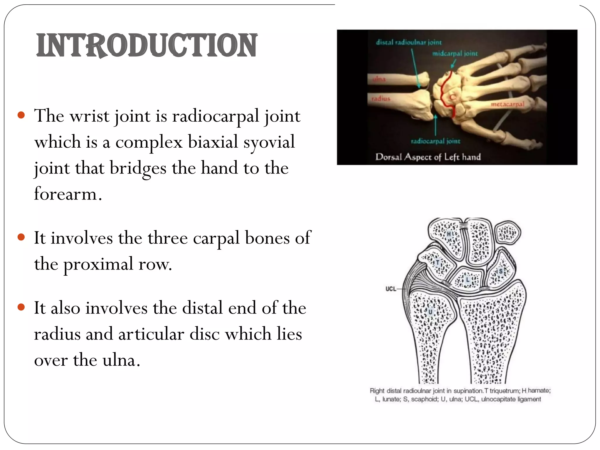
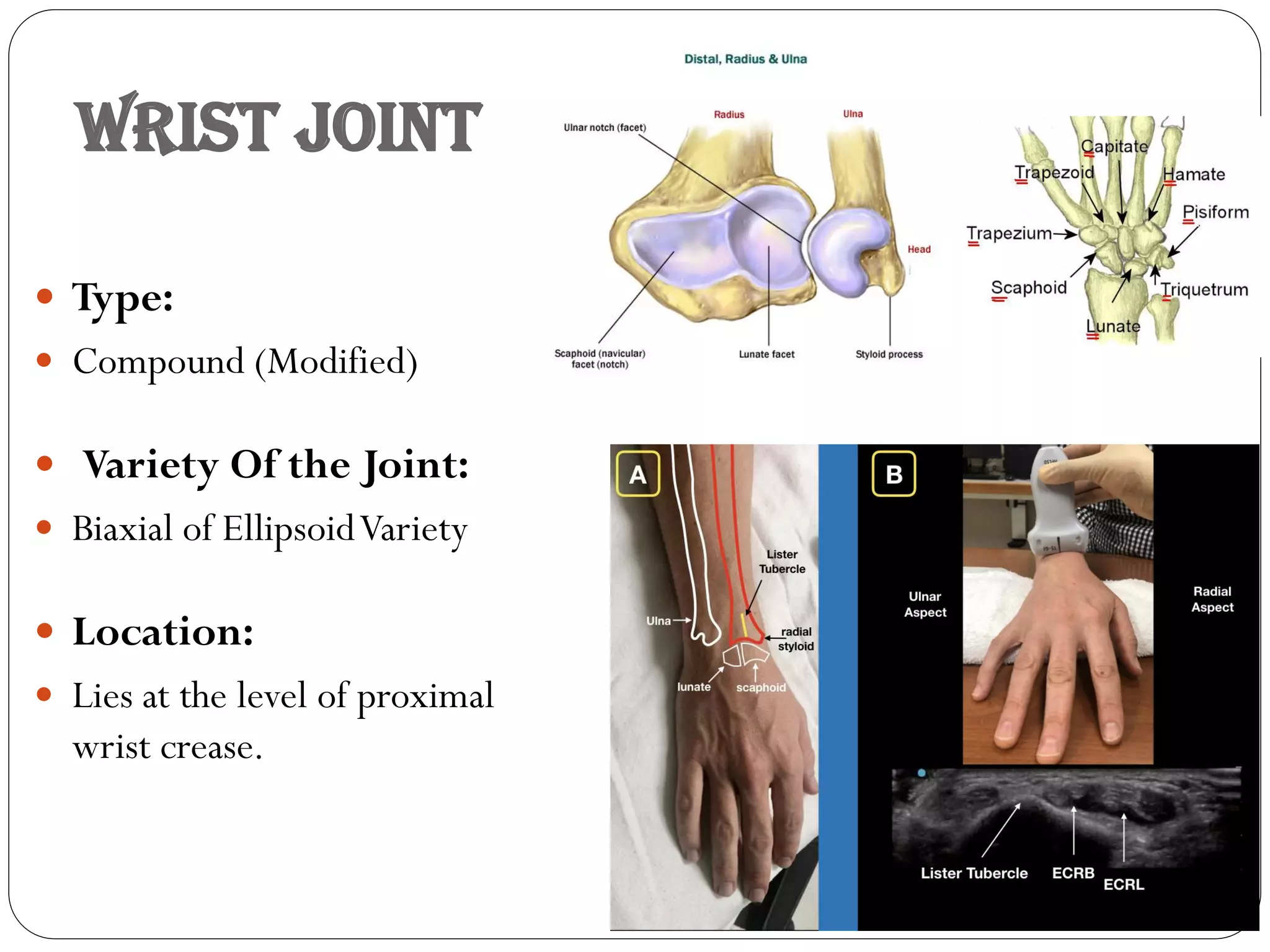
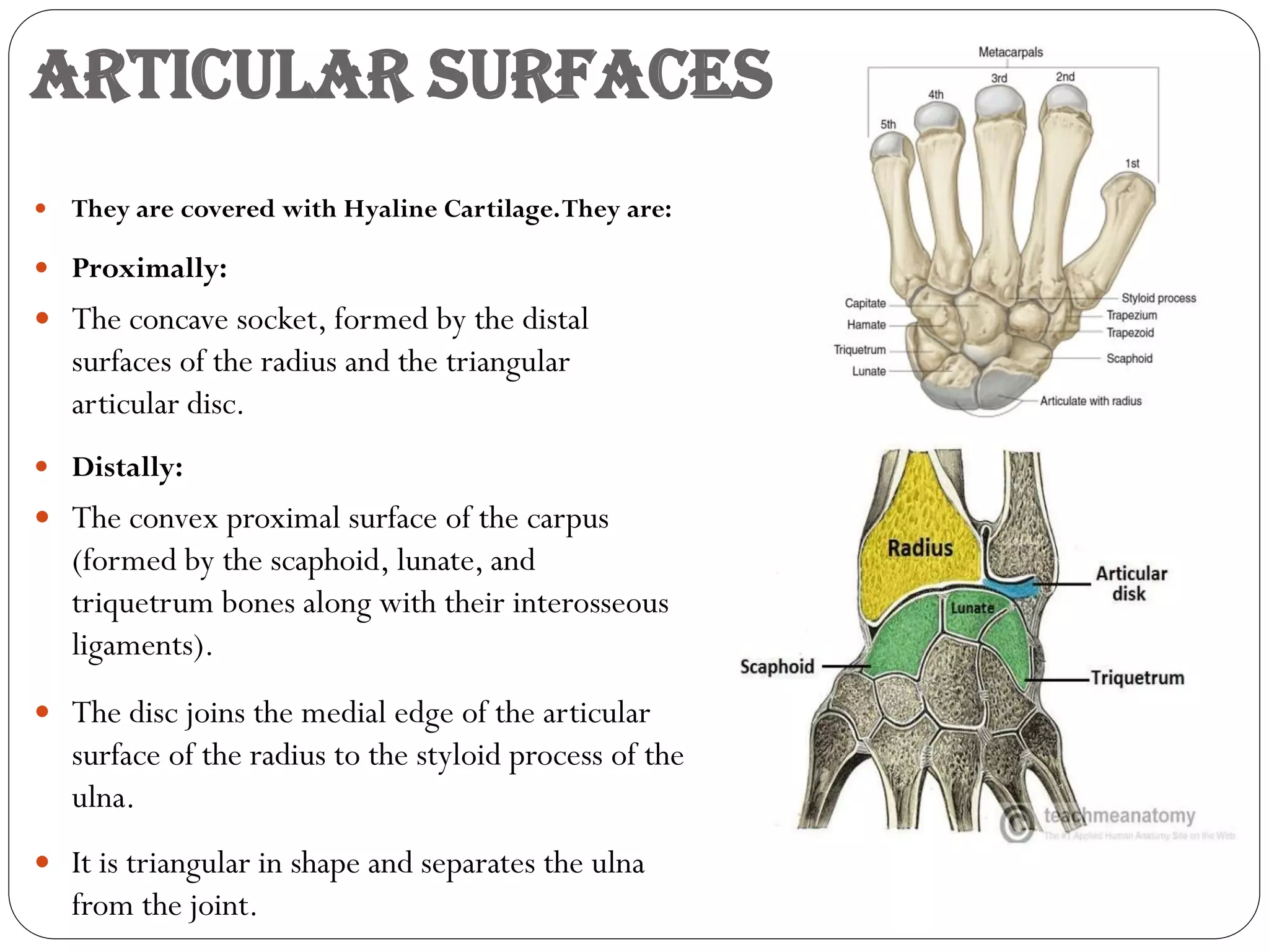
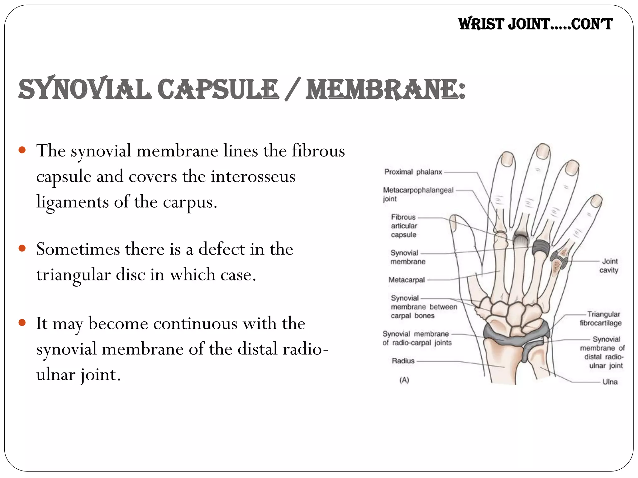
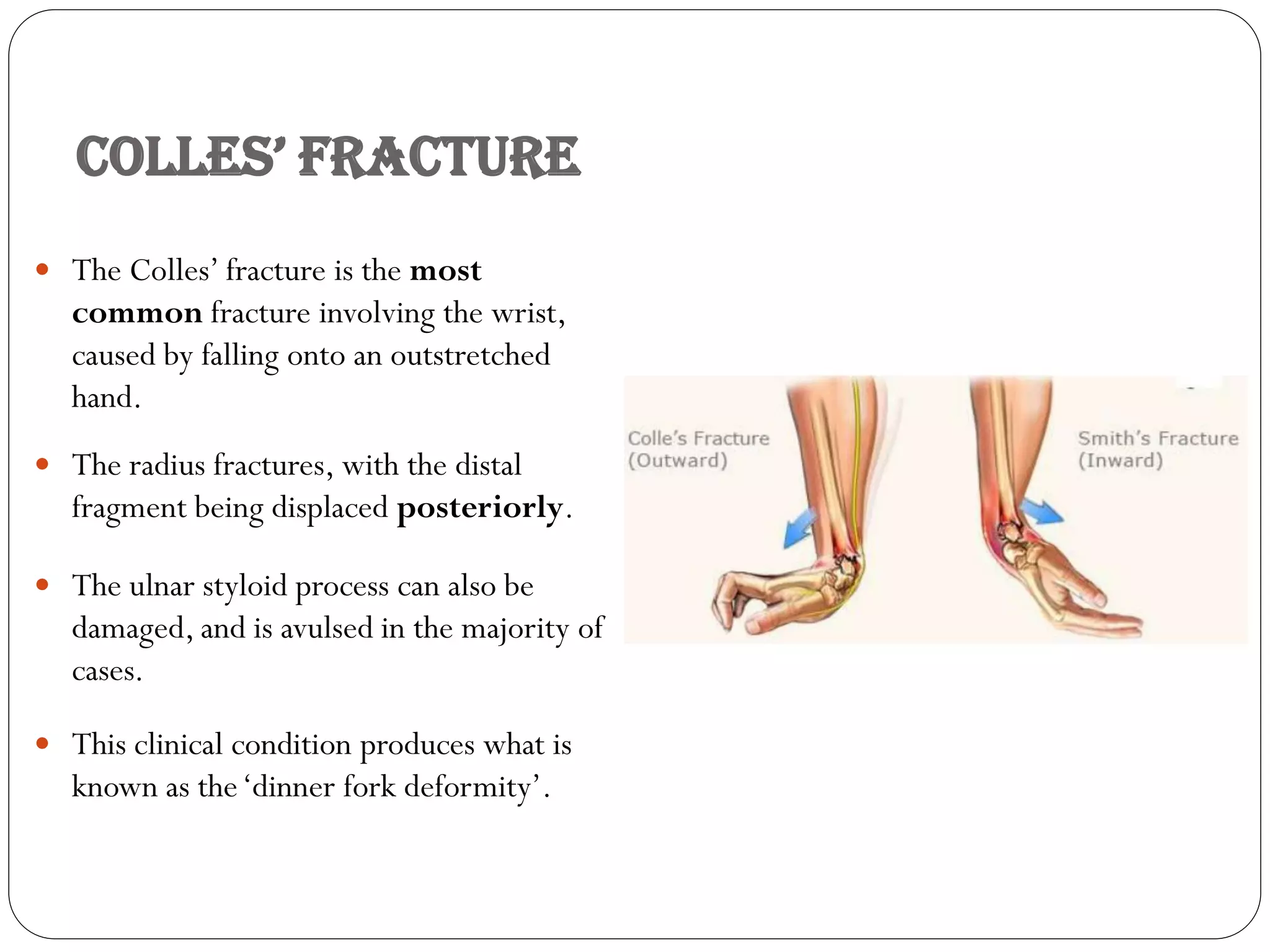
Lecture 09
Wrist Joint
By:
Associate Professor
Dr Farooq Aman Ullah Khan
PMC
Email: Farooq_jmc@yahoo.com
Date: 09th July 2021
INTRODUCTION
 The wrist joint is radiocarpal joint
which is a complex biaxial syovial
joint that bridges the hand to the
forearm.
 It involves the three carpal bones of
the proximal row.
 It also involves the distal end of the
radius and articular disc which lies
over the ulna.
WRIST JOINT
 Type:
 Compound (Modified)
 Variety Of the Joint:
 Biaxial of EllipsoidVariety
 Location:
 Lies at the level of proximal
wrist crease.
Articular Surfaces
 They are covered with Hyaline Cartilage.They are:
 Proximally:
 The concave socket, formed by the distal
surfaces of the radius and the triangular
articular disc.
 Distally:
 The convex proximal surface of the carpus
(formed by the scaphoid, lunate, and
triquetrum bones along with their interosseous
ligaments).
 The disc joins the medial edge of the articular
surface of the radius to the styloid process of the
ulna.
 It is triangular in shape and separates the ulna
from the joint.
ArticulAr SurfAceS…..con’t
 In the resting position of hand only the
scaphoid and lateral part of the lunate
articulate with the two shallow fossae on
the distal surface of the radius.
 (The lateralTriangular facet is for the
Scaphoid and the medial Quadrangular
facet for the Lunate).
 The remainder of the lunate is in contact
with the articular disc while the
triquetrum is applied to the medial part of
the capsule of the joint.
 Note:
 The Ulna is not a part of the wrist joint.
Joint Capsule
 Like any other synovial Joint, the capsule
is double layered.
 The fibrous outer layer attaches to the
distal end of radius, ulna and the
proximal row of the carpal bones.
 The internal layer, synovial membrane
extends up to the margins of the
articular surfaces.
WriSt joint…..con’t
Fibrous Capsule:
 It passes from the margins of the
distal ends of the radius and ulna
 Forms the margins of the articular
disc to the proximal row of carpal
bones, excluding the Pisiform.
 The anterior and posterior parts of
the fibrous capsule contain fibers
which pass obliquely downwards
and medially.
WriSt joint…..con’t
Synovial Capsule / Membrane:
 The synovial membrane lines the fibrous
capsule and covers the interosseus
ligaments of the carpus.
 Sometimes there is a defect in the
triangular disc in which case.
 It may become continuous with the
synovial membrane of the distal radio-
ulnar joint.
WriSt joint…..con’t
LIGAMENTS OF THE WRIST COMPLEX
 The ligamentous structure of carpus is responsible for articular
stability as well as guiding and checking motions between and among
the carpals.
 In general, dorsal ligaments are thin and numerous volar ligaments
are thicker and stronger.
Ligaments
Extrinsic Intrinsic
Connect carpals to
radius ulna proximally
or metacarpals distally
Interconnects the
carpals.
Ligaments of the joint
 Only Extra capsular Ligaments are
present in this joint.
 They comprise of slightly thickened
portions of the capsule
 It is attached to the styloid processes
of the radius and ulna and passing to
the scaphoid and triquetrum
respectively.
 They are called the radial and
ulnar collateral ligaments
respectively.
ligAmentS of the joint…..con’t
 There are four ligaments in the wrist joint, one
for each side of the joint.
 Palmar (Anterior) Radiocarpal:
 It passes from the radius to both rows of carpal
bones.
 Its function, apart from increasing stability, is to
ensure that the hand follows the forearm during
Supination.
 Dorsal(Posterior) Radiocarpal:
 It passes from the radius to both rows of carpal
bones.
 It contributes to the stability of wrist, but also
ensure that the hand follows the forearm during
pronation.
ligAmentS of the joint…..con’t
 Ulnar Collateral:
 Runs from the ulnar styloid process to the
triquetrum and pisiform.
 Work in union with the other collateral
ligament to prevent excessive lateral joint
displacement.
 Radial Collateral:
 Runs from the radial styloid process to the
Scaphoid andTrapezium.
 Work in union with the other collateral
ligament to prevent excessive lateral joint
displacement.
 Palmer ulnocarpal Ligament:
 It is formed due to thickening of the medial
part of the anterior aspect of the fibrous
capsule.
NEUROVASCULAR SUPPLY
of wrist joint
 The wrist joint receive blood from branches
of the dorsal and palmar carpal arches,
which are derived from the ulnar and radial
arteries.
 Innervations to the wrist is delivered
by branches of three nerves:
 Median:
 Anterior interosseus branch.
 Radial :
 Posterior interosseus branch.
 Ulnar:
 Deep and dorsal branches.
Relation
 Anterior:
 Median and ulnar nerve and vessels
 Tendons of Flexor digitorum superficialis , FDF
and associated synivial sheath (Ulnar bursa)
 Tendons of FPL, FCR.
 POSTERIOR:
 Superficial:
 Dorsal cutaneous branch of ulnar nerve, Basilic
Cephalic vein, superficial branch of radial nerve
 Deep:
 Extensor tendons of wrist and finger, and
associated synovial sheath.
relAtion….con’t
 LATERAL:
 Radial artery (across the radial
collateral ligament).
 Tendons ofAbductor pollices
longus, Extensor pollices brevis.
 MEDIAL:
 Dorsal cutaneous branch of
ulnar nerve.
Movements of wrist joint
 The wrist complex consists of radio carpal joint
and midcarpal joint.
 Flexion and extension occur along the transverse
axis, and abduction and adduction occur along
the anteroposterior axis.
 The movements at the wrist joint are usually
associated with movements at the midcarpal
joint.
 The wrist and midcarpal joints togather are
considered as link joint.
 Rotation is not possible at the wrist joint because
the articular surfaces are ellipsoid in shape.
 The lack of rotation at wrist is compensated by
the movements of pronation and supination of
the forearm.
Movements of wrist joint
 Flexion:
 Flexor carpi radialis, flexor carpi
ulnaris.
 Palmaris longus
 Extension:
 Extensor carpi radialis longus,
 Extensor carpi radialis brevis
 Extensor carpi ulnaris.
 Adduction:
 Flexor carpi ulnaris.
 Extensor carpi ulnaris.
 Abduction:
 Flexor carpi radialis,
 Extensor carpi radialis longus,
 Extensor carpi radialis brevis
 Abductor pollicis longus.
Range of movements of the wrist joint
 Flexion
 Extension
 Abduction
 Adduction
Movements Range
 0-60°
 0-50°
 0-15°
 0-25°
Stability Of the joint:
 Bony Factors:
 The lateral and dorsal margins of the radius
extend further distally than its other margins.
 This reduces the likelihood of posterior
dislocation of any of the carpal bones.
 Ligamentous Factors:
 Only the collateral ligaments are strong.
 The anterior and posterior ligaments are
merely thickened fibers of the capsule.
 Muscular Factors:
 The tendons of long flexors of the fingers and
thumb stabilize the joint anteriorly
 The tendons of long extensors of the fingers
and thumb stabilize the joint posteriorly
Clinical Relevance:
Injuries to the Wrist Joint
 The scaphoid bone of the hand is the most
commonly fractured carpal bone – typically by
falling on an oustretched hand (FOOSH).
 In a fracture of the scaphoid, the characteristic
clinical feature is pain and tenderness in
the anatomical snuffbox.
 The scaphoid is at particular risk of avascular
necrosis after fracture because of its so-called
‘retrograde blood supply’ which enters at its
distal end.
 This means that a fracture to the middle (or
‘waist’) of the scaphoid may interrupt the blood
supply to the proximal part of the scaphoid bone
rendering it avascular.
 Patients with a missed scaphoid fracture are
likely to develop osteoarthritis of the wrist in
later life
Scaphoid Fracture
Anterior Dislocation of the Lunate
 This can occur by falling on
a dorsiflexed wrist.
 The lunate is forced anteriorly, and
compresses the carpal tunnel, causing the
symptoms of carpal tunnel syndrome.
 This manifests clinically as paraesthesia in
the sensory distribution of the median nerve
and weakness of thenar muscles.
 The lunate can also undergo avascular
necrosis, so immediate clinical attention to
the fracture is needed.
colleS’ frActure
 The Colles’ fracture is the most
common fracture involving the wrist,
caused by falling onto an outstretched
hand.
 The radius fractures, with the distal
fragment being displaced posteriorly.
 The ulnar styloid process can also be
damaged, and is avulsed in the majority of
cases.
 This clinical condition produces what is
known as the‘dinner fork deformity’.
Ganglion
 Greek word….Swelling or Knot.
 It is a non tender cystic swelling, which
sometimes appears on wrist most
commonly on its dorsal aspect.
 Its size varies from a small grape to a plum.
 It usually occurs due to mucoid de
generation of synovial sheath around the
tendon.
 The cyst is thin walled and contains clear
mucinous fluid.
 The flexion of wrist makes the cyst to
enlarged and it may be become painful.
thAnk You……

Wrist Joint.....2021.pdf

  • 1.
    Lecture 09 Wrist Joint By: AssociateProfessor Dr Farooq Aman Ullah Khan PMC Email: Farooq_jmc@yahoo.com Date: 09th July 2021
  • 2.
    INTRODUCTION  The wristjoint is radiocarpal joint which is a complex biaxial syovial joint that bridges the hand to the forearm.  It involves the three carpal bones of the proximal row.  It also involves the distal end of the radius and articular disc which lies over the ulna.
  • 3.
    WRIST JOINT  Type: Compound (Modified)  Variety Of the Joint:  Biaxial of EllipsoidVariety  Location:  Lies at the level of proximal wrist crease.
  • 4.
    Articular Surfaces  Theyare covered with Hyaline Cartilage.They are:  Proximally:  The concave socket, formed by the distal surfaces of the radius and the triangular articular disc.  Distally:  The convex proximal surface of the carpus (formed by the scaphoid, lunate, and triquetrum bones along with their interosseous ligaments).  The disc joins the medial edge of the articular surface of the radius to the styloid process of the ulna.  It is triangular in shape and separates the ulna from the joint.
  • 5.
    ArticulAr SurfAceS…..con’t  Inthe resting position of hand only the scaphoid and lateral part of the lunate articulate with the two shallow fossae on the distal surface of the radius.  (The lateralTriangular facet is for the Scaphoid and the medial Quadrangular facet for the Lunate).  The remainder of the lunate is in contact with the articular disc while the triquetrum is applied to the medial part of the capsule of the joint.  Note:  The Ulna is not a part of the wrist joint.
  • 6.
    Joint Capsule  Likeany other synovial Joint, the capsule is double layered.  The fibrous outer layer attaches to the distal end of radius, ulna and the proximal row of the carpal bones.  The internal layer, synovial membrane extends up to the margins of the articular surfaces. WriSt joint…..con’t
  • 7.
    Fibrous Capsule:  Itpasses from the margins of the distal ends of the radius and ulna  Forms the margins of the articular disc to the proximal row of carpal bones, excluding the Pisiform.  The anterior and posterior parts of the fibrous capsule contain fibers which pass obliquely downwards and medially. WriSt joint…..con’t
  • 8.
    Synovial Capsule /Membrane:  The synovial membrane lines the fibrous capsule and covers the interosseus ligaments of the carpus.  Sometimes there is a defect in the triangular disc in which case.  It may become continuous with the synovial membrane of the distal radio- ulnar joint. WriSt joint…..con’t
  • 9.
    LIGAMENTS OF THEWRIST COMPLEX  The ligamentous structure of carpus is responsible for articular stability as well as guiding and checking motions between and among the carpals.  In general, dorsal ligaments are thin and numerous volar ligaments are thicker and stronger. Ligaments Extrinsic Intrinsic Connect carpals to radius ulna proximally or metacarpals distally Interconnects the carpals.
  • 10.
    Ligaments of thejoint  Only Extra capsular Ligaments are present in this joint.  They comprise of slightly thickened portions of the capsule  It is attached to the styloid processes of the radius and ulna and passing to the scaphoid and triquetrum respectively.  They are called the radial and ulnar collateral ligaments respectively.
  • 11.
    ligAmentS of thejoint…..con’t  There are four ligaments in the wrist joint, one for each side of the joint.  Palmar (Anterior) Radiocarpal:  It passes from the radius to both rows of carpal bones.  Its function, apart from increasing stability, is to ensure that the hand follows the forearm during Supination.  Dorsal(Posterior) Radiocarpal:  It passes from the radius to both rows of carpal bones.  It contributes to the stability of wrist, but also ensure that the hand follows the forearm during pronation.
  • 12.
    ligAmentS of thejoint…..con’t  Ulnar Collateral:  Runs from the ulnar styloid process to the triquetrum and pisiform.  Work in union with the other collateral ligament to prevent excessive lateral joint displacement.  Radial Collateral:  Runs from the radial styloid process to the Scaphoid andTrapezium.  Work in union with the other collateral ligament to prevent excessive lateral joint displacement.  Palmer ulnocarpal Ligament:  It is formed due to thickening of the medial part of the anterior aspect of the fibrous capsule.
  • 13.
    NEUROVASCULAR SUPPLY of wristjoint  The wrist joint receive blood from branches of the dorsal and palmar carpal arches, which are derived from the ulnar and radial arteries.  Innervations to the wrist is delivered by branches of three nerves:  Median:  Anterior interosseus branch.  Radial :  Posterior interosseus branch.  Ulnar:  Deep and dorsal branches.
  • 14.
    Relation  Anterior:  Medianand ulnar nerve and vessels  Tendons of Flexor digitorum superficialis , FDF and associated synivial sheath (Ulnar bursa)  Tendons of FPL, FCR.  POSTERIOR:  Superficial:  Dorsal cutaneous branch of ulnar nerve, Basilic Cephalic vein, superficial branch of radial nerve  Deep:  Extensor tendons of wrist and finger, and associated synovial sheath.
  • 15.
    relAtion….con’t  LATERAL:  Radialartery (across the radial collateral ligament).  Tendons ofAbductor pollices longus, Extensor pollices brevis.  MEDIAL:  Dorsal cutaneous branch of ulnar nerve.
  • 16.
    Movements of wristjoint  The wrist complex consists of radio carpal joint and midcarpal joint.  Flexion and extension occur along the transverse axis, and abduction and adduction occur along the anteroposterior axis.  The movements at the wrist joint are usually associated with movements at the midcarpal joint.  The wrist and midcarpal joints togather are considered as link joint.  Rotation is not possible at the wrist joint because the articular surfaces are ellipsoid in shape.  The lack of rotation at wrist is compensated by the movements of pronation and supination of the forearm.
  • 17.
    Movements of wristjoint  Flexion:  Flexor carpi radialis, flexor carpi ulnaris.  Palmaris longus  Extension:  Extensor carpi radialis longus,  Extensor carpi radialis brevis  Extensor carpi ulnaris.  Adduction:  Flexor carpi ulnaris.  Extensor carpi ulnaris.  Abduction:  Flexor carpi radialis,  Extensor carpi radialis longus,  Extensor carpi radialis brevis  Abductor pollicis longus.
  • 18.
    Range of movementsof the wrist joint  Flexion  Extension  Abduction  Adduction Movements Range  0-60°  0-50°  0-15°  0-25°
  • 19.
    Stability Of thejoint:  Bony Factors:  The lateral and dorsal margins of the radius extend further distally than its other margins.  This reduces the likelihood of posterior dislocation of any of the carpal bones.  Ligamentous Factors:  Only the collateral ligaments are strong.  The anterior and posterior ligaments are merely thickened fibers of the capsule.  Muscular Factors:  The tendons of long flexors of the fingers and thumb stabilize the joint anteriorly  The tendons of long extensors of the fingers and thumb stabilize the joint posteriorly
  • 20.
    Clinical Relevance: Injuries tothe Wrist Joint  The scaphoid bone of the hand is the most commonly fractured carpal bone – typically by falling on an oustretched hand (FOOSH).  In a fracture of the scaphoid, the characteristic clinical feature is pain and tenderness in the anatomical snuffbox.  The scaphoid is at particular risk of avascular necrosis after fracture because of its so-called ‘retrograde blood supply’ which enters at its distal end.  This means that a fracture to the middle (or ‘waist’) of the scaphoid may interrupt the blood supply to the proximal part of the scaphoid bone rendering it avascular.  Patients with a missed scaphoid fracture are likely to develop osteoarthritis of the wrist in later life Scaphoid Fracture
  • 21.
    Anterior Dislocation ofthe Lunate  This can occur by falling on a dorsiflexed wrist.  The lunate is forced anteriorly, and compresses the carpal tunnel, causing the symptoms of carpal tunnel syndrome.  This manifests clinically as paraesthesia in the sensory distribution of the median nerve and weakness of thenar muscles.  The lunate can also undergo avascular necrosis, so immediate clinical attention to the fracture is needed.
  • 22.
    colleS’ frActure  TheColles’ fracture is the most common fracture involving the wrist, caused by falling onto an outstretched hand.  The radius fractures, with the distal fragment being displaced posteriorly.  The ulnar styloid process can also be damaged, and is avulsed in the majority of cases.  This clinical condition produces what is known as the‘dinner fork deformity’.
  • 23.
    Ganglion  Greek word….Swellingor Knot.  It is a non tender cystic swelling, which sometimes appears on wrist most commonly on its dorsal aspect.  Its size varies from a small grape to a plum.  It usually occurs due to mucoid de generation of synovial sheath around the tendon.  The cyst is thin walled and contains clear mucinous fluid.  The flexion of wrist makes the cyst to enlarged and it may be become painful.
  • 24.