Surgical anatomy of the hand copy - copy
•Download as PPTX, PDF•
3 likes•374 views

IT contains bone,muscle, neurovascular and clinical cases

Recommended
More Related Content
What's hot
What's hot (20)
Similar to Surgical anatomy of the hand copy - copy
Similar to Surgical anatomy of the hand copy - copy (20)
Recently uploaded
Recently uploaded (20)
Surgical anatomy of the hand copy - copy
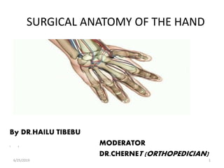
- 1. SURGICAL ANATOMY OF THE HAND MODERATOR DR.CHERNET (ORTHOPEDICIAN) By DR.HAILU TIBEBU • ) 6/25/2019 1
- 2. OUTLINE OF PRESENTATION DEFINITION FUNCTIONS SKIN BONY SKELETON AND JOINTS MUSCLES AND SOFT TISSUES VESSELS AND NERVES CLINICAL CORRELATION 6/25/2019 2
- 3. DEFNITION The hand is the region of the upper limb distal to the wrist joint. It is subdivided into three parts: the wrist(containing carpal bones. the metacarpus. the digits (five fingers including the thumb). The hand has an anterior surface (palm) and a dorsal surface (dorsum ) of the hand. 6/25/2019 3
- 4. 6/25/2019 4
- 5. FUNCTIONS Hand is both mechanical and sensory tool. Humans are endowed with fine control and precision movements unlike other primates. The hand accounts for about 90% of upper limb function: The thumb is involved in 40-50% of hand function The index finger is involved in about 20% of hand function The middle finger, which accounts for about 20% of all hand function, is the strongest finger, and is important for both precision and power functions 6/25/2019 5
- 6. THE BONY SKELETON The distal radius is characterized by the presence of a large dorsal tubercle, which acts as a pulley for the extensor pollicis longus facet for articulation with the distal end of the ulna, two facets for articulation with two carpal bones (the scaphoid and lunate). styloid process. 6/25/2019 6
- 7. Cont.. • The distal end of the ulna is small and characterized by a rounded head and the ulnar styloid process. • The anterolateral and distal part of the head is covered by articular cartilage. • The ulnar styloid process originates from the dorsomedial aspect of the ulna and projects distally. 6/25/2019 7
- 8. CARPAL BONES • Proximal row from lateral to medial: – Scaphoid: boat-shaped with scaphoid tubercle – Lunate: crescent shape – Triquetrum: the three-sided – Pisiform: pea-shaped; lies on palmar surface of triquetrum • Distal row from lateral to medial: – Trapezium: irregular four sided ,articulates with metacarpal bone of the thumb – Trapezoid: the four-sided – Capitate: has head ; largest – Hamate: has hooked process (hook of hamate) 6/25/2019 8
- 10. Articular surfaces • All carpal bone articulate with each other. • carpal bones in the distal row articulate with the metacarpals of the digits. • Only metacarpal of thumb,move over carpal bone. • proximal surfaces of the scaphoid and lunate articulate with the radius to form the wrist joint. 6/25/2019 10
- 11. Carpal arch • Carpal bones do not lie in a flat plane; rather, they form an arch, whose base is directed anteriorly. • The lateral side of this base is formed by tubercles of the scaphoid and trapezium. • The medial side is formed by the pisiform and the hook of the hamate. 6/25/2019 11
- 12. 6/25/2019 12
- 13. The flexor retinaculum attaches to, and spans the distance between base to form anterior wall called carpal tunnel. 6/25/2019 13
- 14. • Free movement of the tendons in the carpal tunnel is facilitated by synovial sheaths, which surround the tendons. • All flexor digitorum profundus and flexor digitorum superficialis surrounded by a single synovial sheath. • separate sheath surrounds the tendon of the flexor pollicis longus. • The median nerve is anterior to the tendons in the carpal tunnel. 6/25/2019 14
- 15. CARPAL TUNNEL • Serves as a conduit for the median nerve and nine flexor tendons,gate way to hand. • The roof of the tunnel is formed by the flexor retinaculum- Fibrous band which bridges the anterior concavity of carpus. • Carpal bones and palmar ligament complex form the base. • Contents-The four tendons of the flexor digitorum profundus, the four tendons of the flexor digitorum superficialis, and the tendon of the flexor pollicis longus and the median nerve. 6/25/2019 15
- 16. Carpal Tunnel Syndrome • Carpal tunnel syndrome is an entrapment syndrome due to pressure on the median nerve with in the carpal tunnel. • Etiology obscure, direct effect of increased pressure on median nerve Overuse swelling of the tendons and tendon sheaths (e.g.RA) cysts arising from the carpal joints. • pain and pins-and-needles sensations in the distribution of the median nerve. • Weakness and loss of muscle bulk of thenar muscles may occur. • Tinel's sign +ve 6/25/2019 16
- 17. Avascular necrosis of the proximal scaphoid • fracture across the waist of the scaphoid bone • common • In 10% of individuals, the scaphoid bone blood supply from the radial artery which supply the proximal portion 6/25/2019 17
- 19. JOINTS OF WRIST AND HAND • Wrist joint formed by distal radius, articular disc over ulna and scaphoid, lunate & triquetrum. – Flexion , Extension ,adduction &abduct. – the radial styloid extends further distally than does the ulnar styloid , the hand can be adducted to a greater degree than it can be abducted. 6/25/2019 19
- 20. The capsule of the wrist joint is reinforced by palmar radiocarpal palmar ulnocarpal and dorsal radiocarpal ligaments. radial and ulnar collateral ligaments of the wrist joint span the distance between the styloid processes of the radius and ulna and the adjacent carpal bones. 6/25/2019 20
- 21. CONT… CARPAL JOINTS • The synovial joints between the carpal bones, share a common articular cavity. • Gliding type of joints allowing slight side to side movements CARPOMETACRPAL JOINTS • Type: gliding synovial joints, except for CMC joint of thumb (saddle shaped synovial joint). • Movements: Flexion ,extension ,abduction and adduction of CMC joint of 1st digit, the rest allowing only limited gliding movements. • Movement of this joints increases medially so metacarpal V slides to the greatest degree • can be best observed on the dorsal surface of the hand as it makes a fist. 6/25/2019 21
- 22. CONT.. • METACARPOPHALANGEAL JOINTS The joints between the distal heads of the metacarpals and the proximal phalanges of the digits are condylar joints. MOVEMENTS which allow flexion, extension, abduction, adduction, circumduction, and limited rotation . The capsule of each joint is reinforced by the palmar ligament and by medial and lateral collateral ligaments. • INTERPHALANGEAL JOINTS • are hinge joints that allow mainly flexion and extension. They are reinforced by medial and lateral collateral ligaments and palmar ligaments. 6/25/2019 22
- 23. Deep transverse metacarpal ligament • 3 thick bands of connective tissue • connecting the palmar ligaments of the metacarpophalangeal joints of the fingers to each other. 6/25/2019 23
- 24. SKIN • Dorsum skin is thin,pliable • Palmar skin is thick, strongly atached to underlying fascia • Most firmly anchored to deep structure at palmar crease • Palmar surface contain high concentration of sensory n. 6/25/2019 24
- 25. • Nails are specialized skin appendage derived from epidermis • 90% nail plate is produced by germinal matrix and 10% by sterile matrix which adds squamous component 6/25/2019 25
- 26. SOFT TISSUES • Palmar Aponeurosis • is a triangular condensation of deep fascia that covers the palm • anchored to the skin in distal regions • The apex is continuous with the palmaris longus tendon or it is anchored to the flexor retinaculum. • Vessels, nerves, and long flexor tendons lie deep to the palmar aponeurosis in the palm. . 6/25/2019 26
- 27. • deep spaces of the hand include midpalmar space • Thenar space • interdigital web spaces • less well-defined • hypothenar space, • Parona space, and 6/25/2019 27
- 30. CONT.. Flexor Pulleys 1. annular pulleys position the tendons close to the underlying bone. 2. Cruciate pulleys are interdigitated between the annular pulleys and are less critical for function. Pulleys prevent bowstringing of the flexor tendons during flexion. There are five annular (A). • A1—metacarpophalangeal joint; • A2—proximal portion of the proximal phalanx • A3—proximal interphalangeal joint • A4—middle portion of the middle phalanx • A5—distal interphalangeal joint • A2 and A4 are essential to prevent bowstringing 6/25/2019 30
- 31. nutrition • Tendon nutrition is believed from two basic sources: • 1) the synovial fluid produced within the tenosynovial sheath and • (2) the blood supply provided through longitudinal vessels in the paratenon, intraosseous vessels at the tendon insertion, and vincular circulation 6/25/2019 31
- 32. CONT… • Extensor Hood – A complex tendon, formed by expansions of tendons of extensor digitorum & extensor pollicis longus muscles on the dorsal aspect of proximal phalanges. – Creates a ‘cable’ system and by inserting on it the intrinsic muscles of the hand provide a mechanism for extending the IPJ while flexing the MCP joints, allowing the hand to execute precision movements. 6/25/2019 32
- 33. Muscles of the hand subdivided into two groups based on their origin: – Intrinsic muscles- that arise and insert within the hand, and – Extrinsic muscles -that originate in the forearm and insert within the hand. • extrinsic muscles function in forcefully gripping ('power grip') with the hand, the intrinsic muscles mainly execute precision movements ('precision grip') with the fingers and thumb. • The extrinsic muscles are again subdivided in to two: – The flexors, located in the anterior compartment,arise from medial epicondyle – The extensors, located in the posterior compartment,arise from lateral epicondyle 6/25/2019 33
- 34. EXTRINSIC MUSCLES – Anterior compartement • The anterior compartment ms occur in three layers: superficial, intermediate, and deep. • Generally, these muscles are associated with: movements of the wrist joint; flexion of the fingers including the thumb; pronation. Superficial muscles • Pronator teres-pronation. • Flexor carpi radialis - Flexes and abducts the wrist. • Palmaris longus-flexes wrist. • Flexor carpi ulnaris - Flexes and adducts the wrist joint 6/25/2019 34
- 35. CONT.. • Anterior Compartment Intermediate Muscle • Flexor Digitorum superficialis • Insertion middle phalanges of digits 2 - 5 • Action Flexes middle phalanges at proximal interphalangeal joints also flexes proximal phalanges at metacarpophalangeal joints of hand. • Flex wrist jnt 6/25/2019 35
- 36. CONT.. DEEP MUSCLES • Flexor pollicis longus insertion Base of distal phalanx of thumb Action Flexes thumb Flexor digitorum profundus Insertion Base of distal phalanx of digits 2 – 5 Action Flexes MCP, distal and proximal interphalangeal joints. Flex wrist Pronator quadratus origion-Lower end of ulna Insertion-1/4 of anterior surface of distal radius Action Pronates forearm; 6/25/2019 36
- 37. Cont.. • All muscles in the anterior compartment of the forearm are innervated by the median nerve, except for the flexor carpi ulnaris muscle and the medial half of the flexor digitorum profundus muscle, which are innervated by the ulnar nerve. • The digital flexors flexor digitorum superficialis flexor digitorum profundus flexor pollicis longus pass through the carpal tunnel to provide dual flexion to the fingers and single flexion to the thumb. 6/25/2019 37
- 38. POSTERIOR COMPARTEMENT • Muscles in the posterior compartment of the forearm occur in two layers: 1. superficial and 2. deep layer. The muscles are associated with: movement of the wrist joint; extension of the fingers and thumb; supination. 1)Superficial muscles • Extensor carpi radialis longus • Extensor carpi radialis brevis • Extensor digitorum and Extensor digiti minimi • Extensor carpi ulnaris • Brachioradials 6/25/2019 38
- 39. CONT.. – Deep muscles • Abductor pollicis longus (APL) • Extensor pollicis brevis (EPB) • Extensor pollicis longus (EPL) • Extensor indicis (EI) • supinator All muscles in the posterior compartment of the forearm are innervated by the radial nerve. 6/25/2019 39
- 40. INTRINSIC MUSCLES OF THE HAND The intrinsic muscles of the hand are located in five compartments Thenar compartment Hypothenar compartment Adductor compartment Central compartment (Lumbricals) Interosseous compartment • All of the intrinsic muscles of the hand are innervated by the deep branch of the ulnar nerve except for the three thenar and two lateral lumbrical muscles, which are innervated by the median nerve. 6/25/2019 40
- 41. CONT.. THENAR MUSCLES include abductor pollicis brevis-abducts thumb at MCPJ flexor pollicis brevis –flexes thumb at MCP opponens pollicis-largest,medialy rotates thumb and flex metcarpal 1 are associated with opposition of the thumb and with delicate movements of the thumb responsible for the thenar eminence on the lateral side of the palm at the base of the thumb. innervated by the recurrent branch of the median nerve. 6/25/2019 41
- 42. CONT.. • Hypothenar muscles – abductor digiti minimi- Abducts little finger at MCPJ – flexor digiti minimi brevis- Flexes little finger at MCPJ – opponens digiti minimi- Laterally rotates metacarpal V • The hypothenar muscles are similar to the thenar muscles in name and in organization. • But are innervated by the deep branch of the ulnar nerve. • NB. Palmaris Brevis (in subcutaneous space) 6/25/2019 42
- 43. CONT.. Adductor Pollicis • Origin 2nd and 3rd metacarpals, capitate • Insertion Medial side of base of proximal phalanx of thumb • Action a powerful adductor of the thumb and opposes the thumb to the rest of the digits in gripping. • Innervation-ulnar nerve 6/25/2019 43
- 44. CONT..LUMBRICALS • There are four lumbrical (worm- like) muscles. • the medial two lumbricals are bipennate and lateral two lumbricals are unipennate ms. • The medial two are innervated by the ulnar nerve; the lateral two are innervated by the median nerve. • Flex MCPJ and extend IPJ. 6/25/2019 44
- 45. THE INTEROSSEI • Muscles between and attached to the metacarpals. • Are divided into two 1. the dorsal interossei – • four bipennate muscles • most dorsal. • They are major abductors of the index, middle, and ring fingers, at the MCP joints. 6/25/2019 45
- 46. • the palmar interossei. • anterior to dorsal interossei • 4 unipennate ms . • Adduct the thumb, index, ring, and little fingers 6/25/2019 46
- 47. ARTERIAL ANATOMY OF THE HAND • The brachial artery bifurcates at the elbow into radial and ulnar branches, which are the main arterial branches to the hand. ULNAR ARTERY Enters the hand anterior to the flexor retinaculum between the pisiform and the hook of hamate via the ulnar canal (Guyon canal) • Lies lateral to the ulnar nerve. 6/25/2019 47
- 48. SUPERFICIAL PALMAR ARCH • . • Direct Continuation of ulnar artery • Completed on lateral side by superficial branch of radial artery. 6/25/2019 48
- 49. CONT… RADIAL ARTERY • Curves dorsally around lateral side of wrist and passes over the floor of anatomical snuff box and enters the palm by passing between the heads of 1st dorsal interosseous muscle. • It then turns medially and passes between the heads of the adductor pollicis. • Ends by anastomosing with the deep branch of the ulnar artery to form the deep palmar arch. 6/25/2019 49
- 50. Cont.. • dorsal carpal branch, which passes medially as the dorsal carpal arch, across the wrist and gives rise to dorsal metacarpal arteries, which subsequently divide to become small dorsal digital arteries, which enter the fingers; • the first dorsal metacarpal artery, which supplies adjacent sides of the index finger and thumb. 6/25/2019 50
- 51. DEEP PALMAR ARCH • Direct continuation of radial artery • Completed on the medial side by deep branch of ulnar artery • Deep to long flexor tendons, in contact with base of metacarpals. • Gives rise to palmar metacarpal branches which join common palmar digital arteries and three perforating branches that join dorsal metacarpal arteries from dorsal carpal arch. 6/25/2019 51
- 52. ALLEN’S TEST • Allen’s test determines if there are any vascular anomalies between the radial and ulnar arteries prior to any vascular procedures. • Occluding both the radial and ulnar vessels while the patient flexes and extends the fingers performs the test. • This creates pallor in palm and digits. Release of pressure on either radial or ulnar vessel should allow return of normal color within 2 to 5 seconds. • only the thumb and lateral side of the index finger will fill with blood (become red) when pressure on the radial artery alone is released and vice versa. 6/25/2019 52
- 53. VEINS OF THE HAND • The dorsal digital veins drain into three dorsal metacarpal veins, which unite to form a dorsal venous network • Superficial to the metacarpus, this network is prolonged proximally on the lateral side as the cephalic vein. • The basilic vein arises from the medial side of the dorsal venous network. 6/25/2019 53
- 54. NERVES • The nerves in the hand arise from the brachial plexus. • The brachial plexus is formed by the union of the anterior rami of the last four cervical (C5-C8) and the first thoracic (T1) nerves that constitute the roots of the plexus. The hand is supplied by three nerves Ulnar(C7-T1) median(C5-T1) radial nerves(C5-T1) • All three nerves contribute to cutaneous or general sensory innervation. 6/25/2019 54
- 55. CONT.. • The ulnar nerve innervates all intrinsic muscles of the hand except for the three thenar muscles and the two lateral lumbricals, which are innervated by the median nerve. • The radial nerve only innervates skin on the dorsolateral side of the hand. 6/25/2019 55
- 56. ULNAR NERVE • Arises directly from the medial cord of brachial plexus (C8-T1) • In cubital fossa ulnar nerve passes through fibro-osseous tunnel • As it leaves the canal it lies between the flexor corpi ulnaris and flexor digitorum profundus till middle of forearm • The ulnar nerve enters the hand lateral to the pisiform and dorsomedially to the ulnar artery . Immediately distal to the pisiform, it divides into a deep branch, which is mainly motor and a superficial branch, which is mainly sensory. 6/25/2019 56
- 57. CONT.. DEEP BRANCH • passes with the deep branch of the ulnar artery • supplies the interossei, adductor pollicis, and the two medial lumbricals and hypothenar muscles. SUPREFICIAL BRANCH innervates the palmaris brevis muscle and continues across the palm to supply skin on the palmar surface of the little finger and the medial half of the ring finger. 6/25/2019 57
- 58. MEDIAN NERVE • Arises from lateral & medial cords of brachial plexus • Contain fibers C5 –T1 • In the arm it descends post. to pectoralis major muscle, lateral to brachial artery • The nerve enters forearm between humeral and ulnar head of pronater teres • Then runs between fl digitorum superficialis and fl digitorum profundus, later emerges 5 cm above wrist radial to tendon of palmaris longus. 6/25/2019 58
- 59. CONT… • Enters the hand through carpal tunnel. • divides into a recurrent branch and palmar digital branches. • The recurrent branch of the median nerve innervates the three thenar muscles • The palmar digital nerves innervate skin on the palmar surfaces of the lateral three and one-half digits and cutaneous regions over the dorsal aspects of the distal phalanges (nailbeds) of the same digits. • the digital nerves aslo supply the lateral two lumbrical muscles. 6/25/2019 59
- 60. RADIAL NERVE • Arises from the post cords of the brachial plexus behind 3rd part of axillary artery. • Neural element of C5 – C8 • At junction of upper and middle 1/3 of arm deviates dorsolaterally between medial and long head of triceps lying adjacent to spiral groove of humerus,where it is commonly injured in humeral shaft fracture. • Supplies no hand muscles. • Branches to superficial and deep branches at the level of elbow joint. • Only the superficial part enters the hand. 6/25/2019 60
- 62. CLINICAL CORRELATIONS Ulnar nerve injury –ocurs at two sites:the elbow and the wrist: • at the elbow, the nerve lies posterior to the medial epicondyle; • at the wrist, the ulnar nerve passes superficial to the flexor retinaculum and lies lateral to the pisiform bone. • lesions are characterized by 'clawing' of the hand . • metacarpophalangeal joints of the fingers are hyperextended and the interphalangeal joints are flexed because the function of most of the intrinsic muscles of the hand is lost. • Clawing is most pronounced in the medial fingers because the function of all intrinsic muscles of these digits is lost while in the lateral two digits, the lumbricals are innervated by the median nerve. 6/25/2019 62
- 63. 6/25/2019 63
- 64. CONT… MEDIAN NERVE INJURY • lesion at the elbow-resulting in “Hand of Benediction” or “obstetricians hand”. • The lumbricals and flexor digitorum profundus tendons to digits 2 and 3 are paralysed. This leads to a loss of flexion at the MCP joint and the DIP joints. If the patient is asked to make a fist, they will be able to flex digit 4 and 5 but not digits 2 and 3. 6/25/2019 64
- 65. CONT.. RADIAL NERVE INJURY • The most common radial nerve injury is damage to the nerve in the radial groove of the humerus, which produces a global paralysis of the muscles of the posterior compartment resulting in “wrist drop”. • Radial nerve damage can result from fracture of the shaft of the humerus as the radial nerve spirals around in the radial groove. 6/25/2019 65
- 66. REFERENCES • Gray’s Anatomy, 39th ed, Elsevier • Frank- h –Netter atlas of anatomy • Campbell’s operative orthopedic 20th ed • Schwartz`s principles of surgery, 10th edition • Internet 6/25/2019 66
- 67. 6/25/2019 67
Editor's Notes
- An ischemic area is present in the flexor digitorum superficialis beneath the A2 pulley at the proximal phalanx. Two zones of ischemia are present in the flexor digitorum profundus—beneath the A2 pulley and beneath the A4 pulley. Distaltransversedigital arteryIntermediatetransversedigitalarteryProximaltransversedigital arteryBranch toVLSCommondigitalarteryVLP
- Vascular supply to flexor tendons is by four transverse communicating branches of digital arteries.
- Before penetrating the back of the hand, the radial artery gives rise to two vessels: a dorsal carpal branch, which passes medially as the dorsal carpal arch, across the wrist and gives rise to dorsal metacarpal arteries, which subsequently divide to become small dorsal digital arteries, which enter the fingers; the first dorsal metacarpal artery, which supplies adjacent sides of the index finger and thumb
- .