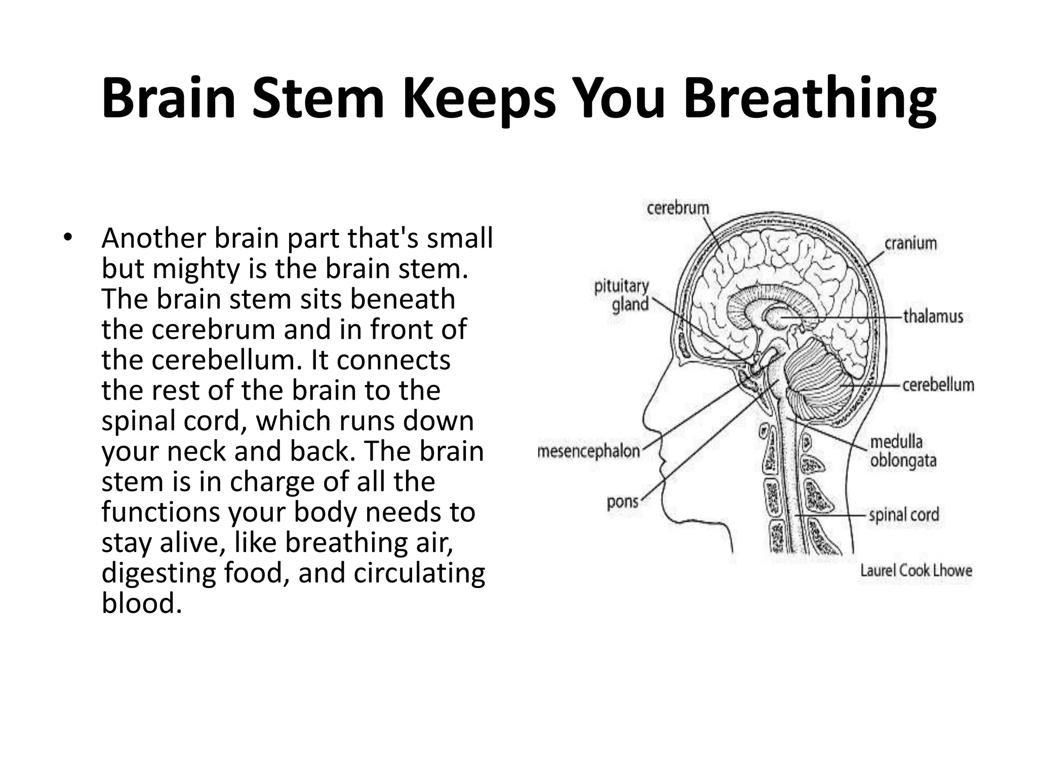
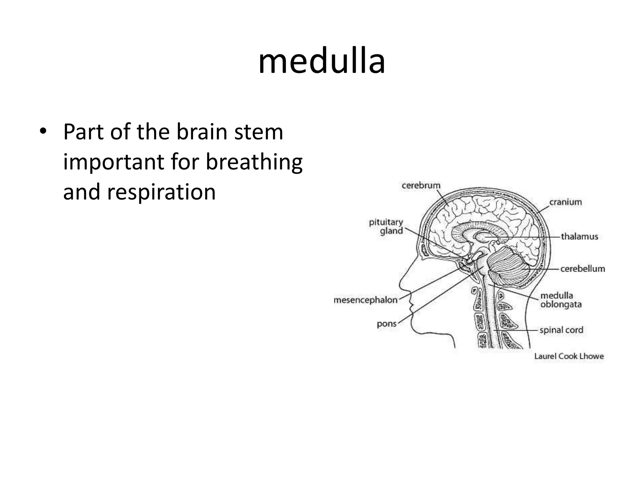
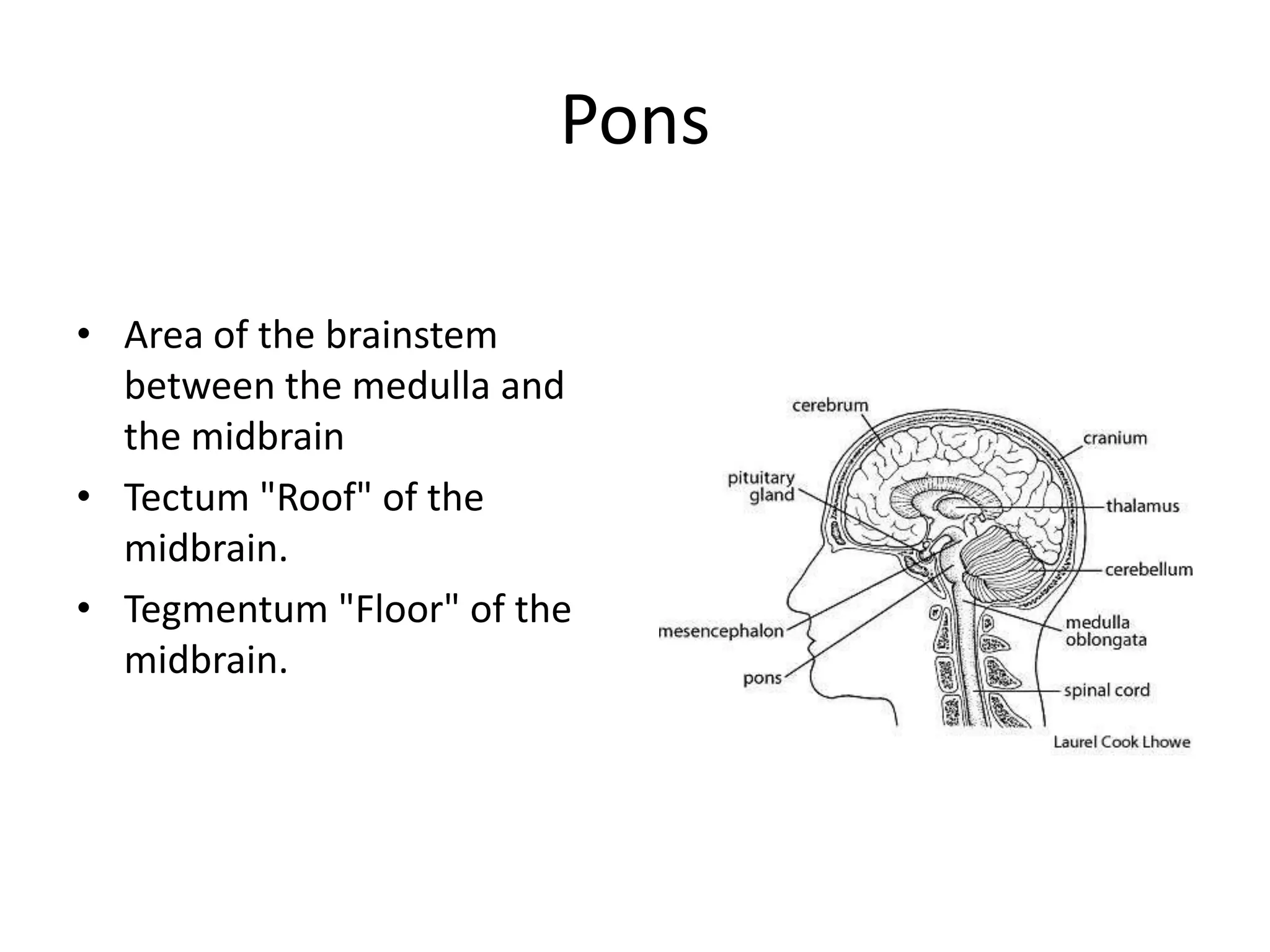
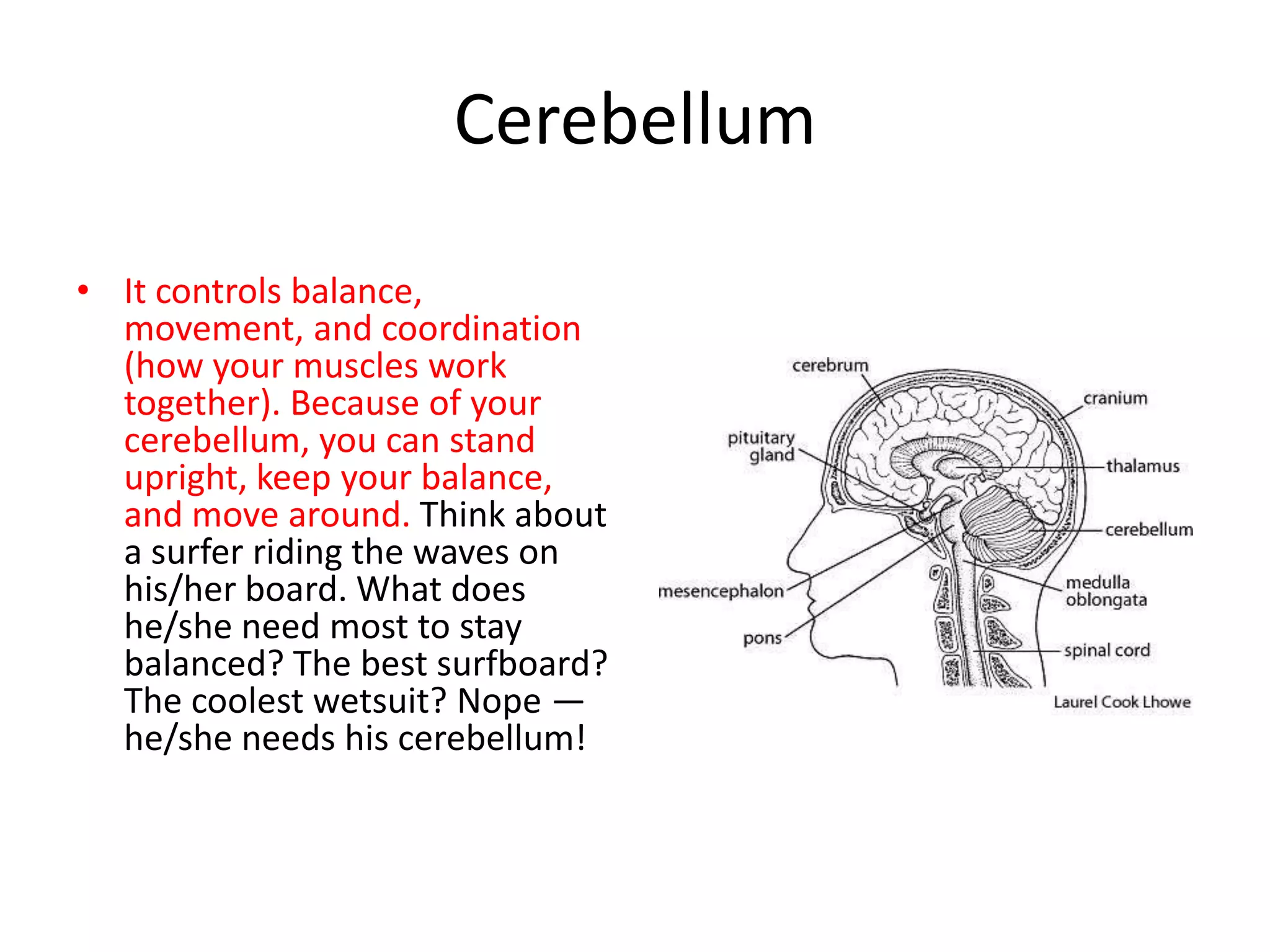
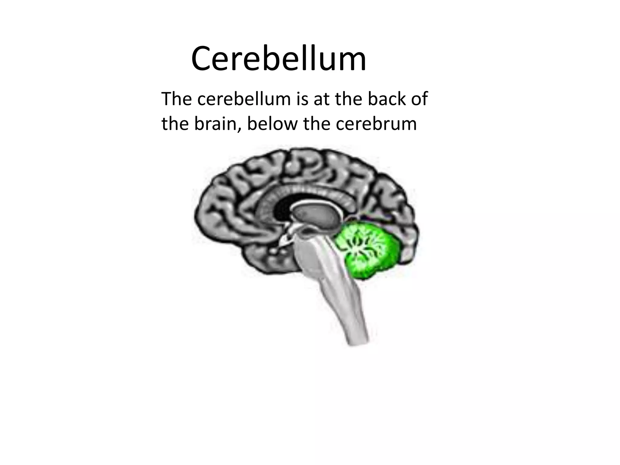
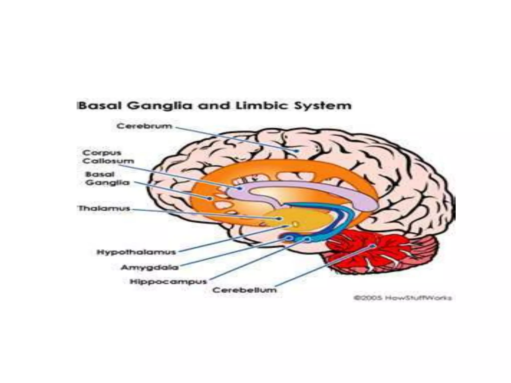
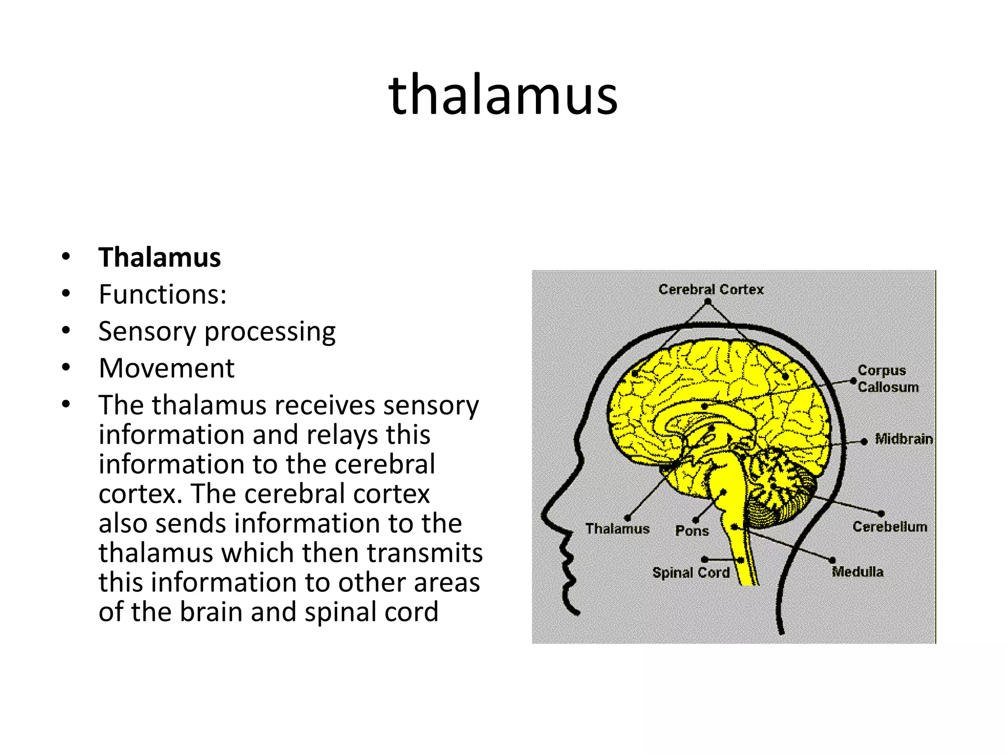
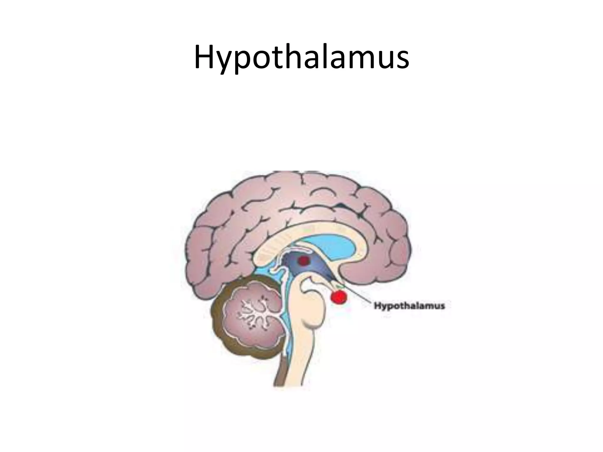
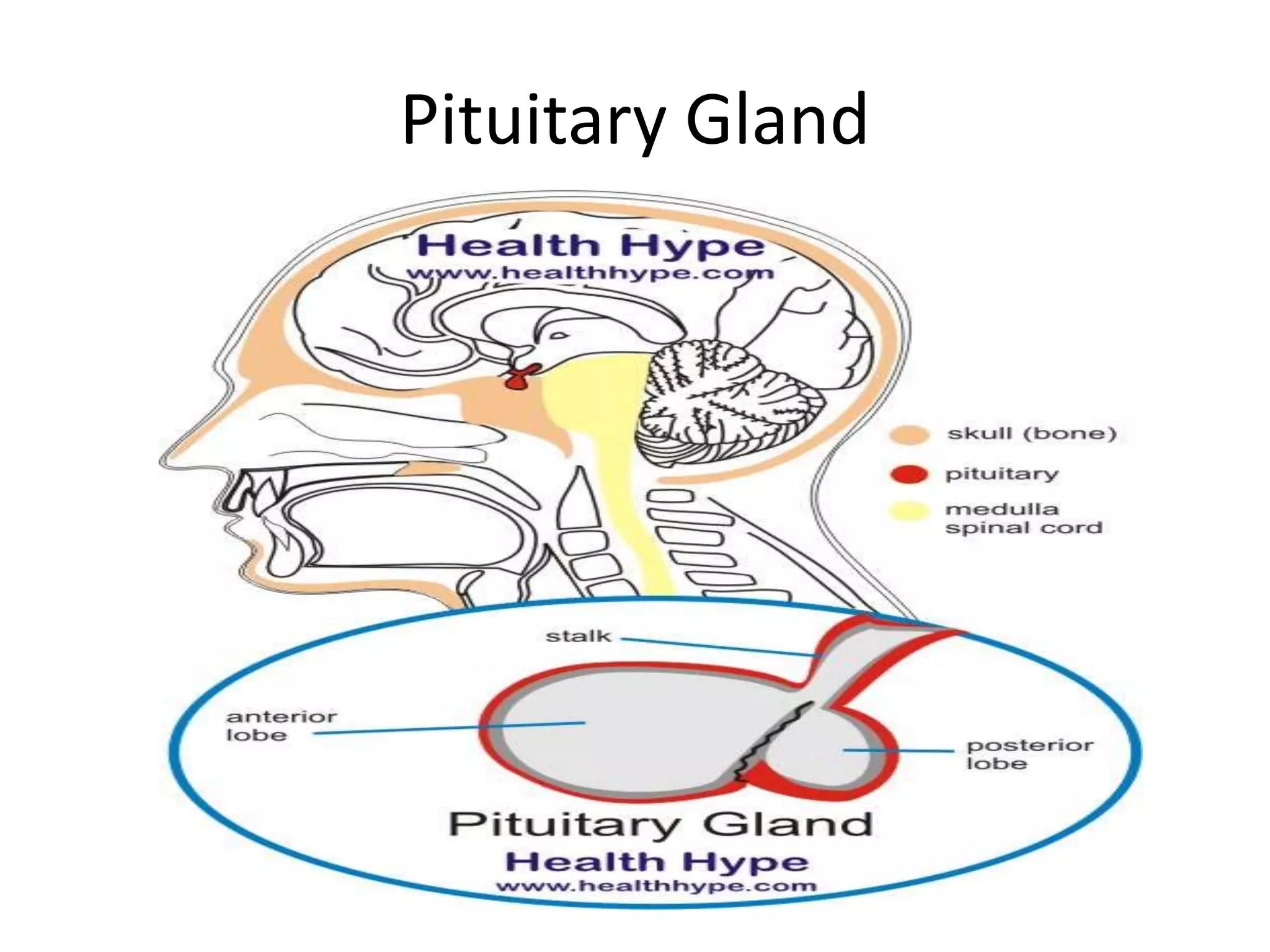
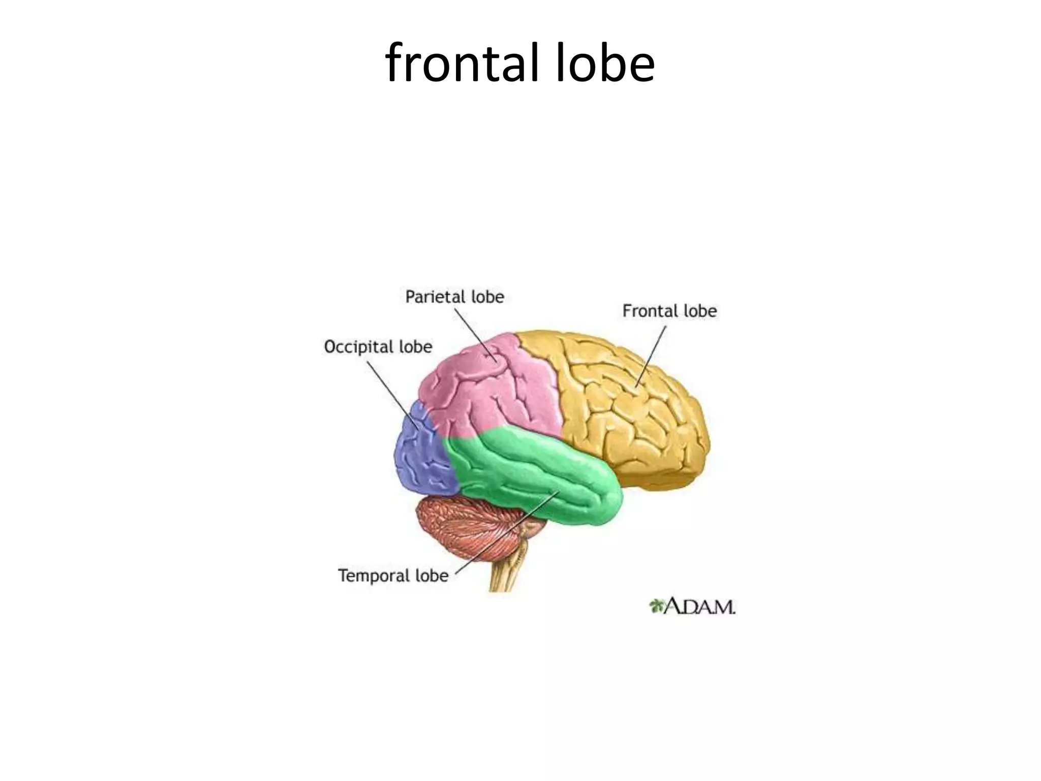
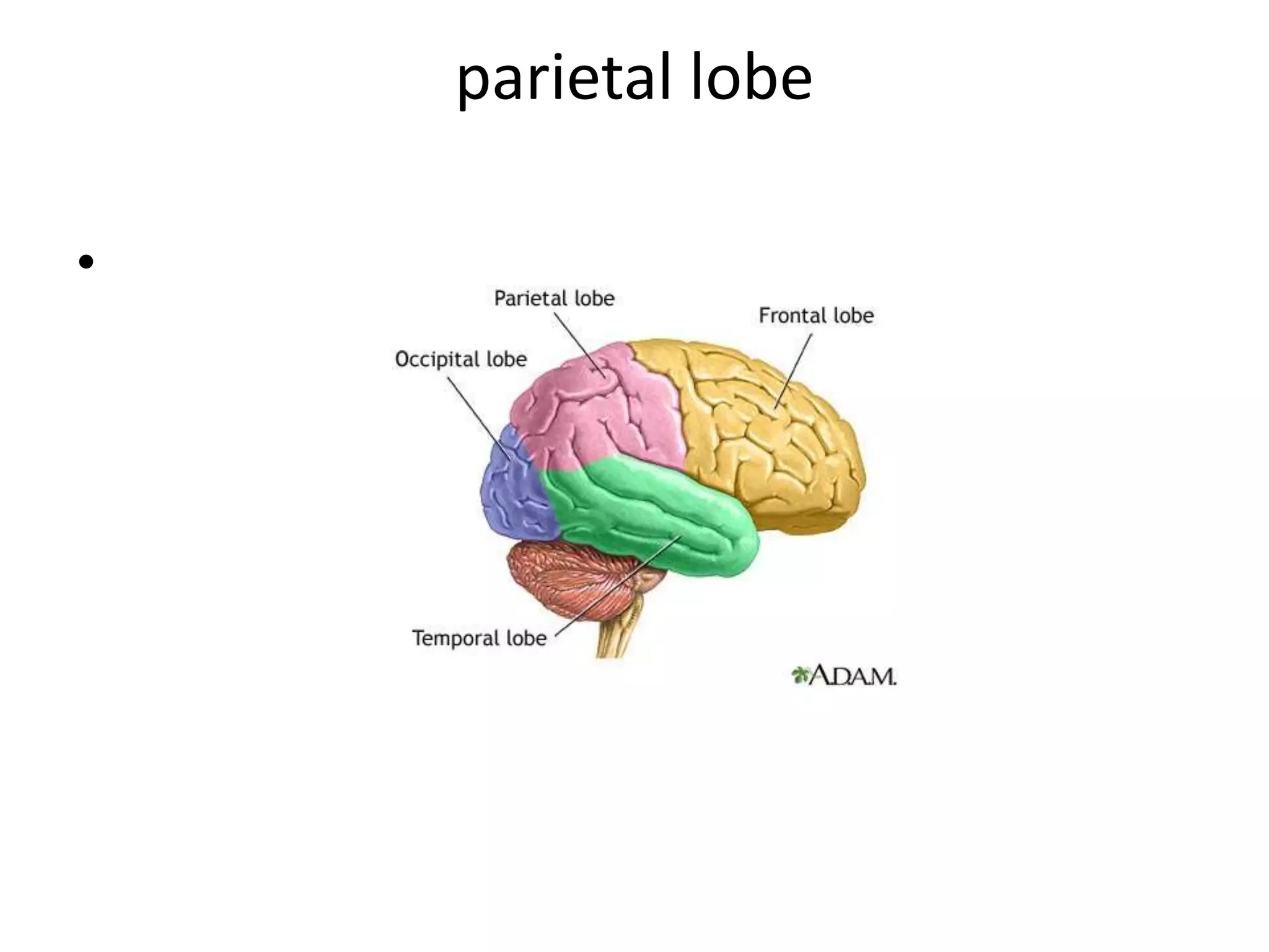
The Brain
November 2nd and 5th
The Brain
Brain Stem Keeps You Breathing

• Another brain part that's small
  but mighty is the brain stem.
  The brain stem sits beneath
  the cerebrum and in front of
  the cerebellum. It connects
  the rest of the brain to the
  spinal cord, which runs down
  your neck and back. The brain
  stem is in charge of all the
  functions your body needs to
  stay alive, like breathing air,
  digesting food, and circulating
  blood.
Lateral(side)view of Brainstem
Brain Stem
•   Functions:
•   Breathing
•   Heart Rate
•   Blood Pressure
•   The brain stem is a general term for the area of
    the brain between the thalamus and spinal cord.
    Structures within the brain stem include the
    medulla, tectum, tegmentum, and pons. Areas
    responsible for the most basic functions of life
    such as breathing, heart rate and blood pressure.
medulla
• Part of the brain stem
  important for breathing
  and respiration
Pons

• Area of the brainstem
  between the medulla and
  the midbrain
• Tectum "Roof" of the
  midbrain.
• Tegmentum "Floor" of the
  midbrain.
Cerebellum

• It controls balance,
  movement, and coordination
  (how your muscles work
  together). Because of your
  cerebellum, you can stand
  upright, keep your balance,
  and move around. Think about
  a surfer riding the waves on
  his/her board. What does
  he/she need most to stay
  balanced? The best surfboard?
  The coolest wetsuit? Nope —
  he/she needs his cerebellum!
Cerebellum
The cerebellum is at the back of
the brain, below the cerebrum
thalamus

•   Thalamus
•   Functions:
•   Sensory processing
•   Movement
•   The thalamus receives sensory
    information and relays this
    information to the cerebral
    cortex. The cerebral cortex
    also sends information to the
    thalamus which then transmits
    this information to other areas
    of the brain and spinal cord
limbic system
•   Functions:
•   Emotions
•   Memory
•   The limbic system (or the limbic areas) is a group
    of structures that includes the amygdala, the
    hippocampus, mammillary bodies and cingulate
    gyrus. These areas are important for controlling
    the emotional response to a given situation. The
    hippocampus is also important for memory
Amygdala

Your brain has a little bunch of cells on each side
called the amygdala. The word amygdala is Latin for
almond, and that's what this area looks like.
Scientists believe that the amygdala is responsible
for emotion. It's normal to feel all different kinds of
emotions, good and bad. Sometimes you might feel
a little sad, and other times you might feel scared,
or silly, or glad.
Amygdala
• A group of 90 healthy gay and heterosexual
  adults, men and women, were scanned by the
  Karolinska Institute scientists to measure the
  volume of both sides, or hemispheres, of their
  brain.
• When these results were collected, it was found
  that lesbians and heterosexual men shared a
  particular "asymmetry" in their hemisphere size,
  while heterosexual women and gay men had no
  difference between the size of the different
  halves of their brain.
Amygdala
• In other words, structurally, at least, the brains of gay
  men were more like heterosexual women, and gay
  women more like heterosexual men.
• A further experiment found that in one particular area
  of the brain, the amygdala, there were other significant
  differences.
• In heterosexual men and gay women, there were more
  nerve "connections" in the right side of the amygdala,
  compared with the left.
• The reverse, with more neural connections in the left
  amygdala, was the case in homosexual men and
  heterosexual women.
hippocampus
•   Hippocampus
•   Functions:
•   Learning
•   Memory
•   The hippocampus is one part of the limbic
    system that is important for memory and
    learning
Hypothalamus
Hypothalamus
• The hypothalamus is composed of several different
  areas and is located at the base of the brain. Although
  it is the size of only a pea (about 1/300 of the total
  brain weight), the hypothalamus is responsible for
  some very important functions. One important
  function of the hypothalamus is the control of body
  temperature. The hypothalamus acts as a "thermostat"
  by sensing changes in body temperature and then
  sending signals to adjust the temperature. For
  example, if you are too hot, the hypothalamus detects
  this and then sends a signal to expand the capillaries in
  your skin. This causes blood to be cooled faster. The
  hypothalamus also controls the pituitary.
Hypothalamus
 Functions:
• Body Temperature
• Emotions
• Hunger
• Thirst
• Circadian Rhythms These are daily rhythms to many of our
   physiological functions and activities....our sleep, body
   temperature, alertness, neurotransmitter levels. Many of
   these rhythms run on a cycle of about 24 hours. Rhythms that
   run on this 24 cycle are called Circadian Rhythms.
• This little gland also plays a role with lots of
  other hormones, like ones that control the
  amount of sugars and water in your body. And
  it helps keep your metabolism going. Your
  metabolism is everything that goes on in your
  body to keep it alive and growing and supplied
  with energy, like breathing, digesting food,
  and moving your blood around.
Pituitary Gland
Pituitary Gland
• The pituitary gland is a small round organ that is about
  1 centimeter in diameter and occupies a small groove
  in the base of the skull.
• Weighs 0.5 to 1 gram, this small pea-sized gland is able
  to influence every other endocrine gland in the body
  and therefore known as the “master gland”.
• The pituitary gland is controlled to a large extent by the
  hypothalamus. We will see it next.
• The hypothalamus is able to send stimulatory or
  inhibitory hormones to the pituitary gland thereby
  regulating its action on other endocrine glands and the
  body as a whole.
Pituitary gland
• Pituitary Gland Controls Growth
• The pituitary gland is very small — only about the
  size of a pea! Its job is to produce and release
  hormones into your body. If your clothes from
  last year are too small, it's because your pituitary
  gland released special hormones that made you
  grow. This gland is a big player in puberty too.
  This is the time when boys' and girls' bodies go
  through major changes as they slowly become
  men and women, all thanks to hormones
  released by the pituitary gland.
Corpus callosum
        • From a top view, notice
          how the brain is divided
          into two halves, called
          hemispheres. Each
          hemisphere
          communicates with the
          other through the
          corpus callosum, a
          bundle of nerve fibers.
Cerebral Cortex
Cerebral Cortex
•   Thought
•   Voluntary movement
•   Language
•   Reasoning
•   Perception
•   The cortex is a sheet of tissue that makes up the outer layer of the
    brain. The thickness of the cerebral cortex varies from 2 to 6 mm.
    The right and left sides of the cerebral cortex are connected by a
    thick band of nerve fibers called the "corpus callosum.“ In higher
    mammals such as humans, the cerebral cortex looks like it has
    many bumps and grooves. A bump or bulge on the cortex is called a
    gyrus (the plural of the word gyrus is "gyri") and a groove is called a
    sulcus (the plural of the word sulcus is "sulci"). Lower mammals,
    such as rats and mice, have very few gyri and sulci.
Cerebral hemispheres
• The cerebrum has two halves, with one on
  either side of the head. Some scientists think
  that the right half helps you think about
  abstract things like music, colors, and shapes.
  The left half is said to be more analytical,
  helping you with math, logic, and speech.
  Scientists do know for sure that the right half
  of the cerebrum controls the left side of your
  body, and the left half controls the right side.
Cerebrum: synopsis
The biggest part of the brain is the cerebrum. The cerebrum
makes up 85% of the brain's weight.
The cerebrum is the thinking part of the brain and it controls
your voluntary muscles, i.e. the ones that move when you want
them to. When you kick a ball; when you have directed thought;
when you need it to solve math problems, figure out a video
game, and draw a picture.
Your memory lives in the cerebrum — both short-term memory.
The cerebrum also helps you reason and order your life, i.e.
you'd better do your homework now because you won’t have
time to get sufficient sleep if you wait; I shouldn’t buy this
because I will need the money later in the month.
frontal lobe
Motor Cortex; Broca’s area

• The frontal lobes are involved in motor function,
  problem solving, spontaneity, memory, language,
  initiation, judgment, impulse control, and social
  and sexual behavior.
• The frontal lobes are extremely vulnerable to
  injury due to their location at the front of the
  cranium.
• MRI studies have shown that the frontal area is
  the most common region of injury following mild
  to moderate traumatic brain injury (Levin et al.,
  1987).
Frontal Lobe
• There are important asymmetrical differences in the frontal
  lobes. The left frontal lobe is involved in controlling
  language related movement, whereas the right frontal lobe
  plays a role in non-verbal abilities. Some researchers
  emphasize that this rule is not absolute and that with many
  people, both lobes are involved in nearly all behavior.
• Disturbance of motor function is typically characterized by
  loss of fine movements and strength of the arms, hands
  and fingers (Kuypers, 1981).
• Patients with frontal lobe damage exhibit little spontaneous
  facial expression, which points to the role of the frontal
  lobes in facial expression (Kolb & Milner, 1981). Broca's
  Aphasia, or difficulty in speaking, has been associated with
  frontal damage by Brown (1972).
Frontal Lobe
• One of the most common characteristics of
  frontal lobe damage is difficulty in interpreting
  feedback from the environment. Perseverating on
  a response (Milner, 1964), risk taking, and non-
  compliance with rules (Miller, 1985), and
  impaired associated learning (using external cues
  to help guide behavior) (Drewe, 1975) are a few
  examples of this type of deficit.
• The frontal lobes are also thought to play a part
  in our spatial orientation, including our body's
  orientation in space (Semmes et al., 1963).
Frontal Lobe
• One of the most common effects of frontal damage can
  be a dramatic change in social behavior. A person's
  personality can undergo significant changes after an
  injury to the frontal lobes, especially when both lobes
  are involved. There are some differences in the left
  versus right frontal lobes in this area. Left frontal
  damage usually manifests as pseudodepression and
  right frontal damage as pseudopsychopathic (Blumer
  and Benson, 1975).
• Sexual behavior can also be effected by frontal lesions.
  Orbital frontal damage can introduce abnormal sexual
  behavior, while dorolateral lesions may reduce sexual
  interest (Walker and Blummer, 1975).
parietal lobe

•
Somatosensory Cortex:somato=body
• The parietal lobes can be divided into two functional regions. One
  involves sensation and perception and the other is concerned with
  integrating sensory input, primarily with the visual system.
• The first function integrates sensory information to form a single
  perception (cognition). The second function constructs a spatial
  coordinate system to represent the world around us.
• Individuals with damage to the parietal lobes often show striking
  deficits, such as abnormalities in body image and spatial relations
  (Kandel, Schwartz & Jessel, 1991).

• Damage to the left parietal lobe can result in what is called
  "Gerstmann's Syndrome." It includes right-left confusion, difficulty
  with writing (agraphia) and difficulty with mathematics (acalculia).
  It can also produce disorders of language (aphasia) and the inability
  to perceive objects normally (agnosia).
Parietal Lobe
Damage to the right parietal lobe can result in neglecting part
of the body or space (contralateral neglect), which can impair
many self-care skills such as dressing and washing. Right side
damage can also cause difficulty in making things
(constructional apraxia), denial of deficits and drawing ability.

Bi-lateral damage (large lesions to both sides) can cause a
visual attention and motor syndrome. This is characterized by
the inability to voluntarily control the gaze (ocular apraxia),
inability to integrate components of a visual scene
(simultanagnosia), and the inability to accurately reach for an
object with visual guidance (optic ataxia) (Westmoreland et
al., 1994).
Parietal Lobe
• Special deficits (primarily to memory and personality) can
  occur if there is damage to the area between the parietal
  and temporal lobes.

• Left parietal-temporal lesions can effect verbal memory
  and the ability to recall strings of digits (Warrington &
  Weiskrantz, 1977). The right parietal-temporal lobe is
  concerned with non-verbal memory.

• Right parietal-temporal lesions can produce significant
  changes in personality.
temporal lobe
Primary Auditory Cortex; Wernicke’s
                 Area
• Kolb & Wishaw (1990) have identified eight principle symptoms of
  temporal lobe damage: 1) disturbance of auditory sensation and
  perception, 2) disturbance of selective attention of auditory and
  visual input, 3) disorders of visual perception, 4) impaired
  organization and categorization of verbal material, 5) disturbance of
  language comprehension, 6) impaired long-term memory, 7)
  altered personality and affective behavior, 8) altered sexual
  behavior.
• Selective attention to visual or auditory input is common with
  damage to the temporal lobes (Milner, 1968). Left side lesions
  result in decreased recall of verbal and visual content, including
  speech perception. Right side lesions result in decreased
  recognition of tonal sequences and many musical abilities. Right
  side lesions can also effect recognition of visual content (e.g. recall
  of faces).
Temporal Lobe
• The temporal lobes are involved in the primary
  organization of sensory input (Read, 1981). Individuals
  with temporal lobes lesions have difficulty placing
  words or pictures into categories.
• Language can be effected by temporal lobe damage.
  Left temporal lesions disturb recognition of words.
  Right temporal damage can cause a loss of inhibition of
  talking.
• The temporal lobes are highly associated with memory
  skills. Left temporal lesions result in impaired memory
  for verbal material. Right side lesions result in recall of
  non-verbal material, such as music and drawings.
Temporal Lobe
• Seizures of the temporal lobe can have
  dramatic effects on an individual's personality.
  Temporal lobe epilepsy can cause
  perseverative speech, paranoia and aggressive
  rages (Blumer and Benson, 1975). Severe
  damage to the temporal lobes can also alter
  sexual behavior (e.g. increase in activity)
  (Blumer and Walker, 1975).
Occipital Lobe
Primary Visual Cortex

• The occipital lobes are the center of our visual
  perception system. They are not particularly
  vulnerable to injury because of their location
  at the back of the brain, although any
  significant trauma to the brain could produce
  subtle changes to our visual-perceptual
  system, such as visual field defects, e.g.
  visuospatial processing, discrimination of
  movement and color discrimination
  (Westmoreland et al., 1994).
• Damage to one side of the occipital lobe causes loss of
  vision with exactly the same "field cut" in both eyes.
• Disorders of the occipital lobe can cause visual
  hallucinations and illusions. Visual hallucinations (visual
  images with no external stimuli) can be caused by lesions to
  the occipital region or temporal lobe seizures. Visual
  illusions (distorted perceptions) can take the form of
  objects appearing larger or smaller than they actually are,
  objects lacking color or objects having abnormal coloring.
• Lesions in the parietal-temporal-occipital association area
  can cause word blindness with writing impairments (alexia
  and agraphia) (Kandel, Schwartz & Jessell, 1991).
Plasticity
• Plasticity, or neuroplasticity, is the lifelong ability
  of the brain to reorganize neural pathways based
  on new experiences. As we learn, we acquire new
  knowledge and skills through instruction or
  experience. In order to learn or memorize a fact
  or skill, there must be persistent functional
  changes in the brain that represent the new
  knowledge. The ability of the brain to change
  with learning is what is known as neuroplasticity.
  To illustrate the concept of plasticity, imagine the
  film of a camera.
• Pretend that the film represents your brain. Now
  imagine using the camera to take a picture of a
  tree. When a picture is taken, the film is exposed
  to new information -- that of the image of a tree.
  In order for the image to be retained, the film
  must react to the light and ?change? to record
  the image of the tree. Similarly, in order for new
  knowledge to be retained in memory, changes in
  the brain representing the new knowledge must
  occur.
• To illustrate plasticity in another way, imagine
  making an impression of a coin in a lump of
  clay. In order for the impression of the coin to
  appear in the clay, changes must occur in the
  clay -- the shape of the clay changes as the
  coin is pressed into the clay. Similarly, the
  neural circuitry in the brain must reorganize in
  response to experience or sensory
  stimulation.

The brain

  • 1.
  • 2.
  • 3.
    Brain Stem KeepsYou Breathing • Another brain part that's small but mighty is the brain stem. The brain stem sits beneath the cerebrum and in front of the cerebellum. It connects the rest of the brain to the spinal cord, which runs down your neck and back. The brain stem is in charge of all the functions your body needs to stay alive, like breathing air, digesting food, and circulating blood.
  • 4.
  • 5.
    Brain Stem • Functions: • Breathing • Heart Rate • Blood Pressure • The brain stem is a general term for the area of the brain between the thalamus and spinal cord. Structures within the brain stem include the medulla, tectum, tegmentum, and pons. Areas responsible for the most basic functions of life such as breathing, heart rate and blood pressure.
  • 6.
    medulla • Part ofthe brain stem important for breathing and respiration
  • 7.
    Pons • Area ofthe brainstem between the medulla and the midbrain • Tectum "Roof" of the midbrain. • Tegmentum "Floor" of the midbrain.
  • 8.
    Cerebellum • It controlsbalance, movement, and coordination (how your muscles work together). Because of your cerebellum, you can stand upright, keep your balance, and move around. Think about a surfer riding the waves on his/her board. What does he/she need most to stay balanced? The best surfboard? The coolest wetsuit? Nope — he/she needs his cerebellum!
  • 9.
    Cerebellum The cerebellum isat the back of the brain, below the cerebrum
  • 11.
    thalamus • Thalamus • Functions: • Sensory processing • Movement • The thalamus receives sensory information and relays this information to the cerebral cortex. The cerebral cortex also sends information to the thalamus which then transmits this information to other areas of the brain and spinal cord
  • 12.
    limbic system • Functions: • Emotions • Memory • The limbic system (or the limbic areas) is a group of structures that includes the amygdala, the hippocampus, mammillary bodies and cingulate gyrus. These areas are important for controlling the emotional response to a given situation. The hippocampus is also important for memory
  • 13.
    Amygdala Your brain hasa little bunch of cells on each side called the amygdala. The word amygdala is Latin for almond, and that's what this area looks like. Scientists believe that the amygdala is responsible for emotion. It's normal to feel all different kinds of emotions, good and bad. Sometimes you might feel a little sad, and other times you might feel scared, or silly, or glad.
  • 14.
    Amygdala • A groupof 90 healthy gay and heterosexual adults, men and women, were scanned by the Karolinska Institute scientists to measure the volume of both sides, or hemispheres, of their brain. • When these results were collected, it was found that lesbians and heterosexual men shared a particular "asymmetry" in their hemisphere size, while heterosexual women and gay men had no difference between the size of the different halves of their brain.
  • 15.
    Amygdala • In otherwords, structurally, at least, the brains of gay men were more like heterosexual women, and gay women more like heterosexual men. • A further experiment found that in one particular area of the brain, the amygdala, there were other significant differences. • In heterosexual men and gay women, there were more nerve "connections" in the right side of the amygdala, compared with the left. • The reverse, with more neural connections in the left amygdala, was the case in homosexual men and heterosexual women.
  • 16.
    hippocampus • Hippocampus • Functions: • Learning • Memory • The hippocampus is one part of the limbic system that is important for memory and learning
  • 17.
  • 18.
    Hypothalamus • The hypothalamusis composed of several different areas and is located at the base of the brain. Although it is the size of only a pea (about 1/300 of the total brain weight), the hypothalamus is responsible for some very important functions. One important function of the hypothalamus is the control of body temperature. The hypothalamus acts as a "thermostat" by sensing changes in body temperature and then sending signals to adjust the temperature. For example, if you are too hot, the hypothalamus detects this and then sends a signal to expand the capillaries in your skin. This causes blood to be cooled faster. The hypothalamus also controls the pituitary.
  • 19.
    Hypothalamus Functions: • BodyTemperature • Emotions • Hunger • Thirst • Circadian Rhythms These are daily rhythms to many of our physiological functions and activities....our sleep, body temperature, alertness, neurotransmitter levels. Many of these rhythms run on a cycle of about 24 hours. Rhythms that run on this 24 cycle are called Circadian Rhythms.
  • 20.
    • This littlegland also plays a role with lots of other hormones, like ones that control the amount of sugars and water in your body. And it helps keep your metabolism going. Your metabolism is everything that goes on in your body to keep it alive and growing and supplied with energy, like breathing, digesting food, and moving your blood around.
  • 21.
  • 22.
    Pituitary Gland • Thepituitary gland is a small round organ that is about 1 centimeter in diameter and occupies a small groove in the base of the skull. • Weighs 0.5 to 1 gram, this small pea-sized gland is able to influence every other endocrine gland in the body and therefore known as the “master gland”. • The pituitary gland is controlled to a large extent by the hypothalamus. We will see it next. • The hypothalamus is able to send stimulatory or inhibitory hormones to the pituitary gland thereby regulating its action on other endocrine glands and the body as a whole.
  • 23.
    Pituitary gland • PituitaryGland Controls Growth • The pituitary gland is very small — only about the size of a pea! Its job is to produce and release hormones into your body. If your clothes from last year are too small, it's because your pituitary gland released special hormones that made you grow. This gland is a big player in puberty too. This is the time when boys' and girls' bodies go through major changes as they slowly become men and women, all thanks to hormones released by the pituitary gland.
  • 24.
    Corpus callosum • From a top view, notice how the brain is divided into two halves, called hemispheres. Each hemisphere communicates with the other through the corpus callosum, a bundle of nerve fibers.
  • 25.
  • 26.
    Cerebral Cortex • Thought • Voluntary movement • Language • Reasoning • Perception • The cortex is a sheet of tissue that makes up the outer layer of the brain. The thickness of the cerebral cortex varies from 2 to 6 mm. The right and left sides of the cerebral cortex are connected by a thick band of nerve fibers called the "corpus callosum.“ In higher mammals such as humans, the cerebral cortex looks like it has many bumps and grooves. A bump or bulge on the cortex is called a gyrus (the plural of the word gyrus is "gyri") and a groove is called a sulcus (the plural of the word sulcus is "sulci"). Lower mammals, such as rats and mice, have very few gyri and sulci.
  • 27.
    Cerebral hemispheres • Thecerebrum has two halves, with one on either side of the head. Some scientists think that the right half helps you think about abstract things like music, colors, and shapes. The left half is said to be more analytical, helping you with math, logic, and speech. Scientists do know for sure that the right half of the cerebrum controls the left side of your body, and the left half controls the right side.
  • 28.
    Cerebrum: synopsis The biggestpart of the brain is the cerebrum. The cerebrum makes up 85% of the brain's weight. The cerebrum is the thinking part of the brain and it controls your voluntary muscles, i.e. the ones that move when you want them to. When you kick a ball; when you have directed thought; when you need it to solve math problems, figure out a video game, and draw a picture. Your memory lives in the cerebrum — both short-term memory. The cerebrum also helps you reason and order your life, i.e. you'd better do your homework now because you won’t have time to get sufficient sleep if you wait; I shouldn’t buy this because I will need the money later in the month.
  • 29.
  • 30.
    Motor Cortex; Broca’sarea • The frontal lobes are involved in motor function, problem solving, spontaneity, memory, language, initiation, judgment, impulse control, and social and sexual behavior. • The frontal lobes are extremely vulnerable to injury due to their location at the front of the cranium. • MRI studies have shown that the frontal area is the most common region of injury following mild to moderate traumatic brain injury (Levin et al., 1987).
  • 31.
    Frontal Lobe • Thereare important asymmetrical differences in the frontal lobes. The left frontal lobe is involved in controlling language related movement, whereas the right frontal lobe plays a role in non-verbal abilities. Some researchers emphasize that this rule is not absolute and that with many people, both lobes are involved in nearly all behavior. • Disturbance of motor function is typically characterized by loss of fine movements and strength of the arms, hands and fingers (Kuypers, 1981). • Patients with frontal lobe damage exhibit little spontaneous facial expression, which points to the role of the frontal lobes in facial expression (Kolb & Milner, 1981). Broca's Aphasia, or difficulty in speaking, has been associated with frontal damage by Brown (1972).
  • 32.
    Frontal Lobe • Oneof the most common characteristics of frontal lobe damage is difficulty in interpreting feedback from the environment. Perseverating on a response (Milner, 1964), risk taking, and non- compliance with rules (Miller, 1985), and impaired associated learning (using external cues to help guide behavior) (Drewe, 1975) are a few examples of this type of deficit. • The frontal lobes are also thought to play a part in our spatial orientation, including our body's orientation in space (Semmes et al., 1963).
  • 33.
    Frontal Lobe • Oneof the most common effects of frontal damage can be a dramatic change in social behavior. A person's personality can undergo significant changes after an injury to the frontal lobes, especially when both lobes are involved. There are some differences in the left versus right frontal lobes in this area. Left frontal damage usually manifests as pseudodepression and right frontal damage as pseudopsychopathic (Blumer and Benson, 1975). • Sexual behavior can also be effected by frontal lesions. Orbital frontal damage can introduce abnormal sexual behavior, while dorolateral lesions may reduce sexual interest (Walker and Blummer, 1975).
  • 34.
  • 35.
    Somatosensory Cortex:somato=body • Theparietal lobes can be divided into two functional regions. One involves sensation and perception and the other is concerned with integrating sensory input, primarily with the visual system. • The first function integrates sensory information to form a single perception (cognition). The second function constructs a spatial coordinate system to represent the world around us. • Individuals with damage to the parietal lobes often show striking deficits, such as abnormalities in body image and spatial relations (Kandel, Schwartz & Jessel, 1991). • Damage to the left parietal lobe can result in what is called "Gerstmann's Syndrome." It includes right-left confusion, difficulty with writing (agraphia) and difficulty with mathematics (acalculia). It can also produce disorders of language (aphasia) and the inability to perceive objects normally (agnosia).
  • 36.
    Parietal Lobe Damage tothe right parietal lobe can result in neglecting part of the body or space (contralateral neglect), which can impair many self-care skills such as dressing and washing. Right side damage can also cause difficulty in making things (constructional apraxia), denial of deficits and drawing ability. Bi-lateral damage (large lesions to both sides) can cause a visual attention and motor syndrome. This is characterized by the inability to voluntarily control the gaze (ocular apraxia), inability to integrate components of a visual scene (simultanagnosia), and the inability to accurately reach for an object with visual guidance (optic ataxia) (Westmoreland et al., 1994).
  • 37.
    Parietal Lobe • Specialdeficits (primarily to memory and personality) can occur if there is damage to the area between the parietal and temporal lobes. • Left parietal-temporal lesions can effect verbal memory and the ability to recall strings of digits (Warrington & Weiskrantz, 1977). The right parietal-temporal lobe is concerned with non-verbal memory. • Right parietal-temporal lesions can produce significant changes in personality.
  • 38.
  • 39.
    Primary Auditory Cortex;Wernicke’s Area • Kolb & Wishaw (1990) have identified eight principle symptoms of temporal lobe damage: 1) disturbance of auditory sensation and perception, 2) disturbance of selective attention of auditory and visual input, 3) disorders of visual perception, 4) impaired organization and categorization of verbal material, 5) disturbance of language comprehension, 6) impaired long-term memory, 7) altered personality and affective behavior, 8) altered sexual behavior. • Selective attention to visual or auditory input is common with damage to the temporal lobes (Milner, 1968). Left side lesions result in decreased recall of verbal and visual content, including speech perception. Right side lesions result in decreased recognition of tonal sequences and many musical abilities. Right side lesions can also effect recognition of visual content (e.g. recall of faces).
  • 40.
    Temporal Lobe • Thetemporal lobes are involved in the primary organization of sensory input (Read, 1981). Individuals with temporal lobes lesions have difficulty placing words or pictures into categories. • Language can be effected by temporal lobe damage. Left temporal lesions disturb recognition of words. Right temporal damage can cause a loss of inhibition of talking. • The temporal lobes are highly associated with memory skills. Left temporal lesions result in impaired memory for verbal material. Right side lesions result in recall of non-verbal material, such as music and drawings.
  • 41.
    Temporal Lobe • Seizuresof the temporal lobe can have dramatic effects on an individual's personality. Temporal lobe epilepsy can cause perseverative speech, paranoia and aggressive rages (Blumer and Benson, 1975). Severe damage to the temporal lobes can also alter sexual behavior (e.g. increase in activity) (Blumer and Walker, 1975).
  • 42.
  • 43.
    Primary Visual Cortex •The occipital lobes are the center of our visual perception system. They are not particularly vulnerable to injury because of their location at the back of the brain, although any significant trauma to the brain could produce subtle changes to our visual-perceptual system, such as visual field defects, e.g. visuospatial processing, discrimination of movement and color discrimination (Westmoreland et al., 1994).
  • 44.
    • Damage toone side of the occipital lobe causes loss of vision with exactly the same "field cut" in both eyes. • Disorders of the occipital lobe can cause visual hallucinations and illusions. Visual hallucinations (visual images with no external stimuli) can be caused by lesions to the occipital region or temporal lobe seizures. Visual illusions (distorted perceptions) can take the form of objects appearing larger or smaller than they actually are, objects lacking color or objects having abnormal coloring. • Lesions in the parietal-temporal-occipital association area can cause word blindness with writing impairments (alexia and agraphia) (Kandel, Schwartz & Jessell, 1991).
  • 45.
    Plasticity • Plasticity, orneuroplasticity, is the lifelong ability of the brain to reorganize neural pathways based on new experiences. As we learn, we acquire new knowledge and skills through instruction or experience. In order to learn or memorize a fact or skill, there must be persistent functional changes in the brain that represent the new knowledge. The ability of the brain to change with learning is what is known as neuroplasticity. To illustrate the concept of plasticity, imagine the film of a camera.
  • 46.
    • Pretend thatthe film represents your brain. Now imagine using the camera to take a picture of a tree. When a picture is taken, the film is exposed to new information -- that of the image of a tree. In order for the image to be retained, the film must react to the light and ?change? to record the image of the tree. Similarly, in order for new knowledge to be retained in memory, changes in the brain representing the new knowledge must occur.
  • 47.
    • To illustrateplasticity in another way, imagine making an impression of a coin in a lump of clay. In order for the impression of the coin to appear in the clay, changes must occur in the clay -- the shape of the clay changes as the coin is pressed into the clay. Similarly, the neural circuitry in the brain must reorganize in response to experience or sensory stimulation.