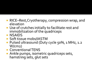
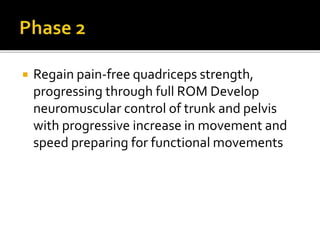
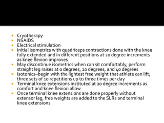
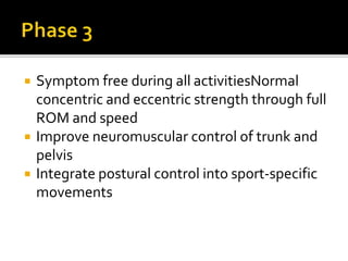
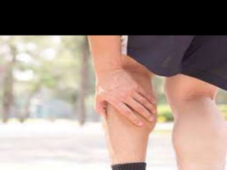
The document discusses various types of muscle and bone injuries, particularly contusions and strains, outlining definitions, mechanisms, and management protocols. It details specific types of injuries such as hamstring, calf, and quadriceps strains, discussing their causes, symptoms, grading, and rehabilitation processes. Additionally, the document highlights the importance of diagnostic tools and treatment strategies to promote recovery and prevent re-injury.
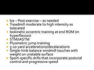
![ Various factors are seen in hamstring injury such
as
Older age
Previous hamstring injury
Limited hamstring flexibility]
Increased fatigue
Poor core stability
Strength imbalance
Previous calf injury
Previous substantial knee injury
Osteitis pubis](https://image.slidesharecdn.com/contusions-210523182107/85/Contusions-strain-in-hamstrings-quadriceps-calf-24-320.jpg)

![ During sporting activities such as sprinting,
these long, bi-articular muscles have to cope
with high internal forces and rapid changes in
muscle length and mode of contraction
leading to a higher risk of strain.
despite this, calf muscle strains have also
been reported to occur during slow-
lengthening muscle actions such as those
performed by ballet dancers, but also during
common daily activities.[9]](https://image.slidesharecdn.com/contusions-210523182107/85/Contusions-strain-in-hamstrings-quadriceps-calf-65-320.jpg)

![ Tape or a compressive wrap can be applied
and the leg elevated where possible. [23]
If major bleeding has occurred, the use of
NSAIDs has to be carefully controlled as they
have an anti-platelet effect
Gentle passive stretching exercises without
pain to maintain range of motion in the
plantar flexors.](https://image.slidesharecdn.com/contusions-210523182107/85/Contusions-strain-in-hamstrings-quadriceps-calf-84-320.jpg)