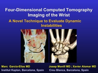
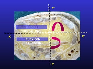
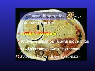
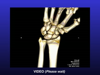
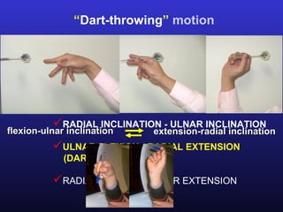
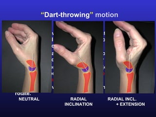
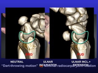
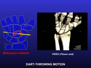
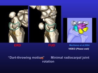
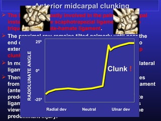
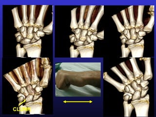
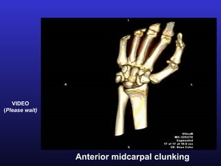
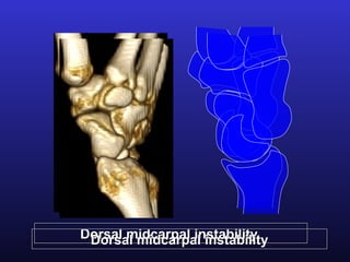
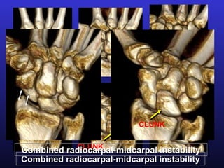
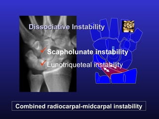
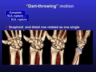
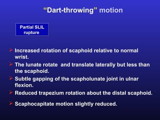
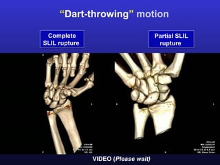
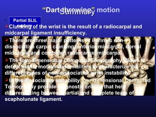
Four-Dimensional Computed Tomography
           Imaging of the Wrist
     A Novel Technique to Evaluate Dynamic
                     Instabilities


Marc Garcia-Elias MD                Josep Monill MD ; Xavier Alomar MD
Institut Kaplan, Barcelona, Spain   Creu Blanca, Barcelona, Spain
Technical parameters in 320-MDCT
arpal Aquilion One, Toshiba Medical Systems
      kinematics

      0.5 mm detectors- 16 cm of z-axis volume
      Gantry rotation time: 350 milliseconds
         FLEXION - EXTENSION
      Minimum temporal resolution: 175 milliseconds
         RADIAL INCLINATION - ULNAR INCLINATION
       100 kVp-80 mA
          ULNAR FLEXION - RADIAL EXTENSION
            (DART-THROWING)
      14 image series obtained for 14 seconds
          RADIAL FLEXION – ULNAR EXTENSION
      DLP 240mGy. cm
Y’


      RADIAL
    CUBITAL INCL.
X                        X’
      FLEXION-
     EXTENSION


                    Y
Carpal kinematics     ECRL
    ULNAR FLEXION –
   RADIAL EXTENSIÓN


   DART-THROWING
 FLEXION - EXTENSION
 RADIAL INCLINATION - ULNAR INCLINATION
 ULNAR FLEXION - RADIAL EXTENSION
 RADIAL FLEXION - ULNAR EXTENSION
FCU
The proximal row hasn´t
tendinous attachments


 The movement always
 begins in the distal row

 The fibers of midcarpal
    Flexion
 ligaments increase in          Neutral   Extension
 tension

 The forces of compression
 generated on the bone
 force it to move


 In the central movements the
radiocarpal joint is slightly active
RADIAL INCLINATION         ULNAR INCLINATION
          Carpal kinematics
   FLEXION                    EXTENSION


    FLEXION - EXTENSION
    RADIAL INCLINATION - ULNAR INCLINATION
    ULNAR FLEXION - RADIAL EXTENSION
    RADIAL FLEXION - ULNAR EXTENSION
The proximal row, from a flexed position in radial
 deviation becomes extended in ulnar deviation


  RADIAL INCLINATION-ULNAR INCLINATION
VIDEO (Please wait)
In radial deviation the           In ulnar deviation it‘s the
trapezium is pushing the          triquetral-hamate joint the
scaphoid towards flexion curve from
                    Smooth        one forcing the proximal
and ulnar translation.            row towards extension
                    flexion to extension
                                  and radial translation.
                           25º
       RADIOLUNATE ANGLE


                           0º


                           -25º

                                  Radial dev   Neutral   Ulnar dev
Which structures ensure a smooth progression
          from flexion to extension?

 The most important ligament inducing progressive
  extension of the proximal row in ulnar deviation is the
  palmar triquetrum-capitate-hamate ligament that
  increase in tension. The triquetrum is pulled by this
  ligament against the proximal pole of the hamate with
  which the triquetrum extends.
 The scaphotrapezial ligament causes extension and
1 Scaphoid-Trapezium scaphoid.
  pronation to the 2 Dorsal Intercarpal  3 Triquetrum-Capitate
 If the proximal row extends too much the capitate
  would sublux dorsally. This is prevented by the
  portion of the dorsal intercarpal ligament.
1

2

    3
The contraction of the flexor carpi ulnaris generates a
 dorsal directed vector on the triquetrum that helps
   extending the proximal row in ulnar deviation.


                 VIDEO (Please wait)
“Dart-throwing” motion

                  Carpal kinematics


          FLEXION - EXTENSION
          RADIAL INCLINATION - ULNAR INCLINATION
flexion-ulnar inclination      extension-radial inclination
          ULNAR FLEXION - RADIAL EXTENSION
             (DART-THROWING)

          RADIAL FLEXION - ULNAR EXTENSION
“Dart-throwing” motion

 In the dart-throwing motion the wrist rotates from an
  extended radial deviation position to a flexed ulnar
  deviated position.
 This oblique plane of motion is the one most
  commonly used in activities of daily living.
 The contribution of the radiocarpal joint to dart-
  throwing motion is minimal, most rotation occurring
  at the midcarpal level.
 In the dart-throwing plane the proximal row does not
  rotate.
   NEUTRAL              RADIAL         RADIAL INCL.
                      INCLINATION         + EXTENSION
ERD                              FUD

     NEUTRAL              ULNAR             ULNAR INCL.+
“Dart-throwing motion” INCLINATION radiocarpal joint rotation
                           Minimal           EXTENSION
Midcarpal rotation          VIDEO (Please wait)


               DART-THROWING MOTION
ERD                   FUD           Moritomo et al,2004
                                      VIDEO (Please wait)


“Dart-throwing motion”     Minimal radiocarpal joint
                     rotation
Non-dissociative Instability- Pathomechanics

          Non-dissociative Instability
 The wrist exhibits a radiocarpal and/or a midcarpal
  subluxation of the entire proximal row during non-
  resisted wrist Pathomechanics
                 motion without injury of the interosseous
  ligaments of the proximal row.
                 Clinical forms
 This result in a clunking wrist. Clunking is a low-pitched
  dull sound produced by sudden subluxation and/or
  reduction of a partially or totally dislocated carpal bone.
 In the clunking non-dissociative wrist the proximal row
  remains flexed until the wrist is ulnarly deviated at
  which point it suddenly jumps into extension.
Non-dissociative Instability- Clinical forms
 Non-dissociative Instability- Clinical forms
 There are two major types of non-dissociated carpal
  clunking: extrinsec and intrinsec.
                      Extrinsic Clunking
 The extrinsec clunking results from injury or bone
  alteration outside the carpal area (dorsal malunited
  radial fractures). Intrinsic Clunking
 The intrinsec clunking derives from insufficiency or
  injury of one, or several, carpal ligaments:
  scaphotrapezoid, triquetrum-capitate-hamate, dorsal
             Anterior        Dorsal       Radiocarpal-
  scaphotriquetral, palmar radiolunate ormidcarpal
            midcarpal       midcarpal
                                            ulnolunate.
 There are three major patterns of intrinsic carpal
  clunking: anterior midcarpal, dorsal midcarpal and
  combined radiocarpal-midcarpal.
Anterior midcarpal clunking
 The ligaments mostly involved in the palmar midcarpal
  instability are the scaphotrapezial ligament, and the
  triquetrum-capitate-hamate ligament.
 The proximal row remains tilted palmarly until near the
  end of ulnar deviation, where it suddenly rotates into
               25º
  extension, sometimes with a palpable thud (catch-up
         RADIOLUNATE ANGLE


  clunk).
 In most cases there is a combination of medial and lateral
  ligament insufficiency.
               0º                           Clunk !
 There are cases where the dysfunction clearly derives
  from a predominant injury at the scaphotrapezial ligament
  (anterolateral midcarpal instability) or from a
  predominant injury ot the triquetrum-capitate-hamate
              -25º
  ligament (anteromedial midcarpal instability). Stress
  views are recommended to assess the location of the
                   Radial dev   Neutral      Ulnar dev
  predominant injury.
CLUNK
VIDEO
(Please wait)


                Anterior midcarpal clunking
Anteromedial midcarpal instability


         H           C

        1
             2

   Tq                                1: Triquetrum - Hamate
                                                 2: Triquetrum -
                                     Capitate


 Anterior midcarpal clunking after
injury ot the triquetrum-capitate-
hamate ligament
  Palmar triqutetrum-capitate-hamate ligament
Anterolateral midcarpal instability
 Anterolateral midcarpal instability


   Anterior
 “drawer” test


STT subluxation
(“open mouth” sign)


 Scaphoid-Trapezium ligament (STT)
Dorsal midcarpal clunking


 The ligaments mostly involved in the dorsal midcarpal
  instability are the radioscaphocapitate ligament, and
  the dorsal scaphotriquetral ligament.
 As the wrist rotates toward ulnardevation the capitate
  subluxes over the edge of the scapholunate socket
  inducing hyperextension of the proximal row.
 Once the capitate is subluxed dorsally there is a
  reactive contraction of wrist extensors and the distal
  row tends to return abruptly to its normal alignment
       Dorsal Intercarpal
  often with an audible clunk.
       Ligament
Dorsal midcarpal instability
 Dorsal midcarpal instability
Radiocarpal- midcarpal clunking

 The pattern of clunking is similar to the anterior
  midcarpal instability but adding an increased mobility at
  the RC joint implying an abnormally flexed and ulnarly
  translocated proximal row in radial deviation.
 This form of clunking is frequent among teenagers with
  hyperlax radio-ulno-carpal ligaments.
 In the radiocarpal (or proximal) type of clunking, the
  ligaments mostly involved are the palmar long and
  short radiolunate and the dorsal radiotriquetral.


        Palmar-dorsal radiocarpal ligaments
CLUNK

            CLUNK
Combined radiocarpal-midcarpal instability
Dissociative Instability


        Scapholunate instability
        Lunotriqueteal instability


Combined radiocarpal-midcarpal instability
Dissociative Instability- Pathomechanics

              Scapholunate instability
 This instability is secondary to rupture of the linkage
  between the bones of the proximal row.
 In the scapholunate instability the scaphoid has lost its
  ligament connections and exhibits dorsoradial
  subluxation over the edge of the radius ligament
      Scapholunate inteosseous during radial
  deviation.               (SLIL)
 It is important to distinguish between partial and
  complete scapholunate interosseous ligament (SLIL)
  tears. Partial tears are benign, often asymptomatic.
  Complete disruptions evolve intocomplete from
         Can we differentiate progressive carpal
  collapse and joint degeneration.
                       partial rupture?
 The dart-throwing plane of motion allows easy
  discrimination between partial and total SLIL injury.
“Dart-throwing” motion
  Complete
 SLIL Complete
      rupture
    SLIL rupture

 Scaphoid and distal row rotated as one single
  functional unit.
 Reduced position of scaphoid in radial extesion.
 Dorsolateral subluxation of scaphoid in ulnar flexion.
 Wide gap between the scaphoid and lunate in ulnar
  flexion.
 Trapezium does not rotate about the distal scaphoid.
 No scaphocapitate motion.
“Dart-throwing” motion

    Partial SLIL
     rupture


 Increased rotation of scaphoid relative to normal
  wrist.
 The lunate rotate and translate laterally but less than
  the scaphoid.
 Subtle gapping of the scapholunate joint in ulnar
  flexion.
 Reduced trapezium rotation about the distal scaphoid.
 Scaphocapitate motion slightly reduced.
“Dart-throwing” motion

 Complete                            Partial SLIL
SLIL rupture                          rupture


               VIDEO (Please wait)
“Dart-throwing” motion
                   Summary
. Partial SLIL
      rupture
Clunking of the wrist is the result of a radiocarpal and
midcarpal ligament insufficiency.
There are three major patterns of intrinsic non-
dissociative carpal clunking: anterior midcarpal, dorsal
midcarpal and combined radiocarpal-midcarpal.
The four-dimensional Computed Tomography allows to
detect subtle motion abnormalities to characterize the
different types of non-dissociative wrist instability.
In the dissociative instability four-dimensional Computed
Tomography provide diagnostic criteria that help
differentiating between partial and complete tears of
scapholunate ligament.

Tac 4D de la muñeca

  • 1.
    Four-Dimensional Computed Tomography Imaging of the Wrist A Novel Technique to Evaluate Dynamic Instabilities Marc Garcia-Elias MD Josep Monill MD ; Xavier Alomar MD Institut Kaplan, Barcelona, Spain Creu Blanca, Barcelona, Spain
  • 2.
    Technical parameters in320-MDCT arpal Aquilion One, Toshiba Medical Systems kinematics 0.5 mm detectors- 16 cm of z-axis volume Gantry rotation time: 350 milliseconds FLEXION - EXTENSION Minimum temporal resolution: 175 milliseconds RADIAL INCLINATION - ULNAR INCLINATION  100 kVp-80 mA ULNAR FLEXION - RADIAL EXTENSION (DART-THROWING) 14 image series obtained for 14 seconds RADIAL FLEXION – ULNAR EXTENSION DLP 240mGy. cm
  • 3.
    Y’ RADIAL CUBITAL INCL. X X’ FLEXION- EXTENSION Y
  • 4.
    Carpal kinematics ECRL ULNAR FLEXION – RADIAL EXTENSIÓN DART-THROWING FLEXION - EXTENSION RADIAL INCLINATION - ULNAR INCLINATION ULNAR FLEXION - RADIAL EXTENSION RADIAL FLEXION - ULNAR EXTENSION FCU
  • 5.
    The proximal rowhasn´t tendinous attachments The movement always begins in the distal row The fibers of midcarpal Flexion ligaments increase in Neutral Extension tension The forces of compression generated on the bone force it to move In the central movements the radiocarpal joint is slightly active
  • 6.
    RADIAL INCLINATION ULNAR INCLINATION Carpal kinematics FLEXION EXTENSION FLEXION - EXTENSION RADIAL INCLINATION - ULNAR INCLINATION ULNAR FLEXION - RADIAL EXTENSION RADIAL FLEXION - ULNAR EXTENSION
  • 7.
    The proximal row,from a flexed position in radial deviation becomes extended in ulnar deviation RADIAL INCLINATION-ULNAR INCLINATION
  • 8.
  • 9.
    In radial deviationthe In ulnar deviation it‘s the trapezium is pushing the triquetral-hamate joint the scaphoid towards flexion curve from Smooth one forcing the proximal and ulnar translation. row towards extension flexion to extension and radial translation. 25º RADIOLUNATE ANGLE 0º -25º Radial dev Neutral Ulnar dev
  • 10.
    Which structures ensurea smooth progression from flexion to extension?  The most important ligament inducing progressive extension of the proximal row in ulnar deviation is the palmar triquetrum-capitate-hamate ligament that increase in tension. The triquetrum is pulled by this ligament against the proximal pole of the hamate with which the triquetrum extends.  The scaphotrapezial ligament causes extension and 1 Scaphoid-Trapezium scaphoid. pronation to the 2 Dorsal Intercarpal 3 Triquetrum-Capitate  If the proximal row extends too much the capitate would sublux dorsally. This is prevented by the portion of the dorsal intercarpal ligament.
  • 11.
  • 12.
    The contraction ofthe flexor carpi ulnaris generates a dorsal directed vector on the triquetrum that helps extending the proximal row in ulnar deviation. VIDEO (Please wait)
  • 13.
    “Dart-throwing” motion Carpal kinematics FLEXION - EXTENSION RADIAL INCLINATION - ULNAR INCLINATION flexion-ulnar inclination extension-radial inclination ULNAR FLEXION - RADIAL EXTENSION (DART-THROWING) RADIAL FLEXION - ULNAR EXTENSION
  • 14.
    “Dart-throwing” motion  Inthe dart-throwing motion the wrist rotates from an extended radial deviation position to a flexed ulnar deviated position.  This oblique plane of motion is the one most commonly used in activities of daily living.  The contribution of the radiocarpal joint to dart- throwing motion is minimal, most rotation occurring at the midcarpal level.  In the dart-throwing plane the proximal row does not rotate. NEUTRAL RADIAL RADIAL INCL. INCLINATION + EXTENSION
  • 15.
    ERD FUD NEUTRAL ULNAR ULNAR INCL.+ “Dart-throwing motion” INCLINATION radiocarpal joint rotation Minimal EXTENSION
  • 16.
    Midcarpal rotation VIDEO (Please wait) DART-THROWING MOTION
  • 17.
    ERD FUD Moritomo et al,2004 VIDEO (Please wait) “Dart-throwing motion” Minimal radiocarpal joint rotation
  • 18.
    Non-dissociative Instability- Pathomechanics Non-dissociative Instability  The wrist exhibits a radiocarpal and/or a midcarpal subluxation of the entire proximal row during non- resisted wrist Pathomechanics motion without injury of the interosseous ligaments of the proximal row. Clinical forms  This result in a clunking wrist. Clunking is a low-pitched dull sound produced by sudden subluxation and/or reduction of a partially or totally dislocated carpal bone.  In the clunking non-dissociative wrist the proximal row remains flexed until the wrist is ulnarly deviated at which point it suddenly jumps into extension.
  • 19.
    Non-dissociative Instability- Clinicalforms Non-dissociative Instability- Clinical forms  There are two major types of non-dissociated carpal clunking: extrinsec and intrinsec. Extrinsic Clunking  The extrinsec clunking results from injury or bone alteration outside the carpal area (dorsal malunited radial fractures). Intrinsic Clunking  The intrinsec clunking derives from insufficiency or injury of one, or several, carpal ligaments: scaphotrapezoid, triquetrum-capitate-hamate, dorsal Anterior Dorsal Radiocarpal- scaphotriquetral, palmar radiolunate ormidcarpal midcarpal midcarpal ulnolunate.  There are three major patterns of intrinsic carpal clunking: anterior midcarpal, dorsal midcarpal and combined radiocarpal-midcarpal.
  • 20.
    Anterior midcarpal clunking The ligaments mostly involved in the palmar midcarpal instability are the scaphotrapezial ligament, and the triquetrum-capitate-hamate ligament.  The proximal row remains tilted palmarly until near the end of ulnar deviation, where it suddenly rotates into 25º extension, sometimes with a palpable thud (catch-up RADIOLUNATE ANGLE clunk).  In most cases there is a combination of medial and lateral ligament insufficiency. 0º Clunk !  There are cases where the dysfunction clearly derives from a predominant injury at the scaphotrapezial ligament (anterolateral midcarpal instability) or from a predominant injury ot the triquetrum-capitate-hamate -25º ligament (anteromedial midcarpal instability). Stress views are recommended to assess the location of the Radial dev Neutral Ulnar dev predominant injury.
  • 21.
  • 22.
    VIDEO (Please wait) Anterior midcarpal clunking
  • 23.
    Anteromedial midcarpal instability H C 1 2 Tq 1: Triquetrum - Hamate 2: Triquetrum - Capitate Anterior midcarpal clunking after injury ot the triquetrum-capitate- hamate ligament Palmar triqutetrum-capitate-hamate ligament
  • 24.
    Anterolateral midcarpal instability Anterolateral midcarpal instability Anterior “drawer” test STT subluxation (“open mouth” sign) Scaphoid-Trapezium ligament (STT)
  • 26.
    Dorsal midcarpal clunking The ligaments mostly involved in the dorsal midcarpal instability are the radioscaphocapitate ligament, and the dorsal scaphotriquetral ligament.  As the wrist rotates toward ulnardevation the capitate subluxes over the edge of the scapholunate socket inducing hyperextension of the proximal row.  Once the capitate is subluxed dorsally there is a reactive contraction of wrist extensors and the distal row tends to return abruptly to its normal alignment Dorsal Intercarpal often with an audible clunk. Ligament
  • 27.
    Dorsal midcarpal instability Dorsal midcarpal instability
  • 28.
    Radiocarpal- midcarpal clunking The pattern of clunking is similar to the anterior midcarpal instability but adding an increased mobility at the RC joint implying an abnormally flexed and ulnarly translocated proximal row in radial deviation.  This form of clunking is frequent among teenagers with hyperlax radio-ulno-carpal ligaments.  In the radiocarpal (or proximal) type of clunking, the ligaments mostly involved are the palmar long and short radiolunate and the dorsal radiotriquetral. Palmar-dorsal radiocarpal ligaments
  • 29.
    CLUNK CLUNK Combined radiocarpal-midcarpal instability Combined radiocarpal-midcarpal instability
  • 30.
    Dissociative Instability Scapholunate instability Lunotriqueteal instability Combined radiocarpal-midcarpal instability
  • 31.
    Dissociative Instability- Pathomechanics Scapholunate instability  This instability is secondary to rupture of the linkage between the bones of the proximal row.  In the scapholunate instability the scaphoid has lost its ligament connections and exhibits dorsoradial subluxation over the edge of the radius ligament Scapholunate inteosseous during radial deviation. (SLIL)  It is important to distinguish between partial and complete scapholunate interosseous ligament (SLIL) tears. Partial tears are benign, often asymptomatic. Complete disruptions evolve intocomplete from Can we differentiate progressive carpal collapse and joint degeneration. partial rupture?  The dart-throwing plane of motion allows easy discrimination between partial and total SLIL injury.
  • 32.
    “Dart-throwing” motion Complete SLIL Complete rupture SLIL rupture  Scaphoid and distal row rotated as one single functional unit.  Reduced position of scaphoid in radial extesion.  Dorsolateral subluxation of scaphoid in ulnar flexion.  Wide gap between the scaphoid and lunate in ulnar flexion.  Trapezium does not rotate about the distal scaphoid.  No scaphocapitate motion.
  • 33.
    “Dart-throwing” motion Partial SLIL rupture  Increased rotation of scaphoid relative to normal wrist.  The lunate rotate and translate laterally but less than the scaphoid.  Subtle gapping of the scapholunate joint in ulnar flexion.  Reduced trapezium rotation about the distal scaphoid.  Scaphocapitate motion slightly reduced.
  • 34.
    “Dart-throwing” motion Complete Partial SLIL SLIL rupture rupture VIDEO (Please wait)
  • 35.
    “Dart-throwing” motion Summary . Partial SLIL rupture Clunking of the wrist is the result of a radiocarpal and midcarpal ligament insufficiency. There are three major patterns of intrinsic non- dissociative carpal clunking: anterior midcarpal, dorsal midcarpal and combined radiocarpal-midcarpal. The four-dimensional Computed Tomography allows to detect subtle motion abnormalities to characterize the different types of non-dissociative wrist instability. In the dissociative instability four-dimensional Computed Tomography provide diagnostic criteria that help differentiating between partial and complete tears of scapholunate ligament.