This study presents the synthesis of bioactive calcium titanate (CaTiO3) coatings via a sol-gel spin-coating process for orthopedic applications. The fabricated coatings were characterized using X-ray diffraction (XRD) and scanning electron microscopy (SEM), confirming the formation of a crack-free, nano-structured layer with promising biocompatibility and osteoconductivity for use in biomedical implants. The results suggest that these CaTiO3 coatings could potentially enhance tissue responses in clinical applications compared to traditional coatings.
![www.ijrasb.com ISSN (ONLINE): 2349-8889
6 Copyright © 2018. IJRASB. All Rights Reserved.
Volume-5, Issue-1, January 2018
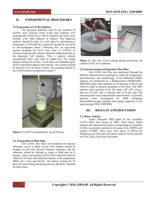
International Journal for Research in Applied Sciences and Biotechnology
Page Number: 6-9
Synthesis and Microstructure CaTiO3 coating by Sol-Gel Spin-Coating
Process
M.R. Sahu1
, P.K.Mallik2
, S. C. Patnaik3
and Ajit Behera4*
1, 2, 3
Department of Metallurgical & Materials Engineering, Indiragandhi Institute of Technology, Sarang, INDIA
4
National Institute of Technology, Rourkela, INDIA
4
Corresponding Author: ajit.behera88@gmail.com
ABSTRACT
Recently, Calcium Titanate has been
introduced as a bioactive bioceramic with acceptable
mechanical and better biological properties compared to
hydroxyapatite for orthopaedic implant applications. In this
study, CaTiO3nano-structure coating was produced by sol-gel
spin-coating route for biomedical applications. Calcium oxide
and titanium isopropoxide were used as a precursor for the sol-
gel spin-coating. After coating process, the specimen was
subjected to heating in oven at 100o
C for 24 hours and the
sample was heated at 800°C for 2 hours. The phase structure
and surface morphology of coating were investigated by X-ray
diffraction (XRD) and scanning electron microscopy (SEM).
Finally, it concluded that the uniform crack-free nano-
structured CaTiO3 coatings could be used for the biomedical
application.
Keywords— Sol-Gel Spin Coating, CaTiO3, Microstructure,
Bioactive coating
I. INTRODUCTION
The main purpose of surface modifications for
biomaterials is to improve tissue responses in a living body
because tissue biomaterial reactions are interfacial
phenomena which are governed by surface properties of the
biomaterial. Ceramic coatings are often applied to facilitate
osteogenesis on metallic biomaterials. Among ceramics,
hydroxyapatite (HA) is the most popular coating material [1–
5]. Many researchers have demonstrated good osteogenesis
on HA-coated metals [6–9], and HA-coated titanium
prepared with a plasma-spraying process has been used
clinically [10–12]. However, fractures at the HA titanium
interface and in the HA layer itself are often degraded after
long-term use in the human body [13]. Accidents caused by
these fractures result in a loss of the biomaterial-bone
fixation. Consequently, clinical use of the HA-coated
titanium has decreased in recent year.
Recently, some of the present authors succeeded in
developing a bioactive calcium titanate (CaTiO3) coating
which can activate osteogenesis on titanium [14-17]. The
bioactive CaTiO3 film was prepared by radiofrequency (RF)
magnetron sputtering with a CaTiO3 target in an argon
atmosphere and post-annealing at 873 K in air [16,17]. The
prepared film was crystallized into perovskite-type CaTiO3,
and the chemical composition of the film was almost in
accordance with that of stoichiometric CaTiO3. A
remarkable feature of the bioactive CaTiO3 film was that the
thickness was about 50 nm [17]. The thickness was 1/1000
that of plasma-sprayed HA coating. This thickness made it
possible to improve the mechanical strength of the film itself.
However, the post-annealing in air yielded not only
crystallization of the CaTiO3 film but also the formation of a
titanium-oxide layer in the interface between the film and the
titanium substrate because of the oxidation of titanium,
resulting in a change in the interface properties.
It was showed that the adhesion strength of the
CaTiO3 film increases with a decrease in the thickness of the
interfacial TiO2 layer [15]. By thinning the TiO2 layer up to
halfits thickness, the adhesion strength estimated by the
tensile test increased by approximately 40%. Likewise,
Kobayashi et al. reported that in the case of sodium titanate
film, the formation of an interfacial TiO2 layer formed by
heating weakened the adhesion strength [18]. Consequently,
in order to obtain a nondestructive bioactive CaTiO3 layer,
development of new coating process without formation of
the thick oxide layer is required.
The objective of the present work is to synthesis of
CaTiO3 thin film using sol-gel process in which calcium
oxide (CaO) and Titanium isopropoxide (C12H2804Ti)as
starting materials, ethanol as the dispersed medium,
ethylene-diamine-tetra-acetic acid (EDTA ) as chilling agent
for the reaction. X-ray diffraction (XRD) and Scanning
Electron Microscopy (SEM) analysis were carried out to
study the microstructural and morphological behavior of
CaTiO3 thin films.](https://image.slidesharecdn.com/synthesisandmicrostructurecatio3coatingbysolgelspincoatingprocess-200419144057/85/Synthesis-and-Microstructure-CaTiO3-coating-by-Sol-Gel-Spin-Coating-Process-1-320.jpg)
![www.ijrasb.com ISSN (ONLINE): 2349-8889
6 Copyright © 2018. IJRASB. All Rights Reserved.
Volume-5, Issue-1, January 2018
International Journal for Research in Applied Sciences and Biotechnology
Page Number: 6-9
Synthesis and Microstructure CaTiO3 coating by Sol-Gel Spin-Coating
Process
M.R. Sahu1
, P.K.Mallik2
, S. C. Patnaik3
and Ajit Behera4*
1, 2, 3
Department of Metallurgical & Materials Engineering, Indiragandhi Institute of Technology, Sarang, INDIA
4
National Institute of Technology, Rourkela, INDIA
4
Corresponding Author: ajit.behera88@gmail.com
ABSTRACT
Recently, Calcium Titanate has been
introduced as a bioactive bioceramic with acceptable
mechanical and better biological properties compared to
hydroxyapatite for orthopaedic implant applications. In this
study, CaTiO3nano-structure coating was produced by sol-gel
spin-coating route for biomedical applications. Calcium oxide
and titanium isopropoxide were used as a precursor for the sol-
gel spin-coating. After coating process, the specimen was
subjected to heating in oven at 100o
C for 24 hours and the
sample was heated at 800°C for 2 hours. The phase structure
and surface morphology of coating were investigated by X-ray
diffraction (XRD) and scanning electron microscopy (SEM).
Finally, it concluded that the uniform crack-free nano-
structured CaTiO3 coatings could be used for the biomedical
application.
Keywords— Sol-Gel Spin Coating, CaTiO3, Microstructure,
Bioactive coating
I. INTRODUCTION
The main purpose of surface modifications for
biomaterials is to improve tissue responses in a living body
because tissue biomaterial reactions are interfacial
phenomena which are governed by surface properties of the
biomaterial. Ceramic coatings are often applied to facilitate
osteogenesis on metallic biomaterials. Among ceramics,
hydroxyapatite (HA) is the most popular coating material [1–
5]. Many researchers have demonstrated good osteogenesis
on HA-coated metals [6–9], and HA-coated titanium
prepared with a plasma-spraying process has been used
clinically [10–12]. However, fractures at the HA titanium
interface and in the HA layer itself are often degraded after
long-term use in the human body [13]. Accidents caused by
these fractures result in a loss of the biomaterial-bone
fixation. Consequently, clinical use of the HA-coated
titanium has decreased in recent year.
Recently, some of the present authors succeeded in
developing a bioactive calcium titanate (CaTiO3) coating
which can activate osteogenesis on titanium [14-17]. The
bioactive CaTiO3 film was prepared by radiofrequency (RF)
magnetron sputtering with a CaTiO3 target in an argon
atmosphere and post-annealing at 873 K in air [16,17]. The
prepared film was crystallized into perovskite-type CaTiO3,
and the chemical composition of the film was almost in
accordance with that of stoichiometric CaTiO3. A
remarkable feature of the bioactive CaTiO3 film was that the
thickness was about 50 nm [17]. The thickness was 1/1000
that of plasma-sprayed HA coating. This thickness made it
possible to improve the mechanical strength of the film itself.
However, the post-annealing in air yielded not only
crystallization of the CaTiO3 film but also the formation of a
titanium-oxide layer in the interface between the film and the
titanium substrate because of the oxidation of titanium,
resulting in a change in the interface properties.
It was showed that the adhesion strength of the
CaTiO3 film increases with a decrease in the thickness of the
interfacial TiO2 layer [15]. By thinning the TiO2 layer up to
halfits thickness, the adhesion strength estimated by the
tensile test increased by approximately 40%. Likewise,
Kobayashi et al. reported that in the case of sodium titanate
film, the formation of an interfacial TiO2 layer formed by
heating weakened the adhesion strength [18]. Consequently,
in order to obtain a nondestructive bioactive CaTiO3 layer,
development of new coating process without formation of
the thick oxide layer is required.
The objective of the present work is to synthesis of
CaTiO3 thin film using sol-gel process in which calcium
oxide (CaO) and Titanium isopropoxide (C12H2804Ti)as
starting materials, ethanol as the dispersed medium,
ethylene-diamine-tetra-acetic acid (EDTA ) as chilling agent
for the reaction. X-ray diffraction (XRD) and Scanning
Electron Microscopy (SEM) analysis were carried out to
study the microstructural and morphological behavior of
CaTiO3 thin films.](https://image.slidesharecdn.com/synthesisandmicrostructurecatio3coatingbysolgelspincoatingprocess-200419144057/75/Synthesis-and-Microstructure-CaTiO3-coating-by-Sol-Gel-Spin-Coating-Process-1-2048.jpg)
![www.ijrasb.com ISSN (ONLINE): 2349-8889
9 Copyright © 2018. IJRASB. All Rights Reserved.
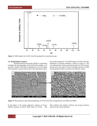
Figure 5- EDS pattern of CaTiO
3
thin film
IV. CONCLUSION
Based on the experimental results and analysis, the
following conclusions have been presented.CaTiO3 thin film
is successfully prepared by sol gel spin coating process.
1. From XRD analysis confirms presence of crystalline
CaTiO3 phases after heat treatment at 900o
C for 1 hour.
It also shows presence of some traces of TiO2 phases.
2. The surface morphology obtained from SEM
micrograph showed that the epitaxial growth grains with
the uniform shape & size of 10µm in length and 5µm in
width.
3. This indicates that the CaTiO3 thin film can be used for
the better biocompatibility and osteoconductivity of
titanium alloy for the biomedical application (Dental and
Hip Implant).
REFERENCES
[1] K. Yamashita, E. Yonehara, X. Ding, M. Nagai, T.
Umegaki, M. Matsuda, J. Biomed. Mater.Res. 43 (1998) 46.
[2] N. Yoshinari, Y. Ohtsuka, T. Derand, Biomaterials 15
(1994) 529.
[3] J.L. Ong, L.C. Lucas, Biomaterials 15 (1994) 337.
[4] K. van Dijk, H.G. Schaeken, J.G. Wolke, J.A. Jansen,
Biomaterials 17 (1996) 405.
[5] Y.C. Tsui, C. Doyle, T.W. Clyne, Biomaterials 19 (1998)
2015.
[6] G. De Lange, C. De Putter, J. Oral Implantol. 19 (1993)
123.
[7] J.A. Jasen, J.P. van derWaerden, K. de Groot K, J.
Biomed. Mater.Res. 25 (1991) 1535.
[8] H.W. Denissen, K. de Groot, P.C. Makkes, A. van den
Hooff, P.J. Klopper, J. Biomed. Mater.Res. 14 (1980) 713.
[9] M. Jarcho, Clin. Orthop.157 (1981) 259.
[10] K. De Groot, R. Geesink, C.P. Klein, P. Serekian, J.
Biomed. Mater.Res. 21 (1987) 1375.
[11] R.G.T. Geesink, K. de Groot, C.P. Klein, J. Bone Jt.
Surg. 70B (1988) 17.
[12] S.R. Radin, P. Ducheyne, J. Mater. Sci., Mater.Med. 3
(1992) 33.
[13] R.G.T. Geesink, K. de Groot, C.P. Klein, Clin.
Orthop.225 (1987) 147.
[14] K. Asami, K. Saito, N. Ohtsu, S. Nagata, T. Hanawa,
Surf. Interface Anal. 35 (2003) 483.
[15] N. Ohtsu, K. Saito, K. Asami, T. Hanawa, Surf.
Coat.Tech. 200 (2006) 5455.
[16] N. Ohtsu, K. Sato, K. Saito, K. Asami, T. Hanawa, J.
Mater. Sci., Mater.Med. (in press).
[17] N. Ohtsu, K. Sato, A. Yanagawa, K. Saito, Y. Imai, T.
Kohgo, A. Yokoyama, K. Asami, T. Hanawa, J. Biomed.
Mater.Res. (in press).
[18] S. Kobayashi, T. Inoue, K. Nakai, Mater. Trans. 46
(2005) 207.
keV
0.00 2.00 4.00 6.00 8.00 10.00 12.00 14.00
Counts[x1.E+3]
0.0
0.2
0.4
0.6
0.8
1.0
1.2
1.4
001
Ti
Ti
O
Ca
Ca
Ti
Ti](https://image.slidesharecdn.com/synthesisandmicrostructurecatio3coatingbysolgelspincoatingprocess-200419144057/85/Synthesis-and-Microstructure-CaTiO3-coating-by-Sol-Gel-Spin-Coating-Process-4-320.jpg)


