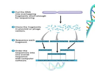
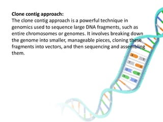
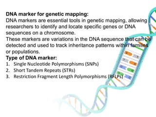
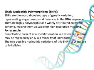
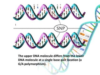
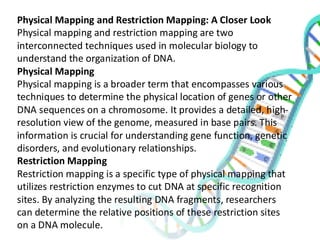
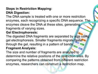
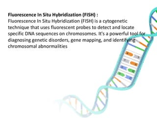
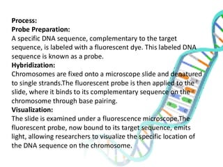
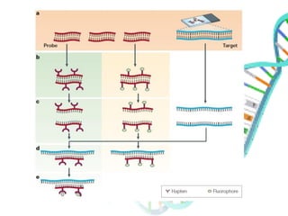
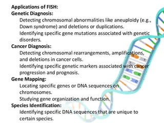
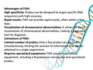
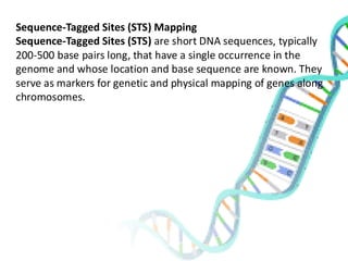
Genome Mapping
Presented By:
Abhishek Gupta
M.Sc Biotechnology
Molecular Genetics
Presented To:
Dr. Shiv Kumar Giri
Department of Biotechnology
Content:-
Genome Mapping
Shot gun approach
Clone contig approach
DNA markers for genetic mapping
RFLP
SNPs
Physical Mapping-Restriction mapping
Florescent in situ hybridization (FISH)
Sequence tagged sites (STS) mapping
Genome mapping :-
Genome mapping is the process of identifying and recording the
location of genes and the distances between genes on a
chromosome.
Types of Genome Mapping:
There are two main types of genome mapping:
Genetic Mapping:
➢ Relies on the principles of genetic inheritance and
recombination.
➢ Tracks the inheritance of genetic markers (like specific DNA
sequences) across generations.
➢ Provides a relative order of genes on a chromosome.
➢ Measured in centimorgans (cM), which represent the
frequency of recombination between two genes.
Physical Mapping:
➢ Directly analyzes DNA molecules to determine the physical
distance between genes.
➢ Uses techniques like restriction enzyme digestion,
hybridization, and sequencing.
➢ Provides a more precise map of gene locations.
➢ Measured in base pairs (bp).
The shotgun approach :-
The shotgun approach is a powerful technique used in
genome mapping to determine the DNA sequence of an
organism.
Clone contig approach:
The clone contig approach is a powerful technique in
genomics used to sequence large DNA fragments, such as
entire chromosomes or genomes. It involves breaking down
the genome into smaller, manageable pieces, cloning these
fragments into vectors, and then sequencing and assembling
them.
Process:
Genome Fragmentation:
•The genomic DNA is fragmented into large pieces, typically using
restriction enzymes or mechanical shearing.
•The size of these fragments is crucial and often ranges from 100 to 200
kilobase pairs (kb).
Cloning into Vectors:
•The fragmented DNA is inserted into cloning vectors, such as Bacterial
Artificial Chromosomes (BACs) or Yeast Artificial Chromosomes (YACs).
•These vectors can accommodate large DNA inserts and are capable of
replicating within host organisms.
Library Construction:
•The cloned DNA fragments are introduced into host cells, creating a
genomic library.
•This library represents a collection of clones, each containing a different
fragment of the genome.
Clone Selection and Mapping:
•Clones are selected from the library and mapped to determine their relative
positions on the genome.
•This mapping can be achieved using various techniques, including
restriction enzyme digestion, hybridization, and sequence-tagged site (STS)
analysis.
Contig Assembly:
•Overlapping clones are identified and assembled into contiguous
sequences, known as contigs.
•This assembly process relies on the identification of shared sequence
regions between adjacent clones.
Sequencing of Clones:
•Individual clones within the contigs are sequenced using high-
throughput sequencing technologies.
•This generates short sequence reads that are aligned and assembled
to create the final sequence of the contig.
Gap Closure:
•Gaps between contigs are filled by additional sequencing or by using
techniques like chromosome walking or PCR.
Advantages of the Clone Contig Approach:
Accuracy: By sequencing large, contiguous fragments, the clone
contig approach reduces the risk of assembly errors.
Long-Range Information: It provides valuable information about
long-range genomic structure, such as gene order and
orientation.
Complexity Handling: It is well-suited for complex genomes with
repetitive sequences and large-scale structural variations.
DNA marker for genetic mapping:
DNA markers are essential tools in genetic mapping, allowing
researchers to identify and locate specific genes or DNA
sequences on a chromosome.
These markers are variations in the DNA sequence that can be
detected and used to track inheritance patterns within families
or populations.
Type of DNA marker:
1. Single Nucleotide Polymorphisms (SNPs)
2. Short Tandem Repeats (STRs)
3. Restriction Fragment Length Polymorphisms (RFLPs)
Single Nucleotide Polymorphisms (SNPs):
SNPs are the most abundant type of genetic variation,
representing single base-pair differences in the DNA sequence.
They are highly polymorphic and widely distributed across the
genome, making them valuable for high-resolution mapping.
For example
G nucleotide present at a specific location in a reference genome
may be replaced by an A in a minority of individuals.
The two possible nucleotide variations of this SNP – G or A – are
called alleles.
The upper DNA molecule differs from the lower
DNA molecule at a single base-pair location (a
G/A polymorphism).
SNP analysis
SNPs can be easily assayed due to only containing two
possible alleles and three possible genotypes involving the
two alleles: homozygous A, homozygous B and
heterozygous AB, leading to many possible techniques for
analysis.
Some include:
DNA sequencing; capillary electrophoresis; mass
spectrometry; single-strand conformation polymorphism
(SSCP); single base extension; electrochemical analysis;
denaturating HPLC and gel electrophoresis; restriction
fragment length polymorphism; and hybridization analysis.
Short Tandem Repeats (STRs):
Also known as microsatellites, STRs consist of short DNA
sequences that are repeated multiple times in tandem. The
number of repeats at a specific STR locus can vary between
individuals, creating polymorphism.
STRs are highly informative and are commonly used in
forensic DNA analysis and paternity testing.
STR testing is used to diagnose conditions that are
predominantly caused by expansions (increases) in the
length of a specific repetitive region of the genome. This
includes well-known conditions such as Huntington disease
(caused by a CAG trinucleotide repeat expansion in the HTT
gene) and fragile X syndrome (caused by a CGG trinucleotide
repeat expansion in the 5’ UTR of the FMR1 gene). Carrier
testing and predictive testing for such conditions is also done
by STR testing.
There are two commonly used methods for sizing repeat expansions.
Fluorescent polymerase chain reaction (PCR) sizing (also known as fragment analysis).
➢ Patient DNA is amplified using PCR.
➢ The primers are located either side of the repeat region, so that the length of the
PCR product reflects the length of the repeat.
➢ One of the primers is fluorescently labelled.
➢ Capillary electrophoresis is used to separate PCR products by size and
fluorescence of the PCR product is detected.
Repeat-primed PCR (can amplify larger expansions).
➢ Patient DNA is amplified using PCR.
➢ One primer is located next to the repeat region and is fluorescently labelled.
➢ A second primer targets the repeat itself. This binds at multiple locations in the
repeat region, generating multiple products, with the largest products reflecting
the size of the entire expansion. This primer is present in limited amounts, so
that a third primer (more abundant) takes over after a few PCR cycles.
➢ This third primer targets the PCR products generated by the previous two,
amplifying all of them.
➢ Capillary electrophoresis is used to separate PCR products by size and
fluorescence of the PCR products is detected.
Restriction Fragment Length Polymorphisms (RFLPs):
RFLPs arise from variations in the DNA sequence that create or
eliminate restriction enzyme recognition sites. This results in
different fragment sizes when DNA is digested with specific
restriction enzymes and analyzed by gel electrophoresis. RFLPs
were among the earliest DNA markers used in genetic mapping
but are now less commonly used due to the development of more
efficient techniques.
Principle
Restriction endonucleases are enzymes that cut lengthy DNA into
short pieces. Each restriction endonuclease targets different
nucleotide sequences in a DNA strand and therefore cuts at
different sites.
The distance between the cleavage sites of a certain restriction
endonuclease differs between individuals. Hence, the length of
the DNA fragments produced by a restriction endonuclease will
differ across both individual organisms and species.
RFLP is performed using a series of steps :
DNA Extraction
To begin with, DNA is extracted from blood, saliva or other samples and purified.
DNA Fragmentation
The purified DNA is digested using restriction endonucleases. The recognition sites of
these enzymes are generally 4 to 6 base pairs in length. The shorter the sequence
recognized, the greater the number of fragments generated from digestion.
For example, if there is a short sequence of GAGC that occurs repeatedly in a sample of
DNA. The restriction endonuclease that recognizes the GAGC sequence cuts the DNA at
every repetition of the GAGC pattern.
If one sample repeats the GAGC sequence 4 times whilst another sample repeats it 2
times, the length of the fragments generated by the enzyme for the two samples will be
different.
Gel Electrophoresis
The restriction fragments produced during DNA fragmentation are analyzed using gel
electrophoresis.
The fragments are negatively charged and can be easily separated by electrophoresis,
which separates molecules based on their size and charge. The fragmented DNA samples
are placed in the chamber containing the electrophoretic gel and two electrodes.
When an electric field is applied, the fragments migrate towards the positive electrode.
Smaller fragments move faster through the gel leaving the larger ones behind and thus
the DNA samples are separated into distinct bands on the gel.
Visualization of Bands
The gel is treated with luminescent dyes in order to make the DNA bands visible
Physical Mapping and Restriction Mapping: A Closer Look
Physical mapping and restriction mapping are two
interconnected techniques used in molecular biology to
understand the organization of DNA.
Physical Mapping
Physical mapping is a broader term that encompasses various
techniques to determine the physical location of genes or other
DNA sequences on a chromosome. It provides a detailed, high-
resolution view of the genome, measured in base pairs. This
information is crucial for understanding gene function, genetic
disorders, and evolutionary relationships.
Restriction Mapping
Restriction mapping is a specific type of physical mapping that
utilizes restriction enzymes to cut DNA at specific recognition
sites. By analyzing the resulting DNA fragments, researchers
can determine the relative positions of these restriction sites
on a DNA molecule.
Steps in Restriction Mapping:
DNA Digestion:
The DNA sample is treated with one or more restriction
enzymes, each recognizing a specific DNA sequence. The
enzymes cleave the DNA at these sites, generating
fragments of varying sizes.
Gel Electrophoresis:
The digested DNA fragments are separated by size using
gel electrophoresis. Smaller fragments migrate faster
through the gel, resulting in a pattern of bands.
Fragment Analysis:
The size and number of fragments are analyzed to
determine the relative positions of the restriction sites. By
comparing the patterns obtained from different restriction
enzymes, researchers can construct a restriction map.
Fluorescence In Situ Hybridization (FISH) :
Fluorescence In Situ Hybridization (FISH) is a cytogenetic
technique that uses fluorescent probes to detect and locate
specific DNA sequences on chromosomes. It's a powerful tool for
diagnosing genetic disorders, gene mapping, and identifying
chromosomal abnormalities
Process:
Probe Preparation:
A specific DNA sequence, complementary to the target
sequence, is labeled with a fluorescent dye. This labeled DNA
sequence is known as a probe.
Hybridization:
Chromosomes are fixed onto a microscope slide and denatured
to single strands.The fluorescent probe is then applied to the
slide, where it binds to its complementary sequence on the
chromosome through base pairing.
Visualization:
The slide is examined under a fluorescence microscope.The
fluorescent probe, now bound to its target sequence, emits
light, allowing researchers to visualize the specific location of
the DNA sequence on the chromosome.
Applications of FISH:
Genetic Diagnosis:
Detecting chromosomal abnormalities like aneuploidy (e.g.,
Down syndrome) and deletions or duplications.
Identifying specific gene mutations associated with genetic
disorders.
Cancer Diagnosis:
Detecting chromosomal rearrangements, amplifications,
and deletions in cancer cells.
Identifying specific genetic markers associated with cancer
progression and prognosis.
Gene Mapping:
Locating specific genes or DNA sequences on
chromosomes.
Studying gene organization and function.
Species Identification:
Identifying specific DNA sequences that are unique to
certain species.
Advantages of FISH:
High specificity: Probes can be designed to target specific DNA
sequences with high accuracy.
Rapid results: FISH can provide rapid results, often within a few
days.
Visualization of chromosomal abnormalities: It allows for direct
visualization of chromosomal abnormalities, making it a valuable
tool for diagnosis.
Limitations of FISH:
Limited number of probes: Only a few probes can be used
simultaneously, limiting the amount of information that can be
obtained in a single experiment.
Requires specialized equipment: FISH requires specialized
equipment, including a fluorescence microscope and specialized
probes.
Sequence-Tagged Sites (STS) Mapping
Sequence-Tagged Sites (STS) are short DNA sequences, typically
200-500 base pairs long, that have a single occurrence in the
genome and whose location and base sequence are known. They
serve as markers for genetic and physical mapping of genes along
chromosomes.
STS Mapping Works
1.Identification of STSs:
•STSs are identified by sequencing random genomic DNA fragments or
by analyzing known gene sequences.
•Unique sequences are selected and primers are designed to amplify
them by PCR.
2.PCR Amplification:
•Genomic DNA from an organism is amplified using the specific primers
for each STS.
•The presence or absence of a PCR product indicates whether the STS is
present in the genome.
3.Mapping STSs:
•STSs are mapped onto chromosomes using various techniques,
including:
•Genetic mapping: By analyzing the inheritance of STSs in a
genetic cross, their relative positions on chromosomes can be
determined.
•Physical mapping: By hybridizing fluorescently labeled STS probes
to chromosomes, their physical location can be visualized.
4.Constructing Genetic and Physical Maps:
•Once the positions of multiple STSs are known, they can be used to
construct high-resolution genetic and physical maps of the genome.
•These maps provide valuable information about gene order, distance,
and orientation.
Advantages of STS Mapping:
High specificity: STSs are unique sequences, ensuring accurate
mapping.
Versatility: STSs can be used for both genetic and physical
mapping.
Efficiency: PCR-based amplification allows for rapid and sensitive
detection of STSs.
Standardization: STSs can be shared and used by different
laboratories, facilitating collaborative research.
Applications of STS Mapping:
Gene mapping: Identifying the chromosomal location of genes
associated with specific traits or diseases.
Genome sequencing: Assisting in the assembly of genome
sequences by providing markers for aligning and ordering DNA
fragments.
Comparative genomics: Comparing the genomes of different
species to identify conserved regions and evolutionary
relationships.
Reference:
I. Principles of Genetics (2006),4th edition.Suustad and Simmons, Wilet.
II. https://www.researchgate.net/figure/Agarose-gel-electrophoresis-of-the-plasmid-
DNA-cleaved-by-restriction-enzymes-a_fig1_346705234
III. https://pmc.ncbi.nlm.nih.gov/articles/PMC7477013/#:~:text=Long%2Dread%20seq
uencing%20technology%20reveals,the%20reference%20human%20genome%20su
ggests.
IV. https://www.news-medical.net/life-sciences/Restriction-Fragment-Length-
Polymorphism-(RFLP)-Technique.aspx
V. https://www.genomicseducation.hee.nhs.uk/genotes/knowledge-hub/short-
tandem-repeat-str-
testing/#:~:text=Short%20tandem%20repeats%20(STRs)%20occur,triplet%20repeat
%20such%20as%20CAG.
VI. https://en.wikipedia.org/wiki/Single-nucleotide_polymorphism
Study about 'Genome mapping' in molecular genetics.pdf

Study about 'Genome mapping' in molecular genetics.pdf

  • 1.
    Genome Mapping Presented By: AbhishekGupta M.Sc Biotechnology Molecular Genetics Presented To: Dr. Shiv Kumar Giri Department of Biotechnology
  • 2.
    Content:- Genome Mapping Shot gunapproach Clone contig approach DNA markers for genetic mapping RFLP SNPs Physical Mapping-Restriction mapping Florescent in situ hybridization (FISH) Sequence tagged sites (STS) mapping
  • 3.
    Genome mapping :- Genomemapping is the process of identifying and recording the location of genes and the distances between genes on a chromosome. Types of Genome Mapping: There are two main types of genome mapping: Genetic Mapping: ➢ Relies on the principles of genetic inheritance and recombination. ➢ Tracks the inheritance of genetic markers (like specific DNA sequences) across generations. ➢ Provides a relative order of genes on a chromosome. ➢ Measured in centimorgans (cM), which represent the frequency of recombination between two genes.
  • 4.
    Physical Mapping: ➢ Directlyanalyzes DNA molecules to determine the physical distance between genes. ➢ Uses techniques like restriction enzyme digestion, hybridization, and sequencing. ➢ Provides a more precise map of gene locations. ➢ Measured in base pairs (bp). The shotgun approach :- The shotgun approach is a powerful technique used in genome mapping to determine the DNA sequence of an organism.
  • 6.
    Clone contig approach: Theclone contig approach is a powerful technique in genomics used to sequence large DNA fragments, such as entire chromosomes or genomes. It involves breaking down the genome into smaller, manageable pieces, cloning these fragments into vectors, and then sequencing and assembling them.
  • 7.
    Process: Genome Fragmentation: •The genomicDNA is fragmented into large pieces, typically using restriction enzymes or mechanical shearing. •The size of these fragments is crucial and often ranges from 100 to 200 kilobase pairs (kb). Cloning into Vectors: •The fragmented DNA is inserted into cloning vectors, such as Bacterial Artificial Chromosomes (BACs) or Yeast Artificial Chromosomes (YACs). •These vectors can accommodate large DNA inserts and are capable of replicating within host organisms. Library Construction: •The cloned DNA fragments are introduced into host cells, creating a genomic library. •This library represents a collection of clones, each containing a different fragment of the genome. Clone Selection and Mapping: •Clones are selected from the library and mapped to determine their relative positions on the genome. •This mapping can be achieved using various techniques, including restriction enzyme digestion, hybridization, and sequence-tagged site (STS) analysis.
  • 8.
    Contig Assembly: •Overlapping clonesare identified and assembled into contiguous sequences, known as contigs. •This assembly process relies on the identification of shared sequence regions between adjacent clones. Sequencing of Clones: •Individual clones within the contigs are sequenced using high- throughput sequencing technologies. •This generates short sequence reads that are aligned and assembled to create the final sequence of the contig. Gap Closure: •Gaps between contigs are filled by additional sequencing or by using techniques like chromosome walking or PCR.
  • 9.
    Advantages of theClone Contig Approach: Accuracy: By sequencing large, contiguous fragments, the clone contig approach reduces the risk of assembly errors. Long-Range Information: It provides valuable information about long-range genomic structure, such as gene order and orientation. Complexity Handling: It is well-suited for complex genomes with repetitive sequences and large-scale structural variations.
  • 10.
    DNA marker forgenetic mapping: DNA markers are essential tools in genetic mapping, allowing researchers to identify and locate specific genes or DNA sequences on a chromosome. These markers are variations in the DNA sequence that can be detected and used to track inheritance patterns within families or populations. Type of DNA marker: 1. Single Nucleotide Polymorphisms (SNPs) 2. Short Tandem Repeats (STRs) 3. Restriction Fragment Length Polymorphisms (RFLPs)
  • 11.
    Single Nucleotide Polymorphisms(SNPs): SNPs are the most abundant type of genetic variation, representing single base-pair differences in the DNA sequence. They are highly polymorphic and widely distributed across the genome, making them valuable for high-resolution mapping. For example G nucleotide present at a specific location in a reference genome may be replaced by an A in a minority of individuals. The two possible nucleotide variations of this SNP – G or A – are called alleles.
  • 12.
    The upper DNAmolecule differs from the lower DNA molecule at a single base-pair location (a G/A polymorphism).
  • 13.
    SNP analysis SNPs canbe easily assayed due to only containing two possible alleles and three possible genotypes involving the two alleles: homozygous A, homozygous B and heterozygous AB, leading to many possible techniques for analysis. Some include: DNA sequencing; capillary electrophoresis; mass spectrometry; single-strand conformation polymorphism (SSCP); single base extension; electrochemical analysis; denaturating HPLC and gel electrophoresis; restriction fragment length polymorphism; and hybridization analysis.
  • 14.
    Short Tandem Repeats(STRs): Also known as microsatellites, STRs consist of short DNA sequences that are repeated multiple times in tandem. The number of repeats at a specific STR locus can vary between individuals, creating polymorphism. STRs are highly informative and are commonly used in forensic DNA analysis and paternity testing. STR testing is used to diagnose conditions that are predominantly caused by expansions (increases) in the length of a specific repetitive region of the genome. This includes well-known conditions such as Huntington disease (caused by a CAG trinucleotide repeat expansion in the HTT gene) and fragile X syndrome (caused by a CGG trinucleotide repeat expansion in the 5’ UTR of the FMR1 gene). Carrier testing and predictive testing for such conditions is also done by STR testing.
  • 16.
    There are twocommonly used methods for sizing repeat expansions. Fluorescent polymerase chain reaction (PCR) sizing (also known as fragment analysis). ➢ Patient DNA is amplified using PCR. ➢ The primers are located either side of the repeat region, so that the length of the PCR product reflects the length of the repeat. ➢ One of the primers is fluorescently labelled. ➢ Capillary electrophoresis is used to separate PCR products by size and fluorescence of the PCR product is detected. Repeat-primed PCR (can amplify larger expansions). ➢ Patient DNA is amplified using PCR. ➢ One primer is located next to the repeat region and is fluorescently labelled. ➢ A second primer targets the repeat itself. This binds at multiple locations in the repeat region, generating multiple products, with the largest products reflecting the size of the entire expansion. This primer is present in limited amounts, so that a third primer (more abundant) takes over after a few PCR cycles. ➢ This third primer targets the PCR products generated by the previous two, amplifying all of them. ➢ Capillary electrophoresis is used to separate PCR products by size and fluorescence of the PCR products is detected.
  • 17.
    Restriction Fragment LengthPolymorphisms (RFLPs): RFLPs arise from variations in the DNA sequence that create or eliminate restriction enzyme recognition sites. This results in different fragment sizes when DNA is digested with specific restriction enzymes and analyzed by gel electrophoresis. RFLPs were among the earliest DNA markers used in genetic mapping but are now less commonly used due to the development of more efficient techniques. Principle Restriction endonucleases are enzymes that cut lengthy DNA into short pieces. Each restriction endonuclease targets different nucleotide sequences in a DNA strand and therefore cuts at different sites. The distance between the cleavage sites of a certain restriction endonuclease differs between individuals. Hence, the length of the DNA fragments produced by a restriction endonuclease will differ across both individual organisms and species.
  • 18.
    RFLP is performedusing a series of steps : DNA Extraction To begin with, DNA is extracted from blood, saliva or other samples and purified. DNA Fragmentation The purified DNA is digested using restriction endonucleases. The recognition sites of these enzymes are generally 4 to 6 base pairs in length. The shorter the sequence recognized, the greater the number of fragments generated from digestion. For example, if there is a short sequence of GAGC that occurs repeatedly in a sample of DNA. The restriction endonuclease that recognizes the GAGC sequence cuts the DNA at every repetition of the GAGC pattern. If one sample repeats the GAGC sequence 4 times whilst another sample repeats it 2 times, the length of the fragments generated by the enzyme for the two samples will be different. Gel Electrophoresis The restriction fragments produced during DNA fragmentation are analyzed using gel electrophoresis. The fragments are negatively charged and can be easily separated by electrophoresis, which separates molecules based on their size and charge. The fragmented DNA samples are placed in the chamber containing the electrophoretic gel and two electrodes. When an electric field is applied, the fragments migrate towards the positive electrode. Smaller fragments move faster through the gel leaving the larger ones behind and thus the DNA samples are separated into distinct bands on the gel. Visualization of Bands The gel is treated with luminescent dyes in order to make the DNA bands visible
  • 20.
    Physical Mapping andRestriction Mapping: A Closer Look Physical mapping and restriction mapping are two interconnected techniques used in molecular biology to understand the organization of DNA. Physical Mapping Physical mapping is a broader term that encompasses various techniques to determine the physical location of genes or other DNA sequences on a chromosome. It provides a detailed, high- resolution view of the genome, measured in base pairs. This information is crucial for understanding gene function, genetic disorders, and evolutionary relationships. Restriction Mapping Restriction mapping is a specific type of physical mapping that utilizes restriction enzymes to cut DNA at specific recognition sites. By analyzing the resulting DNA fragments, researchers can determine the relative positions of these restriction sites on a DNA molecule.
  • 21.
    Steps in RestrictionMapping: DNA Digestion: The DNA sample is treated with one or more restriction enzymes, each recognizing a specific DNA sequence. The enzymes cleave the DNA at these sites, generating fragments of varying sizes. Gel Electrophoresis: The digested DNA fragments are separated by size using gel electrophoresis. Smaller fragments migrate faster through the gel, resulting in a pattern of bands. Fragment Analysis: The size and number of fragments are analyzed to determine the relative positions of the restriction sites. By comparing the patterns obtained from different restriction enzymes, researchers can construct a restriction map.
  • 22.
    Fluorescence In SituHybridization (FISH) : Fluorescence In Situ Hybridization (FISH) is a cytogenetic technique that uses fluorescent probes to detect and locate specific DNA sequences on chromosomes. It's a powerful tool for diagnosing genetic disorders, gene mapping, and identifying chromosomal abnormalities
  • 23.
    Process: Probe Preparation: A specificDNA sequence, complementary to the target sequence, is labeled with a fluorescent dye. This labeled DNA sequence is known as a probe. Hybridization: Chromosomes are fixed onto a microscope slide and denatured to single strands.The fluorescent probe is then applied to the slide, where it binds to its complementary sequence on the chromosome through base pairing. Visualization: The slide is examined under a fluorescence microscope.The fluorescent probe, now bound to its target sequence, emits light, allowing researchers to visualize the specific location of the DNA sequence on the chromosome.
  • 25.
    Applications of FISH: GeneticDiagnosis: Detecting chromosomal abnormalities like aneuploidy (e.g., Down syndrome) and deletions or duplications. Identifying specific gene mutations associated with genetic disorders. Cancer Diagnosis: Detecting chromosomal rearrangements, amplifications, and deletions in cancer cells. Identifying specific genetic markers associated with cancer progression and prognosis. Gene Mapping: Locating specific genes or DNA sequences on chromosomes. Studying gene organization and function. Species Identification: Identifying specific DNA sequences that are unique to certain species.
  • 26.
    Advantages of FISH: Highspecificity: Probes can be designed to target specific DNA sequences with high accuracy. Rapid results: FISH can provide rapid results, often within a few days. Visualization of chromosomal abnormalities: It allows for direct visualization of chromosomal abnormalities, making it a valuable tool for diagnosis. Limitations of FISH: Limited number of probes: Only a few probes can be used simultaneously, limiting the amount of information that can be obtained in a single experiment. Requires specialized equipment: FISH requires specialized equipment, including a fluorescence microscope and specialized probes.
  • 27.
    Sequence-Tagged Sites (STS)Mapping Sequence-Tagged Sites (STS) are short DNA sequences, typically 200-500 base pairs long, that have a single occurrence in the genome and whose location and base sequence are known. They serve as markers for genetic and physical mapping of genes along chromosomes.
  • 28.
    STS Mapping Works 1.Identificationof STSs: •STSs are identified by sequencing random genomic DNA fragments or by analyzing known gene sequences. •Unique sequences are selected and primers are designed to amplify them by PCR. 2.PCR Amplification: •Genomic DNA from an organism is amplified using the specific primers for each STS. •The presence or absence of a PCR product indicates whether the STS is present in the genome. 3.Mapping STSs: •STSs are mapped onto chromosomes using various techniques, including: •Genetic mapping: By analyzing the inheritance of STSs in a genetic cross, their relative positions on chromosomes can be determined. •Physical mapping: By hybridizing fluorescently labeled STS probes to chromosomes, their physical location can be visualized. 4.Constructing Genetic and Physical Maps: •Once the positions of multiple STSs are known, they can be used to construct high-resolution genetic and physical maps of the genome. •These maps provide valuable information about gene order, distance, and orientation.
  • 29.
    Advantages of STSMapping: High specificity: STSs are unique sequences, ensuring accurate mapping. Versatility: STSs can be used for both genetic and physical mapping. Efficiency: PCR-based amplification allows for rapid and sensitive detection of STSs. Standardization: STSs can be shared and used by different laboratories, facilitating collaborative research. Applications of STS Mapping: Gene mapping: Identifying the chromosomal location of genes associated with specific traits or diseases. Genome sequencing: Assisting in the assembly of genome sequences by providing markers for aligning and ordering DNA fragments. Comparative genomics: Comparing the genomes of different species to identify conserved regions and evolutionary relationships.
  • 30.
    Reference: I. Principles ofGenetics (2006),4th edition.Suustad and Simmons, Wilet. II. https://www.researchgate.net/figure/Agarose-gel-electrophoresis-of-the-plasmid- DNA-cleaved-by-restriction-enzymes-a_fig1_346705234 III. https://pmc.ncbi.nlm.nih.gov/articles/PMC7477013/#:~:text=Long%2Dread%20seq uencing%20technology%20reveals,the%20reference%20human%20genome%20su ggests. IV. https://www.news-medical.net/life-sciences/Restriction-Fragment-Length- Polymorphism-(RFLP)-Technique.aspx V. https://www.genomicseducation.hee.nhs.uk/genotes/knowledge-hub/short- tandem-repeat-str- testing/#:~:text=Short%20tandem%20repeats%20(STRs)%20occur,triplet%20repeat %20such%20as%20CAG. VI. https://en.wikipedia.org/wiki/Single-nucleotide_polymorphism