
Recommended
Recommended
More Related Content
Similar to structural organization of bacteriophage.pptx
Similar to structural organization of bacteriophage.pptx (20)
Recently uploaded
Recently uploaded (20)
structural organization of bacteriophage.pptx
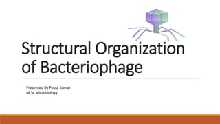
- 1. Structural Organization of Bacteriophage Presented By Pooja Kumari M.Sc Microbiology
- 3. Definition Bacteriophage (phage) are obligate intracellular viruses that specifically infect bacteria and just like other viruses and need a host cell to reproduce. They may transmit genetic information from bacterium to another by the process named transduction.
- 4. Overview Like most viruses, bacteriophages typically carry only the genetic information needed for replication of their nucleic acid and synthesis of their protein coats. They require precursors, energy generation and ribosomes supplied by their bacterial host cell. Like all viruses, phages are simple organisms that consist of a core of genetic material (nucleic acid) surrounded by a protein capsid. The nucleic acid may be either DNA or RNA and may be double-stranded or single-stranded. Bacteriophages that infect Esch. Coli, called the T-even phages (T2, T4, T6), have been extansively studied. T-even phages are tadpole shaped, and posses a head and tail. Ø HEAD: The size of the head varies in different phages from 28nm to 100nm. Ø Hexagonal in shape and consists of tightly packed core of nucleic acid (double stranded DNA) enclosed by a protein coat, called capsid. Ø it protects the nucleic acid from digestion by enzymes. ØTAIL: It is composed of a hollow core surrounded by a contractile sheath, and a terminal base plate which has atteched to it prongs, tail fibres (usually six in numbers) or both.
- 5. Structural organization Phages may have different shapes and sizes. The most studied group is that of tailed phages with a dsDNA genome, and it also represents the largest group . The tailed phages have three major components: a capsid where the genome is packed, a tail that serves as a pipe during infection to secure transfer of genome into host cell and a special adhesive system (adsorption apparatus) at the very end of the tail that will recognize the host cell and penetrate its wall. Cell resources are used for the phage reproduction. The functional phage is a result of a multistep process that starts with all the necessary proteins produced by the host cell after infection: capsid, portal, tail, scaffolding, terminase, etc. The capsids of the dsDNA phages often have fivefold or icosahedral symmetries, which are broken at one of the fivefold axes by the head-to-tail interface (HTI). The main component of the HTI is a dodecameric portal protein (PP) within the capsid.
- 6. Structural organization The PP represents the DNA-packaging motor which is the crucial part of these nano-machines. The HTI also includes oligomeric rings of head completion proteins that play dual roles: making an additional interface to molecules of ATP which provide energy for DNA packaging and then connecting the portal protein and the tail. Some HTIs also serve as valves that close the exit channel preventing leakage of genome from the capsid but opening as soon as the phage is attached to the host cell. The phage tail is the structural component of the phage that is essential during infection. Its adsorption apparatus located on the distal end of the tail recognizes a receptor, or the envelope chemistry of the host cell and ensures genome delivery to the cell cytoplasm.
- 7. Self-assembly pathway of phages. Multiple copies of the capsid/scaffold complex bind the portal protein to form the procapsid; then, the scaffold proteins are ejected, and DNA is packaged into the procapsid, which expands to the size of the mature capsid. The head completion proteins (the stopper and the adaptor) are bound to the portal complex preventing DNA leakage. Next, decoration proteins bind to the capsid, and the tail, assembled separately or after DNA packaging, is attached; thus, the final infectious phage is produced.
- 8. Ø The preassembled tail attaches in Myoviridae and Siphoviridae, while in Podoviridae the tail assembles at the stopper.
- 9. Procapsids During the assembly process ,the capsid of a phage has a precursor formation, named the procapsid. Scaffolding proteins (SPs) drive the assembly process ,by accompanying major capsid protein (MCP) subunits to form an icosahedral procapsid that is eventually filled with dsDNA, The SPs are bound to the portal complex during formation of a procapsid with scaffolding inside. The sequence of conformational changes from a procapsid to the phage capsid where genome has been packed is named as the maturation process and goes through a series of intermediates. As soon as the procapsid is assembled, the scaffolding domain is cleaved off and then like the separate SP will be removed from the capsid to make room for the genome. The spherical capsid shell expands during maturation and becomes thinner due to alterations in the inter- and intra-subunit contacts.
- 10. Capsids Most tailed phages have capsids of an icosahedral shape formed by multiple copies of one or more proteins. The capsid is the protein shell that encloses the nucleic acid; with its enclosed nucleic acid, it is called the nucleocapsid. This shell is composed of a large number of protein subunits (polypeptides) known as capsomers. The capsid has three functions: 1) it protects the nucleic acid from digestion by enzymes, 2) contains special sites on its surface that allow the virion to attach to a host cell, and 3) provides proteins that enable the virion to penetrate the host cell membrane and, in some cases, to inject the infectious nucleic acid into the cell's cytoplasm.
- 11. Connectors In phages and herpesviruses, one of the fivefold vertices of the capsid is replaced by a head-to-tail interface (HTI), which is a multi-protein complex (connector). The HTI consist of a portal complex (PP) and head completion proteins that serve as a valve for closing the channel and keeping the phage genome inside the capsid at high pressure and only opens to allow genome release from the capsid (under natural conditions) as soon as the phage becomes tightly attached to a host cell.
- 12. Tail Tail indicating that the phage is attached to the bacterium, the connector will be open allowing the release of DNA into the bacterium . The tail and its related structures are necessary phage elements securing the entry of the viral nucleic acid into the host bacterium during the infectivity process. The tail serves both as a signal transmitter and subsequently as a pipeline through which DNA is delivered into the host cell during infection. The tails may be short or long, the latter are divided into contractile and noncontractile tails. The long tails are typically composed of many copies of several proteins arranged with helical symmetry. Eg. Siphoviridae have long flexible tails; Podoviridae have very short tails; while Myoviridae have rigid long contractile tails.
- 13. Tail Ø All types of tails have outer appendages attached to the distant end of the tail and often include a baseplate with several fibers and a tip, or a needle that are selective to the membrane receptors of the bacterium. Ø As soon the receptor has been found by the tail needle, which happens during multiple short living reversible attachments to the bacterium, the baseplate and tail fibers are involved in the binding of the phage to the bacterial outer membrane that makes the attachment irreversible. Ø The docking (irreversible attachment) of the phage induces opening of the phage connector and release of the genome through the tail tube into the bacterial cell.
- 14. Adsorption apparatus Most phages have an oligomeric ring formed by distal tail proteins (DTPs), which is attached to the last ring of the tail tube. The DTP ring usually serves as an apparatus to recognize and connect to receptor-binding proteins; sometimes, this interaction is assisted by tail fibers found in T4, T5 and other phages.
- 15. References May 2019 DOI:10.5772/intechopen.85484 In book: Bacteriophages - Biology and Applications [Working Title] Authors: Helen White Birkbeck, University of London E.V. Orlova Birkbeck, University of London BIROn - Birkbeck Institutional Research Online Orlova, Elena (2012) Bacteriophages and their structural organization. In: Kurtboke, I. (ed.) Bacteriophages. Rijeka, Croatia: Intech, pp. 3-30. ISBN 9789535102724. Dr. C P Baveja, Director prof. & Head Microbiology at Maulana Azad Medical Collage, New Delhi
- 16. THANK YOU
Editor's Notes
- whiskers," which extend outward from the collar region of the phage, control retraction and extension of the tail fibers in response to certain environmental conditions.