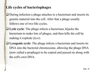
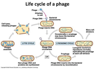
2. Page 2
Introduction
Bacteriophage, also called phage or bacterial virus , any of a
group of viruses that infect bacteria.
Bacteriophages were discovered independently
by Frederick W. Twort in Great Britain (1915) and Félix
d’Hérelle in France (1917).
D’Hérelle coined the
term bacteriophage, meaning
“bacteria eater,” to describe the
agent’s bacteriocidal ability.
3. Page 3
Bacteriophages also infect the single-celled prokaryotic
organisms known as archaea.
Like all viruses, phages are simple organism that consist of
core of genetic material (nuleic acid) surrounded by a protein
capsid.
The nuleic acid either DNA or RNA & may be single stranded
or double stranded.
6. Page 6
T phage
Structure is composed of
icosahedral head, double stranded
DNA and a tail.
Infects E.coli
Main types of T phages are T2,
T4 and T12.
7. Page 7
In the infection linear DNA of the phage is released to the host
cell and becomes circular by replicating that later produces a
long DNA chain known as ‘Concatamen’. This coils into the
phage’s head by headfull mechanism while packaging.
Infection kills the host cell as the new particles are released
outside by bursting the host cell.
8. Page 8
Lambda (λ)
Composed of head and tail.
Head is consisting of double
stranded linear DNA.
Host cell is E.coli.
9. Page 9
At both 5’ ends of the DNA strand, 12 complementary base
pair, single stranded segments are present. These two ends are
known as “cos ends”.
Because of the cos ends, phage chromosome circularizes
before replication. Concatamen is produced during the
replication and during the packaging, Terminase enzyme cuts
off the cos ends.
10. Page 10
Phi×174(ΦX174)
The genome is a circular ssDNA molecule of 5386 nucleotides,
coding for 11 proteins.
After adsorption, synthesizes the complementary strand and
becomes double stranded.
Use as a positive control in DNA sequencing.
11. Page 11
Structure of phage ϕX174.
(a) Virion components.
(b)Reconstruction of capsid showing subunit organization.
12. Page 12
M13 (Filamentous)
About 870 nm in length and 6nm in
width.
Consists of single stranded DNA
(ssDNA).
Three kinds of capsomeres build the
capsid.
13. Page 13
Infects E. coli by adsorbing to the cell and entering through
pilli.
Does not kill the host. Particles are released by budding,
therefore when the particles are released, the host cell is alive.
An efficient vector in gene cloning as it can hold longer pieces
of foreign DNA.
16. Page 16
Phi(Φ)6 phage
Segmented, double-stranded RNA genome, totalling ~13.5 kb
in length.
Φ6 and its relatives have a lipid membrane around their
nucleocapsid, a rare trait among bacteriophages.
Host- pseudomonas
17. Page 17
MS2
Contains the smallest known genome.
Super coiled single stranded DNA.
Infect only through sex pilli. Therefore
male specific.
Infect E.coli.
18. Page 18
Q beta phages
Its genome consists of a circular,
positive sense single-stranded
RNA molecule.
Infects E. coli.
Q beta phage is one of the smallest known viruses, measuring
24 nm in diameter. Its icosahedral capsid is composed of 180
copies of a single coat protein.
19. Page 19
Bacteriophage infections
Bacteriophages, just like other viruses, must infect a host cell
in order to reproduce.
The steps that make up the infection process are collectively
called the lifecycle of the phage.
Some phages can only reproduce via a lytic lifecycle, in which
they burst and kill their host cells. Other phages can alternate
between a lytic lifecycle and a lysogenic lifecycle, in which
they don't kill the host cell (and are instead copied along with
the host DNA each time the cell divides).
20. Page 20
Life cycles of bacteriophages
During infection a phage attaches to a bacterium and inserts its
genetic material into the cell. After that a phage usually
follows one of two life cycles.
Lytic cycle: The phage infects a bacterium, hijacks the
bacterium to make lots of phages, and then kills the cell by
making it explode (lyse).
Lysogenic cycle: The phage infects a bacterium and inserts its
DNA into the bacterial chromosome, allowing the phage DNA
(now called a prophage) to be copied and passed on along with
the cell's own DNA.
21. Page 21
Lytic cycle
In the lytic cycle, a phage acts like a typical virus, its host cell
and uses the cell's resources to make lots of new phages,
causing the cell to lyse (burst) and die in the process.
22. Page 22
The stages of the lytic cycle are:
Attachment: Proteins in the "tail" of the phage bind to a
specific receptor on the surface of the bacterial cell.
Entry: The phage injects its double-stranded DNA genome
into the cytoplasm of the bacterium.
DNA copying and protein synthesis: Phage DNA is copied,
and phage genes are expressed to make proteins, such as
capsid proteins.
Assembly of new phage: Capsids assemble from the capsid
proteins and are stuffed with DNA to make lots of new phage
particles.
23. Page 23
Lysis: Late in the lytic cycle, the phage expresses genes for
proteins that poke holes in the plasma membrane and cell wall.
The holes let water flow in, making the cell expand and burst
like an overfilled water balloon.
Cell bursting, or lysis, releases hundreds of new phages, which
can find and infect other host cells. In this way, a few cycles
of lytic infection can let the phage spread like wildfire through
a bacterial population.
25. Page 25
Lysogenic cycle
The lysogenic cycle allows a phage to reproduce without
killing its host. Some phages can only use the lytic cycle.
In the lysogenic cycle, the first two steps (attachment and
DNA injection) occur just as they do for the lytic cycle.
Attachment: Proteins in the "tail" of the phage bind to a
specific receptor on the surface of the bacterial cell.
Entry: The phage injects its double-stranded DNA genome
into the cytoplasm of the bacterium.
26. Page 26
Once the phage DNA is inside the cell, it is not immediately
copied or expressed to make proteins.
Instead, it recombines with a particular region of the bacterial
chromosome. This causes the phage DNA to be integrated into
the chromosome.
27. Page 27
The integrated phage DNA, called a prophage, is not active: its
genes aren't expressed, and it doesn't drive production of new
phages.
However, each time a host cell divides, the prophage is
copied along with the host DNA, getting a free ride.
The lysogenic cycle is less flashy (and less gory) than the lytic
cycle, but at the end of the day, it's just another way for the
phage to reproduce.
28. Page 28
Under the right conditions, the prophage can become active
and come back out of the bacterial chromosome, triggering the
remaining steps of the lytic cycle (DNA copying and protein
synthesis, phage assembly, and lysis).
31. Page 31
Differences Between Lytic and Lysogenic Cycles
Lytic Cycle Lysogenic Cycle
Viral DNA destroys Cell DNA,
takes over cell functions and
destroys the cell.
Not destroys the cell.
The Virus replicates and produces
progeny phages.
The Virus does not produce
progeny.
There are symptoms of viral
infection.
Virtulant viral infection takes
place.
There are no symptoms of viral
infection.
Temperate viral replication takes
place.
32. Page 32
Applications
Phage Therapy
Use of phages to treat bacterial infections in animals and
humans. treat bacterial dysentery, staphylococcal lung
infections, surgical wound infections, staphylococcal
septicemia Easier to develop new phage than a new antibiotic.
33. Page 33
Phage Lysins as Antimicrobials
Use of phage endolysins as potential therapeutics instead of
whole phage.
Phage endolysins, or lysins, are enzymes that damage the cell
walls' integrity phage lysins composed of at least two
distinctly separate functional domains: C-terminal cell-wall
binding domain, which directs the enzyme to its target N-
terminal catalytic domain.
34. Page 34
Phage Display
1. Insert a diverse group of genes into the phage genome. Each
phage receives a different gene.
2. Create a library of genetically modified phages which are
all related but each of which have a different gene.
3. Isolate a disease causing molecule, such as a receptor or an
enzyme, and expose the phage library to the molecule.
4. Some of the phages will bind to the disease causing
molecule.
35. Page 35
Wash the phage library .All of the phages with proteins that
didn't bind to the disease causing molecule will be removed.
5. Replicate the phages that remain so that they can be
sequenced.
6. Since we know where the new gene was inserted we can
determine the amino acid sequence of the protein that bound to
the disease
36. Page 36
Applications of Phage Display
To select proteins, peptides, or antibodies with affinity to a
molecule or protein of interest To clone antibodies from
unstable hybridoma.
To identify molecules that can be recognized and internalized
by eukaryotic cells.
To identify epitopes, functional and accessible sites from
antigens.
To study protein-protein interaction.
To design vaccines.
37. Page 37
Use of Phage in Vaccination
Whole bacteriophage acts as efficient DNA vaccine delivery
vehicles. Phage-display vaccination. Phage DNA vaccination.
Hybrid phage vaccine.
Bacteriophage is also used in identifying pathogenic bacteria
(also called phage typing) in diagnostic laboratories. One other
use for bacteriophages is for killing specific bacteria found in
food. For example, LISTEX by Micreos is made up of
bacteriophages that can kill the L. monocytogenes bacteria in
cheese.
Prophage containing host bacteria is called lysogen or lysogenic bacterium.
When lysogenic bacterium expose the UV or a chemical the prophage withdraw from the host DNA to under go lytic cycle.