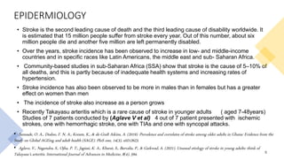
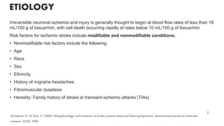
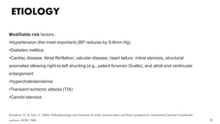
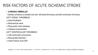
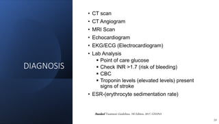
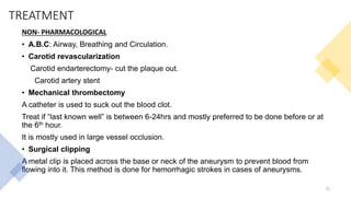
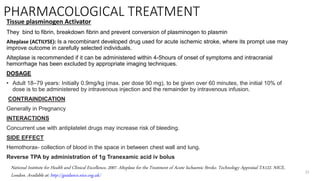
STROKE
PREPARED BY
OBED ADAMS
1
INTRODUCTION
• DEFINITION
• Stroke is defined as the rapid loss of brain function due to disturbance
in blood flow and supply to the brain.
OR
• An acute episode of focal dysfunction of the brain, retina, or spinal
cord.
• It is clinically defined as the rapid onset of cerebral deficit lasting more
than 24hours and are caused by acute vascular injury to parts of the
brain.
2
• Hankey, G. J. (2017). Stroke. Lancet, 389(10069), 641-654.
INTRODUCTION
• BRIEF ANATOMY AND PHYSIOLOGY OF THE BRAIN AND BLOOD SUPPLY
The transfer of blood from the heart to the brain is through Anterior and posterior vessels that are
present.
Anterior circulation involves all arteries that originates from the internal carotid arteries.
The internal carotid artery is responsible for supply of blood to the anterior and middle parts of
the brain.
3
ANTERIOR CIRCULATION POSTERIOR CIRCULATION
internal carotid artery Posterior cerebral artery
Anterior Cerebral artery Basilar artery
Middle cerebral artery Vertebral artery
Anterior communicating artery Posterior communicating artery
Haines, D. (2013). Fundamental neuroscience for basic and clinical applications (4th ed.). Elsevier Saunders
https://www.kenhub.com/en/library/anatomy/arteries-of-the-brain
INTRODUCTION
Anterior cerebral artery- supply blood to the superior parts of the frontal
lobe and the anterior parietal lobe
Middle Cerebral artery- supply blood to the lateral parts of the frontal lobe
and the temporal lobe.
POSTERIOR CIRCULATION
Posterior cerebral artery –supply blood to the occipital lobe, temporal lobe,
thalamus and the midbrain
The basilar artery - found in the pontine cistern. Its branches are
responsible for supplying the pons, cerebellum, internal ear, and other
inferior parts.
The vertebral arteries -runs through the spinal column in the neck to
provide blood to the brain and the upper part of the spinal cord.
4
Menshawi, K., Mohr, J., & Gutierrez, J. (2015). A Functional Perspective on the Embryology and Anatomy of the Cerebral Blood Supply. Journal Of Stroke,
17(2), 144
EPIDERMIOLOGY
• Stroke is the second leading cause of death and the third leading cause of disability worldwide. It
is estimated that 15 million people suffer from stroke every year. Out of this number, about six
million people die and another five million are left permanently disabled.
• Over the years, stroke incidence has been observed to increase in low- and middle-income
countries and in specific races like Latin Americans, the middle east and sub- Saharan Africa.
• Community-based studies in sub-Saharan Africa (SSA) show that stroke is the cause of 5–10% of
all deaths, and this is partly because of inadequate health systems and increasing rates of
hypertension.
• Stroke incidence has also been observed to be more in males than in females but has a greater
effect on women than men
• The incidence of stroke also increase as a person grows
• Recently Takayasu arteritis which is a rare cause of stroke in younger adults ( aged 7-48years)
Studies of 7 patients conducted by (Aglave V et al) 4 out of 7 patient presented with ischemic
strokes, one with hemorrhagic stroke, one with TIAs and one with syncopal attacks.
5
• Sanuade, O. A., Dodoo, F. N. A., Koram, K., & de-Graft Aikins, A. (2019). Prevalence and correlates of stroke among older adults in Ghana: Evidence from the
Study on Global AGEing and adult health (SAGE). PloS one, 14(3), e0212623.
• Aglave, V., Nagendra, S., Ojha, P. T., Jagiasi, K. A., Kharat, S., Barvalia, P., & Gaikwad, A. (2021). Unusual etiology of stroke in young adults: think of
Takayasu’s arteritis. International Journal of Advances in Medicine, 8(4), 594.
INTRODUCTION
6
TYPES OF STROKE
Acute ischemic stroke
Transient ischemic stroke
Intracerebral hemorrhage
Sub-arachnoid stroke
ETIOLOGY
Irreversible neuronal ischemia and injury is generally thought to begin at blood flow rates of less than 18
mL/100 g of tissue/min, with cell death occurring rapidly at rates below 10 mL/100 g of tissue/min
Risk factors for ischemic stroke include modifiable and nonmodifiable conditions.
• Nonmodifiable risk factors include the following:
• Age
• Race
• Sex
• Ethnicity
• History of migraine headaches
• Fibromuscular dysplasia
• Heredity: Family history of stroke or transient ischemic attacks (TIAs)
7
Kuriakose, D., & Xiao, Z. (2020). Pathophysiology and treatment of stroke: present status and future perspectives. International journal of molecular
sciences, 21(20), 7609.
ETIOLOGY
8
Modifiable risk factors :
•Hypertension (the most important) (BP reduces by 5-6mm Hg)
•Diabetes mellitus
•Cardiac disease: Atrial fibrillation, valvular disease, heart failure, mitral stenosis, structural
anomalies allowing right-to-left shunting (e.g., patent foramen Ovalle), and atrial and ventricular
enlargement
•Hypercholesterolemia
•Transient ischemic attacks (TIA)
•Carotid stenosis
Kuriakose, D., & Xiao, Z. (2020). Pathophysiology and treatment of stroke: present status and future perspectives. International journal of molecular
sciences, 21(20), 7609.
ETIOLOGY
• Lifestyle issues: Excessive alcohol intake, tobacco use, illicit drug use, physical inactivity
• Obesity
• Oral contraceptive use/postmenopausal hormone use
• Sickle cell disease
• Aneuyrsyms
• Arteriovenous malformation
• Malignancy
• Cerebral amyloid angiopathy
• Coagulopathy
• Hemorrhagic transformation
9
HEMORRAGHIC STROKE (ICH)
Kuriakose, D., & Xiao, Z. (2020). Pathophysiology and treatment of stroke: present status and future perspectives. International journal of molecular sciences, 21(20), 7609.
PATHOPHYSIOLOGY
CIRCLE OF WILLIS
Atherosclerosis
Constriction of
Vascular
Arteries
Thrombotic stroke
Ischemic
stroke(↓O2supply to
the brain)
Stress
Necrosis
Loss of neuronal
function
stress /internal
injury in brain
tissue.
Ruptured
blood vessel
Infarction
Hemorrhagic stroke
10
Ischemic stroke may also be caused by
inflammation,
energy failure,
loss of homeostasis,
acidosis,
increased intracellular calcium levels,
excitotoxicity,
free radical-mediated toxicity,
cytokine-mediated cytotoxicity,
complement activation,
impairment of the blood–brain barrier,
activation of glial cells,
oxidative stress and infiltration of leukocytes.
Kuriakose, D., & Xiao, Z. (2020). Pathophysiology and treatment of stroke: present status and future perspectives. International journal of molecular
sciences, 21(20), 7609.
ISCHEMIC STROKE
PATHOPHYSIOLOGY
11
stress may result in plaque rupture, collagen exposure, platelet
aggregation, and clot formation.
The clot can remain in the vessel, causing local occlusion, or
travel distally as an embolism, eventually lodging downstream in a
cerebral vessel.
In cardiogenic embolism, stasis of blood in the atria or ventricles
of the heart leads to the formation of local clots that can become
dislodged and travel directly through the aorta to the cerebral
circulation.
Finally, thrombus formation and embolism leads to an arterial
occlusion, decreasing cerebral blood flow and causing ischemia
distal to the occlusion.
Dirnagl U, Iadecola C, Moskowitz MA.
Pathobiology of ischemic stroke: An integrated
view. Trends Neurosci 1999;22:391–397.
PATHOPHYSIOLOGY
HEMORRHAGIC STROKE
The pathophysiology of hemorrhagic stroke specifically intracerebral hemorrhage is not
clearly outlined but studies show that the accumulation of blood in the parenchyma and
neurotoxicity of blood component due to tissue damage and infection leads to stroke.
In sub-arachnoid hemorrhage also known as cerebral aneurysm, blood accumulates in
the sub-arachnoid space due to either trauma, connective tissue disease or Arterio-
veno malformation.
Sub-arachnoid hemorrhage may be sarcular or fusiform.
These cases can easily lead to increase in intracranial pressure with leads to herniation
and ultimately death.
12
Smith, S. D., & Eskey, C. J. (2011). Hemorrhagic stroke. Radiologic Clinics, 49(1), 27-45.
TRANSIENT ISCHEMIC STROKE
Transient ischemic attack is classified as a mini stroke. It happens when the blood supply to
part of the brain is briefly blocked.
TIA occurs due to the development of neuro deficit because of occlusion but there is no
infarction.; it last for less than 24hrs.
TIA is a warning sign for the actual event.
TREATMENT
• Suspected transient ischemic attack
Adult: 300 mg aspirin stat orally until diagnosis established
If it develops into acute ischemic attack, an addition of dipyridamole
13
ACUTE ISCHEMIC STROKE
This type of stroke present with neuro deficits and infarction on CT MRI. It is the
most common type of stroke (87%)
An ischemic stroke happens when blood flows through the artery that supplies
oxygen-rich blood to the brain becomes blocked
CAUSES
HYPOXIC EMBOLIC THROMBOTIC
Decrease amount of O2 to
the brain
A blood clot clogs in a
vessel during blood
circulation in the brain
Blood clot in an artery in
the brain.
In events like Cardiac
arrest, cardiogenic shock,
severe respiratory failure
Atrial Fibrillation and left
ventricular aneurysm
Hypertension ,smoking,
hyperlipidemia, obesity
Age and Sex
Common in large vessels
thrombi, vertebral and
basilar artery
González, R. G. (2011). Acute ischemic stroke. J. A. Hirsch, W. J. Koroshetz, M. H. Lev, & P. W. Schaefer (Eds.). Springer-Verlag Berlin Heidelberg. 14
RISK FACTORS OF ACUTE ISCHEMIC STROKE
• HYPOXIC CAUSES
Global hypoperfusion
Cardiac Arrest
Acute respiratory failure
I.C.A stenosis and ↓O2
Shock
Cardiac surgery
THROMBOTIC CAUSES
15
Modifiable Non-modifiable
Hypotension Age
Diabetes
Mellitus
Sex(more in
males)
Hyperlipidemia Race
Smoking Genetic/family
history
Obesity
González, R. G. (2011). Acute ischemic stroke. J. A. Hirsch, W. J. Koroshetz, M. H. Lev, & P. W. Schaefer (Eds.). Springer-Verlag Berlin Heidelberg.
RISK FACTORS OF ACUTE ISCHEMIC STROKE
• EMBOLIC CAUSES
Stroke that arise due to embolism may be;
1. Arterio- aterio embolus
2. Cardiac embolus
3. Vasculitis
4. Dissection
ARTERIO- ATERIO EMBOLUS
1. I.C.A plaque
2. Aortic arch plaque
16
González, R. G. (2011). Acute ischemic stroke. J. A. Hirsch, W. J. Koroshetz, M. H. Lev, & P. W. Schaefer (Eds.). Springer-Verlag Berlin Heidelberg.
RISK FACTORS OF ACUTE ISCHEMIC STROKE
• CARDIAC EMBOLUS
Cardiac embolus is divided into two: left atrial thrombus and left ventricular thrombus
LEFT ATRIAL THROMBUS
 Atrial fibrillation
 Mechanical valve
 Rheumatic heart disease
 Infection endocarditis
LEFT VENTRICULAR THROMBUS
 Left ventricular aneurysms
 Myocardial infarction
 Heart Failure and CHF
17
González, R. G. (2011). Acute ischemic stroke. J. A. Hirsch, W. J. Koroshetz, M. H. Lev, & P. W. Schaefer (Eds.). Springer-Verlag Berlin Heidelberg.
CLINICAL MANIFESTAION (SIGNS)
• Paralysis of a limb
• Facial paralysis (lower half)
• Initial flaccidity of limbs, but later spasticity and exaggerated reflexes
• Hemianopia (loss of one-half of visual field)
• Hemi-anesthesia (loss of sensation of one-half of body)
• Contralateral weakness in extremities
• Sensory loss
• Dysarthria/dysphasia (alteration of speech)
• Neck stiffness (in subarachnoid haemorrhage)
• Stiffness in muscle (inability to stand or change body posture)
18
Standard Treatment Guidelines, 7th Edition, 2017, GHANA
SYMPTOMS
• Sudden numbness or weakness in the face, arm, or leg,
especially on one side of the body.
• Speech impediment (confusion, trouble speaking, or
difficulty understanding speech.)
• Sudden trouble seeing in one or both eyes.
• Sudden trouble walking, dizziness, loss of balance, or
lack of coordination.
• Unconciousness
• Sudden severe headache with no known cause.
• Seizures
19
Standard Treatment Guidelines, 7th Edition, 2017, GHANA
DIAGNOSIS
• CT scan
• CT Angiogram
• MRI Scan
• Echocardiogram
• EKG/ECG (Electrocardiogram)
• Lab Analysis
 Point of care glucose
 Check INR >1.7 (risk of bleeding)
 CBC
 Troponin levels (elevated levels) present
signs of stroke
• ESR-(erythrocyte sedimentation rate)
20
Standard Treatment Guidelines, 7th Edition, 2017, GHANA
TREATMENT
NON- PHARMACOLOGICAL
• A.B.C: Airway, Breathing and Circulation.
• Carotid revascularization
Carotid endarterectomy- cut the plaque out.
Carotid artery stent
• Mechanical thrombectomy
A catheter is used to suck out the blood clot.
Treat if “last known well” is between 6-24hrs and mostly preferred to be done before or at
the 6th hour.
It is mostly used in large vessel occlusion.
• Surgical clipping
A metal clip is placed across the base or neck of the aneurysm to prevent blood from
flowing into it. This method is done for hemorrhagic strokes in cases of aneurysms.
21
PHARMACOLOGICAL TREATMENT
Tissue plasminogen Activator
They bind to fibrin, breakdown fibrin and prevent conversion of plasminogen to plasmin
Alteplase (ACTILYSE): Is a recombinant developed drug used for acute ischemic stroke, where its prompt use may
improve outcome in carefully selected individuals.
Alteplase is recommended if it can be administered within 4-5hours of onset of symptoms and intracranial
hemorrhage has been excluded by appropriate imaging techniques.
DOSAGE
• Adult 18–79 years: Initially 0.9mg/kg (max. per dose 90 mg), to be given over 60 minutes, the initial 10% of
dose is to be administered by intravenous injection and the remainder by intravenous infusion.
CONTRAINDICATION
Generally in Pregnancy
INTERACTIONS
Concurrent use with antiplatelet drugs may increase risk of bleeding.
SIDE EFFECT
Hemothorax- collection of blood in the space in between chest wall and lung.
Reverse TPA by administration of 1g Tranexamic acid iv bolus
22
National Institute for Health and Clinical Excellence, 2007. Alteplase for the Treatment of Acute Ischaemic Stroke. Technology Appraisal TA122. NICE,
London. Available at: http://guidance.nice.org.uk/
TREATMENT
• ANTIPLATELETS
Aspirin
Patients with a disabling ischemic stroke and atrial fibrillation should receive aspirin for 2 weeks before
being for anticoagulant treatment.
In transient ischemic attack,
Adult: min 75 mg orally (dispersible tablet)
max 300 mg orally once daily until diagnosis established
In Acute ischemic attack,
Adult: 300 mg once daily for 14 days, to be initiated 24 hours after thrombolysis or as soon as possible
within 48 hours of symptom onset in patients not receiving thrombolytic drugs.
300mg stat, then 75mg once daily
23
Walker R., Whittlesea C. Clinical Pharmacy and Therapeutics, Churchill livingstone Elsevier Ltd. 12th Edition 2012 Page 377-389
The British National Fomulary; “Thromboemblisim” Pages 125-128
TREATMENT
Clopidogrel
Adult: 75 mg orally once daily
UNLICENSED USE: 600mg Use in transient ischemic attack or acute ischemic stroke, in patients
with aspirin hypersensitivity or intolerant of aspirin.
• Dipyridamole with aspirin
Used for secondary prevention of ischemic stroke and transient ischemic attacks. Studies confirms
more effectiveness than aspirin alone
Adult: 25/200 mg twice daily orally using modified releasing medicines
• ANTICOAGULANT
This treatment goal is to target the prevention of stroke in cardio embolism
Warfarin
Warfarin is the most effective treatment for the prevention of stroke in patients with atrial
fibrillation
Adult: Initially 5–10 mg orally, to be taken on day 1 subsequent dose is based on INR.
24
Walker R., Whittlesea C. Clinical Pharmacy and Therapeutics, Churchill livingstone Elsevier Ltd. 12th Edition 2012 Page 377-389
TREATMENT
• ANTITHROMBOTIC DRUGS › FACTOR XA INHIBITORS
Apixaban (Eliquis)
Used in Prophylaxis of stroke
Adult: 5 mg twice daily, reduce dose to 2.5 mg twice daily in patients with at least two of the following
characteristics:
I. age 80 years and over,
II. body-weight less than 61 kg,
III. serum creatinine levels > 133 micromol/litre
• Edoxaban
is a direct and reversible inhibitor of activated factor X (factor Xa), which prevents conversion of prothrombin to
thrombin and prolongs clotting time, thereby reducing the risk of thrombus formation.
Used in prophylaxis of stroke in patients with at least one modifiable risk factors.
DOSAGE
Adult (body-weight up to 61 kg): 30 mg once daily
Adult (body-weight 61 kg and above): 60 mg once daily
16
DiPiro T. J. ,Talbert .L R, Pharmacotherapy; A Pathophysiologic Approach, The McGraw-Hill Companies, Inc. 7TH Edition 2008 page
TREATMENT
• Rivaroxaban
is a direct inhibitor of activated factor X (factor Xa).
Prophylaxis of stroke and systemic embolism in patients with non-valvular atrial
fibrillation and with at least one modifiable risk factors:
Adult: 20 mg orally once daily, to be taken with food
• TREATING UNDERLYING DISEASE (RISK MODIFICATION)
This treatment focuses on treating the risk factors of stroke
• Hypertension
Blood pressure should be measured and treatment initiated to achieve a target blood
pressure of <130/80 mmHg
o ACE Inhibitors
NB: Beta blockers are not to be used unless otherwise indicated
26
Tripathi K.D Essentials of Medical Pharmacology, Jaypee Brothers and Medical Publisher LTD. 7th Edition 2013, pages 554-556; 620-632
TREATMENT
• Hyperlipidemia
• Statins
Atorvastatin is recommended and should be initiated 48 hours after stroke symptom
Min dose – 20mg orally once daily
Max dose – 80mg orally once daily
NB: Statins are not to be taken in pregnancy and should be discontinued 3 months before attempting to
conceive as it might cause some fetal defect.
DIABETES
• Insulin
• Dextrose saline
• Normal saline
INFLAMMATION
• Antihistamines
SIEZURES
• Antiepileptic drugs
CEREBRAL EDEMA
• Mannitol
27
Tripathi K.D Essentials of Medical Pharmacology, Jaypee Brothers and Medical Publisher LTD. 7th Edition 2013, pages 554-556; 620-632
CONCLUSION
For patient with suspected stroke, the F.A.S.T simple analysis done
before any further step is taken
• F—Face: Ask the person to smile. Does one side of the face droop?
• A—Arms: Ask the person to raise both arms. Does one arm drift
downward?
• S—Speech: Ask the person to repeat a simple phrase. Is the speech
slurred or strange?
• T—Time: If you see any of these signs, medical attention should be
given.
Patients with acute stroke should be monitored closely for adverse
effect of treatment, infections and/or worsening of neurological defect.
28
REFERENCES
• Hankey, G. J. (2017). Stroke. Lancet, 389(10069), 641-654.
• Walker R., Whittlesea C. Clinical Pharmacy and Therapeutics, Churchill livingstone Elsevier Ltd. 12th Edition 2012 Page 377-389
• The British National Fomulary; “Thromboemblisim” Pages 125-128
• Standard Treatment Guidelines, 7th Edition, 2017, GHANA
• González, R. G. (2011). Acute ischemic stroke. J. A. Hirsch, W. J. Koroshetz, M. H. Lev, & P. W. Schaefer (Eds.). Springer-Verlag
Berlin Heidelberg.
• National Institute for Health and Clinical Excellence, 2007. Alteplase for the Treatment of Acute Ischaemic Stroke. Technology
Appraisal TA122. NICE, London. Available at: http://guidance.nice.org.uk/
• Haines, D. (2013). Fundamental neuroscience for basic and clinical applications (4th ed.). Elsevier Saunders
• https://www.kenhub.com/en/library/anatomy/arteries-of-the-brain
• DiPiro T. J. ,Talbert .L R, Pharmacotherapy; A Pathophysiologic Approach, The McGraw-Hill Companies, Inc. 7TH Edition 2008 page
• Tripathi K.D Essentials of Medical Pharmacology, Jaypee Brothers and Medical Publisher LTD. 7th Edition 2013, pages 554-556; 620-
632
29
THANK YOU
30

Stroke : Introduction, types and treatment.

  • 1.
  • 2.
    INTRODUCTION • DEFINITION • Strokeis defined as the rapid loss of brain function due to disturbance in blood flow and supply to the brain. OR • An acute episode of focal dysfunction of the brain, retina, or spinal cord. • It is clinically defined as the rapid onset of cerebral deficit lasting more than 24hours and are caused by acute vascular injury to parts of the brain. 2 • Hankey, G. J. (2017). Stroke. Lancet, 389(10069), 641-654.
  • 3.
    INTRODUCTION • BRIEF ANATOMYAND PHYSIOLOGY OF THE BRAIN AND BLOOD SUPPLY The transfer of blood from the heart to the brain is through Anterior and posterior vessels that are present. Anterior circulation involves all arteries that originates from the internal carotid arteries. The internal carotid artery is responsible for supply of blood to the anterior and middle parts of the brain. 3 ANTERIOR CIRCULATION POSTERIOR CIRCULATION internal carotid artery Posterior cerebral artery Anterior Cerebral artery Basilar artery Middle cerebral artery Vertebral artery Anterior communicating artery Posterior communicating artery Haines, D. (2013). Fundamental neuroscience for basic and clinical applications (4th ed.). Elsevier Saunders https://www.kenhub.com/en/library/anatomy/arteries-of-the-brain
  • 4.
    INTRODUCTION Anterior cerebral artery-supply blood to the superior parts of the frontal lobe and the anterior parietal lobe Middle Cerebral artery- supply blood to the lateral parts of the frontal lobe and the temporal lobe. POSTERIOR CIRCULATION Posterior cerebral artery –supply blood to the occipital lobe, temporal lobe, thalamus and the midbrain The basilar artery - found in the pontine cistern. Its branches are responsible for supplying the pons, cerebellum, internal ear, and other inferior parts. The vertebral arteries -runs through the spinal column in the neck to provide blood to the brain and the upper part of the spinal cord. 4 Menshawi, K., Mohr, J., & Gutierrez, J. (2015). A Functional Perspective on the Embryology and Anatomy of the Cerebral Blood Supply. Journal Of Stroke, 17(2), 144
  • 5.
    EPIDERMIOLOGY • Stroke isthe second leading cause of death and the third leading cause of disability worldwide. It is estimated that 15 million people suffer from stroke every year. Out of this number, about six million people die and another five million are left permanently disabled. • Over the years, stroke incidence has been observed to increase in low- and middle-income countries and in specific races like Latin Americans, the middle east and sub- Saharan Africa. • Community-based studies in sub-Saharan Africa (SSA) show that stroke is the cause of 5–10% of all deaths, and this is partly because of inadequate health systems and increasing rates of hypertension. • Stroke incidence has also been observed to be more in males than in females but has a greater effect on women than men • The incidence of stroke also increase as a person grows • Recently Takayasu arteritis which is a rare cause of stroke in younger adults ( aged 7-48years) Studies of 7 patients conducted by (Aglave V et al) 4 out of 7 patient presented with ischemic strokes, one with hemorrhagic stroke, one with TIAs and one with syncopal attacks. 5 • Sanuade, O. A., Dodoo, F. N. A., Koram, K., & de-Graft Aikins, A. (2019). Prevalence and correlates of stroke among older adults in Ghana: Evidence from the Study on Global AGEing and adult health (SAGE). PloS one, 14(3), e0212623. • Aglave, V., Nagendra, S., Ojha, P. T., Jagiasi, K. A., Kharat, S., Barvalia, P., & Gaikwad, A. (2021). Unusual etiology of stroke in young adults: think of Takayasu’s arteritis. International Journal of Advances in Medicine, 8(4), 594.
  • 6.
    INTRODUCTION 6 TYPES OF STROKE Acuteischemic stroke Transient ischemic stroke Intracerebral hemorrhage Sub-arachnoid stroke
  • 7.
    ETIOLOGY Irreversible neuronal ischemiaand injury is generally thought to begin at blood flow rates of less than 18 mL/100 g of tissue/min, with cell death occurring rapidly at rates below 10 mL/100 g of tissue/min Risk factors for ischemic stroke include modifiable and nonmodifiable conditions. • Nonmodifiable risk factors include the following: • Age • Race • Sex • Ethnicity • History of migraine headaches • Fibromuscular dysplasia • Heredity: Family history of stroke or transient ischemic attacks (TIAs) 7 Kuriakose, D., & Xiao, Z. (2020). Pathophysiology and treatment of stroke: present status and future perspectives. International journal of molecular sciences, 21(20), 7609.
  • 8.
    ETIOLOGY 8 Modifiable risk factors: •Hypertension (the most important) (BP reduces by 5-6mm Hg) •Diabetes mellitus •Cardiac disease: Atrial fibrillation, valvular disease, heart failure, mitral stenosis, structural anomalies allowing right-to-left shunting (e.g., patent foramen Ovalle), and atrial and ventricular enlargement •Hypercholesterolemia •Transient ischemic attacks (TIA) •Carotid stenosis Kuriakose, D., & Xiao, Z. (2020). Pathophysiology and treatment of stroke: present status and future perspectives. International journal of molecular sciences, 21(20), 7609.
  • 9.
    ETIOLOGY • Lifestyle issues:Excessive alcohol intake, tobacco use, illicit drug use, physical inactivity • Obesity • Oral contraceptive use/postmenopausal hormone use • Sickle cell disease • Aneuyrsyms • Arteriovenous malformation • Malignancy • Cerebral amyloid angiopathy • Coagulopathy • Hemorrhagic transformation 9 HEMORRAGHIC STROKE (ICH) Kuriakose, D., & Xiao, Z. (2020). Pathophysiology and treatment of stroke: present status and future perspectives. International journal of molecular sciences, 21(20), 7609.
  • 10.
    PATHOPHYSIOLOGY CIRCLE OF WILLIS Atherosclerosis Constrictionof Vascular Arteries Thrombotic stroke Ischemic stroke(↓O2supply to the brain) Stress Necrosis Loss of neuronal function stress /internal injury in brain tissue. Ruptured blood vessel Infarction Hemorrhagic stroke 10 Ischemic stroke may also be caused by inflammation, energy failure, loss of homeostasis, acidosis, increased intracellular calcium levels, excitotoxicity, free radical-mediated toxicity, cytokine-mediated cytotoxicity, complement activation, impairment of the blood–brain barrier, activation of glial cells, oxidative stress and infiltration of leukocytes. Kuriakose, D., & Xiao, Z. (2020). Pathophysiology and treatment of stroke: present status and future perspectives. International journal of molecular sciences, 21(20), 7609. ISCHEMIC STROKE
  • 11.
    PATHOPHYSIOLOGY 11 stress may resultin plaque rupture, collagen exposure, platelet aggregation, and clot formation. The clot can remain in the vessel, causing local occlusion, or travel distally as an embolism, eventually lodging downstream in a cerebral vessel. In cardiogenic embolism, stasis of blood in the atria or ventricles of the heart leads to the formation of local clots that can become dislodged and travel directly through the aorta to the cerebral circulation. Finally, thrombus formation and embolism leads to an arterial occlusion, decreasing cerebral blood flow and causing ischemia distal to the occlusion. Dirnagl U, Iadecola C, Moskowitz MA. Pathobiology of ischemic stroke: An integrated view. Trends Neurosci 1999;22:391–397.
  • 12.
    PATHOPHYSIOLOGY HEMORRHAGIC STROKE The pathophysiologyof hemorrhagic stroke specifically intracerebral hemorrhage is not clearly outlined but studies show that the accumulation of blood in the parenchyma and neurotoxicity of blood component due to tissue damage and infection leads to stroke. In sub-arachnoid hemorrhage also known as cerebral aneurysm, blood accumulates in the sub-arachnoid space due to either trauma, connective tissue disease or Arterio- veno malformation. Sub-arachnoid hemorrhage may be sarcular or fusiform. These cases can easily lead to increase in intracranial pressure with leads to herniation and ultimately death. 12 Smith, S. D., & Eskey, C. J. (2011). Hemorrhagic stroke. Radiologic Clinics, 49(1), 27-45.
  • 13.
    TRANSIENT ISCHEMIC STROKE Transientischemic attack is classified as a mini stroke. It happens when the blood supply to part of the brain is briefly blocked. TIA occurs due to the development of neuro deficit because of occlusion but there is no infarction.; it last for less than 24hrs. TIA is a warning sign for the actual event. TREATMENT • Suspected transient ischemic attack Adult: 300 mg aspirin stat orally until diagnosis established If it develops into acute ischemic attack, an addition of dipyridamole 13
  • 14.
    ACUTE ISCHEMIC STROKE Thistype of stroke present with neuro deficits and infarction on CT MRI. It is the most common type of stroke (87%) An ischemic stroke happens when blood flows through the artery that supplies oxygen-rich blood to the brain becomes blocked CAUSES HYPOXIC EMBOLIC THROMBOTIC Decrease amount of O2 to the brain A blood clot clogs in a vessel during blood circulation in the brain Blood clot in an artery in the brain. In events like Cardiac arrest, cardiogenic shock, severe respiratory failure Atrial Fibrillation and left ventricular aneurysm Hypertension ,smoking, hyperlipidemia, obesity Age and Sex Common in large vessels thrombi, vertebral and basilar artery González, R. G. (2011). Acute ischemic stroke. J. A. Hirsch, W. J. Koroshetz, M. H. Lev, & P. W. Schaefer (Eds.). Springer-Verlag Berlin Heidelberg. 14
  • 15.
    RISK FACTORS OFACUTE ISCHEMIC STROKE • HYPOXIC CAUSES Global hypoperfusion Cardiac Arrest Acute respiratory failure I.C.A stenosis and ↓O2 Shock Cardiac surgery THROMBOTIC CAUSES 15 Modifiable Non-modifiable Hypotension Age Diabetes Mellitus Sex(more in males) Hyperlipidemia Race Smoking Genetic/family history Obesity González, R. G. (2011). Acute ischemic stroke. J. A. Hirsch, W. J. Koroshetz, M. H. Lev, & P. W. Schaefer (Eds.). Springer-Verlag Berlin Heidelberg.
  • 16.
    RISK FACTORS OFACUTE ISCHEMIC STROKE • EMBOLIC CAUSES Stroke that arise due to embolism may be; 1. Arterio- aterio embolus 2. Cardiac embolus 3. Vasculitis 4. Dissection ARTERIO- ATERIO EMBOLUS 1. I.C.A plaque 2. Aortic arch plaque 16 González, R. G. (2011). Acute ischemic stroke. J. A. Hirsch, W. J. Koroshetz, M. H. Lev, & P. W. Schaefer (Eds.). Springer-Verlag Berlin Heidelberg.
  • 17.
    RISK FACTORS OFACUTE ISCHEMIC STROKE • CARDIAC EMBOLUS Cardiac embolus is divided into two: left atrial thrombus and left ventricular thrombus LEFT ATRIAL THROMBUS  Atrial fibrillation  Mechanical valve  Rheumatic heart disease  Infection endocarditis LEFT VENTRICULAR THROMBUS  Left ventricular aneurysms  Myocardial infarction  Heart Failure and CHF 17 González, R. G. (2011). Acute ischemic stroke. J. A. Hirsch, W. J. Koroshetz, M. H. Lev, & P. W. Schaefer (Eds.). Springer-Verlag Berlin Heidelberg.
  • 18.
    CLINICAL MANIFESTAION (SIGNS) •Paralysis of a limb • Facial paralysis (lower half) • Initial flaccidity of limbs, but later spasticity and exaggerated reflexes • Hemianopia (loss of one-half of visual field) • Hemi-anesthesia (loss of sensation of one-half of body) • Contralateral weakness in extremities • Sensory loss • Dysarthria/dysphasia (alteration of speech) • Neck stiffness (in subarachnoid haemorrhage) • Stiffness in muscle (inability to stand or change body posture) 18 Standard Treatment Guidelines, 7th Edition, 2017, GHANA
  • 19.
    SYMPTOMS • Sudden numbnessor weakness in the face, arm, or leg, especially on one side of the body. • Speech impediment (confusion, trouble speaking, or difficulty understanding speech.) • Sudden trouble seeing in one or both eyes. • Sudden trouble walking, dizziness, loss of balance, or lack of coordination. • Unconciousness • Sudden severe headache with no known cause. • Seizures 19 Standard Treatment Guidelines, 7th Edition, 2017, GHANA
  • 20.
    DIAGNOSIS • CT scan •CT Angiogram • MRI Scan • Echocardiogram • EKG/ECG (Electrocardiogram) • Lab Analysis  Point of care glucose  Check INR >1.7 (risk of bleeding)  CBC  Troponin levels (elevated levels) present signs of stroke • ESR-(erythrocyte sedimentation rate) 20 Standard Treatment Guidelines, 7th Edition, 2017, GHANA
  • 21.
    TREATMENT NON- PHARMACOLOGICAL • A.B.C:Airway, Breathing and Circulation. • Carotid revascularization Carotid endarterectomy- cut the plaque out. Carotid artery stent • Mechanical thrombectomy A catheter is used to suck out the blood clot. Treat if “last known well” is between 6-24hrs and mostly preferred to be done before or at the 6th hour. It is mostly used in large vessel occlusion. • Surgical clipping A metal clip is placed across the base or neck of the aneurysm to prevent blood from flowing into it. This method is done for hemorrhagic strokes in cases of aneurysms. 21
  • 22.
    PHARMACOLOGICAL TREATMENT Tissue plasminogenActivator They bind to fibrin, breakdown fibrin and prevent conversion of plasminogen to plasmin Alteplase (ACTILYSE): Is a recombinant developed drug used for acute ischemic stroke, where its prompt use may improve outcome in carefully selected individuals. Alteplase is recommended if it can be administered within 4-5hours of onset of symptoms and intracranial hemorrhage has been excluded by appropriate imaging techniques. DOSAGE • Adult 18–79 years: Initially 0.9mg/kg (max. per dose 90 mg), to be given over 60 minutes, the initial 10% of dose is to be administered by intravenous injection and the remainder by intravenous infusion. CONTRAINDICATION Generally in Pregnancy INTERACTIONS Concurrent use with antiplatelet drugs may increase risk of bleeding. SIDE EFFECT Hemothorax- collection of blood in the space in between chest wall and lung. Reverse TPA by administration of 1g Tranexamic acid iv bolus 22 National Institute for Health and Clinical Excellence, 2007. Alteplase for the Treatment of Acute Ischaemic Stroke. Technology Appraisal TA122. NICE, London. Available at: http://guidance.nice.org.uk/
  • 23.
    TREATMENT • ANTIPLATELETS Aspirin Patients witha disabling ischemic stroke and atrial fibrillation should receive aspirin for 2 weeks before being for anticoagulant treatment. In transient ischemic attack, Adult: min 75 mg orally (dispersible tablet) max 300 mg orally once daily until diagnosis established In Acute ischemic attack, Adult: 300 mg once daily for 14 days, to be initiated 24 hours after thrombolysis or as soon as possible within 48 hours of symptom onset in patients not receiving thrombolytic drugs. 300mg stat, then 75mg once daily 23 Walker R., Whittlesea C. Clinical Pharmacy and Therapeutics, Churchill livingstone Elsevier Ltd. 12th Edition 2012 Page 377-389 The British National Fomulary; “Thromboemblisim” Pages 125-128
  • 24.
    TREATMENT Clopidogrel Adult: 75 mgorally once daily UNLICENSED USE: 600mg Use in transient ischemic attack or acute ischemic stroke, in patients with aspirin hypersensitivity or intolerant of aspirin. • Dipyridamole with aspirin Used for secondary prevention of ischemic stroke and transient ischemic attacks. Studies confirms more effectiveness than aspirin alone Adult: 25/200 mg twice daily orally using modified releasing medicines • ANTICOAGULANT This treatment goal is to target the prevention of stroke in cardio embolism Warfarin Warfarin is the most effective treatment for the prevention of stroke in patients with atrial fibrillation Adult: Initially 5–10 mg orally, to be taken on day 1 subsequent dose is based on INR. 24 Walker R., Whittlesea C. Clinical Pharmacy and Therapeutics, Churchill livingstone Elsevier Ltd. 12th Edition 2012 Page 377-389
  • 25.
    TREATMENT • ANTITHROMBOTIC DRUGS› FACTOR XA INHIBITORS Apixaban (Eliquis) Used in Prophylaxis of stroke Adult: 5 mg twice daily, reduce dose to 2.5 mg twice daily in patients with at least two of the following characteristics: I. age 80 years and over, II. body-weight less than 61 kg, III. serum creatinine levels > 133 micromol/litre • Edoxaban is a direct and reversible inhibitor of activated factor X (factor Xa), which prevents conversion of prothrombin to thrombin and prolongs clotting time, thereby reducing the risk of thrombus formation. Used in prophylaxis of stroke in patients with at least one modifiable risk factors. DOSAGE Adult (body-weight up to 61 kg): 30 mg once daily Adult (body-weight 61 kg and above): 60 mg once daily 16 DiPiro T. J. ,Talbert .L R, Pharmacotherapy; A Pathophysiologic Approach, The McGraw-Hill Companies, Inc. 7TH Edition 2008 page
  • 26.
    TREATMENT • Rivaroxaban is adirect inhibitor of activated factor X (factor Xa). Prophylaxis of stroke and systemic embolism in patients with non-valvular atrial fibrillation and with at least one modifiable risk factors: Adult: 20 mg orally once daily, to be taken with food • TREATING UNDERLYING DISEASE (RISK MODIFICATION) This treatment focuses on treating the risk factors of stroke • Hypertension Blood pressure should be measured and treatment initiated to achieve a target blood pressure of <130/80 mmHg o ACE Inhibitors NB: Beta blockers are not to be used unless otherwise indicated 26 Tripathi K.D Essentials of Medical Pharmacology, Jaypee Brothers and Medical Publisher LTD. 7th Edition 2013, pages 554-556; 620-632
  • 27.
    TREATMENT • Hyperlipidemia • Statins Atorvastatinis recommended and should be initiated 48 hours after stroke symptom Min dose – 20mg orally once daily Max dose – 80mg orally once daily NB: Statins are not to be taken in pregnancy and should be discontinued 3 months before attempting to conceive as it might cause some fetal defect. DIABETES • Insulin • Dextrose saline • Normal saline INFLAMMATION • Antihistamines SIEZURES • Antiepileptic drugs CEREBRAL EDEMA • Mannitol 27 Tripathi K.D Essentials of Medical Pharmacology, Jaypee Brothers and Medical Publisher LTD. 7th Edition 2013, pages 554-556; 620-632
  • 28.
    CONCLUSION For patient withsuspected stroke, the F.A.S.T simple analysis done before any further step is taken • F—Face: Ask the person to smile. Does one side of the face droop? • A—Arms: Ask the person to raise both arms. Does one arm drift downward? • S—Speech: Ask the person to repeat a simple phrase. Is the speech slurred or strange? • T—Time: If you see any of these signs, medical attention should be given. Patients with acute stroke should be monitored closely for adverse effect of treatment, infections and/or worsening of neurological defect. 28
  • 29.
    REFERENCES • Hankey, G.J. (2017). Stroke. Lancet, 389(10069), 641-654. • Walker R., Whittlesea C. Clinical Pharmacy and Therapeutics, Churchill livingstone Elsevier Ltd. 12th Edition 2012 Page 377-389 • The British National Fomulary; “Thromboemblisim” Pages 125-128 • Standard Treatment Guidelines, 7th Edition, 2017, GHANA • González, R. G. (2011). Acute ischemic stroke. J. A. Hirsch, W. J. Koroshetz, M. H. Lev, & P. W. Schaefer (Eds.). Springer-Verlag Berlin Heidelberg. • National Institute for Health and Clinical Excellence, 2007. Alteplase for the Treatment of Acute Ischaemic Stroke. Technology Appraisal TA122. NICE, London. Available at: http://guidance.nice.org.uk/ • Haines, D. (2013). Fundamental neuroscience for basic and clinical applications (4th ed.). Elsevier Saunders • https://www.kenhub.com/en/library/anatomy/arteries-of-the-brain • DiPiro T. J. ,Talbert .L R, Pharmacotherapy; A Pathophysiologic Approach, The McGraw-Hill Companies, Inc. 7TH Edition 2008 page • Tripathi K.D Essentials of Medical Pharmacology, Jaypee Brothers and Medical Publisher LTD. 7th Edition 2013, pages 554-556; 620- 632 29
  • 30.