This document provides an overview of larynx physiology including:
- Anatomy of the vocal folds and their layers, nerve supply, and functions such as breathing, swallowing, and coughing.
- The biomechanics and myoelastic-aerodynamic theory of phonation involving vibration of the vocal folds driven by subglottic air pressure.
- Assessment techniques for laryngeal function including videolaryngostroboscopy, contact endoscopy, and narrow band imaging to visualize mucosal waves and vascular patterns.
This is a presentation I used for my seminar on 'Phonosurgery' on 4th November, 2015. I hope they are useful to you. Constructive as well as Destructive criticism welcomed.
Inner ear malformations and ImplantationUtkal Mishra
This slide vividly describes relevant anatomy & embryology of cochlea. It gives the reader insights into various cochlear malformations & implantation.
Spaces of middle ear and their surgical importanceDr Soumya Singh
one of the imp topics in ENT that should be understood very thoroughly if u want to pursue as an otologist.I tried to simplify the topic with simple diagrams and models for better understanding .
Cavity obliteration is a procedure done at the end of Mastoidectomy to get a cavity-less mastoid cavity thus solving the problem of discharging post-operative cavity.
ECochG is a variant of brainstem audio evoked response (ABR) where the recording electrode is placed as close as practical to the cochlea. We will use the abbreviation ECOG and ECochG interchangeably below. ECOG is preferable to us as it is shorter.
ECOG is intended to diagnose Meniere's disease, and particular, hydrops (swelling of the inner ear). ECOG may also be abnormal in perilymph fistula, and in superior canal dehiscence. The common feature connecting these illnesses is an imbalance in pressure between the endolymphatic and perilymphatic compartment of the inner ear.
ECOG can also be used to show that the cochlea is normal, in persons who are deaf. The cochlear microphonic of ECOG may be normal in auditory neuropathy (Santarelli and Arslan 2002) as well as other disorders in which the cochlea is preserved but the auditory nerve is damaged (Yokoyama, Nishida et al. 1999).
Finally, ECOG's have also been used to as a indicator of the temporary threshold shift that may follow noise injury (Nam et al, 2004).
Vocal cord paralysis and evaluation of hoarseness
Slides prepared and compiled by highly experienced ENT teacher, Dr. Krishna Koirala from Nepal, for teaching undergraduate and postgraduate ENT students in the field of otorhinolaryngology.
A clear and concise explanation of the basic concepts in the subject matter concerned.
He is the Head of department with a sound knowledge in the field of ENT to teach both undergraduate and postgraduate ENT students
the ppt includes the anatomy of larynx, the physiology of sound production and pathology of vocal cords explaining the myoelastic aerodynamic theory and bernoulli effect in phonation
Cochlear Fluid is the one of the most important fluid not only for hearing sensation but also for the balance of human body. It is very important to know the embryology, anatomy, and physiology of cochlear fluid mechanism to know the various pathological conditions of inner ear.
Eustachian tube is commonly overlooked even by many physicians as effect of chronic otitis media rather than a cause. this is a humble attempt to explain the role eustachian tube dysfunction and interventions to reduce the same
This presentation explains the working of the ear... It is best for medical students.. It includes all the key points necessary for an exam too... So this presentation can also be used as a notes for your exams...
Similar to Physiology of larynx AND assessment of laryngeal function (20)
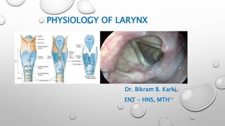
Basavarajeeyam is an important text for ayurvedic physician belonging to andhra pradehs. It is a popular compendium in various parts of our country as well as in andhra pradesh. The content of the text was presented in sanskrit and telugu language (Bilingual). One of the most famous book in ayurvedic pharmaceutics and therapeutics. This book contains 25 chapters called as prakaranas. Many rasaoushadis were explained, pioneer of dhatu druti, nadi pareeksha, mutra pareeksha etc. Belongs to the period of 15-16 century. New diseases like upadamsha, phiranga rogas are explained.
Lung Cancer: Artificial Intelligence, Synergetics, Complex System Analysis, S...Oleg Kshivets
RESULTS: Overall life span (LS) was 2252.1±1742.5 days and cumulative 5-year survival (5YS) reached 73.2%, 10 years – 64.8%, 20 years – 42.5%. 513 LCP lived more than 5 years (LS=3124.6±1525.6 days), 148 LCP – more than 10 years (LS=5054.4±1504.1 days).199 LCP died because of LC (LS=562.7±374.5 days). 5YS of LCP after bi/lobectomies was significantly superior in comparison with LCP after pneumonectomies (78.1% vs.63.7%, P=0.00001 by log-rank test). AT significantly improved 5YS (66.3% vs. 34.8%) (P=0.00000 by log-rank test) only for LCP with N1-2. Cox modeling displayed that 5YS of LCP significantly depended on: phase transition (PT) early-invasive LC in terms of synergetics, PT N0—N12, cell ratio factors (ratio between cancer cells- CC and blood cells subpopulations), G1-3, histology, glucose, AT, blood cell circuit, prothrombin index, heparin tolerance, recalcification time (P=0.000-0.038). Neural networks, genetic algorithm selection and bootstrap simulation revealed relationships between 5YS and PT early-invasive LC (rank=1), PT N0—N12 (rank=2), thrombocytes/CC (3), erythrocytes/CC (4), eosinophils/CC (5), healthy cells/CC (6), lymphocytes/CC (7), segmented neutrophils/CC (8), stick neutrophils/CC (9), monocytes/CC (10); leucocytes/CC (11). Correct prediction of 5YS was 100% by neural networks computing (area under ROC curve=1.0; error=0.0).
CONCLUSIONS: 5YS of LCP after radical procedures significantly depended on: 1) PT early-invasive cancer; 2) PT N0--N12; 3) cell ratio factors; 4) blood cell circuit; 5) biochemical factors; 6) hemostasis system; 7) AT; 8) LC characteristics; 9) LC cell dynamics; 10) surgery type: lobectomy/pneumonectomy; 11) anthropometric data. Optimal diagnosis and treatment strategies for LC are: 1) screening and early detection of LC; 2) availability of experienced thoracic surgeons because of complexity of radical procedures; 3) aggressive en block surgery and adequate lymph node dissection for completeness; 4) precise prediction; 5) adjuvant chemoimmunoradiotherapy for LCP with unfavorable prognosis.
These simplified slides by Dr. Sidra Arshad present an overview of the non-respiratory functions of the respiratory tract.
Learning objectives:
1. Enlist the non-respiratory functions of the respiratory tract
2. Briefly explain how these functions are carried out
3. Discuss the significance of dead space
4. Differentiate between minute ventilation and alveolar ventilation
5. Describe the cough and sneeze reflexes
Study Resources:
1. Chapter 39, Guyton and Hall Textbook of Medical Physiology, 14th edition
2. Chapter 34, Ganong’s Review of Medical Physiology, 26th edition
3. Chapter 17, Human Physiology by Lauralee Sherwood, 9th edition
4. Non-respiratory functions of the lungs https://academic.oup.com/bjaed/article/13/3/98/278874
NVBDCP.pptx Nation vector borne disease control programSapna Thakur
NVBDCP was launched in 2003-2004 . Vector-Borne Disease: Disease that results from an infection transmitted to humans and other animals by blood-feeding arthropods, such as mosquitoes, ticks, and fleas. Examples of vector-borne diseases include Dengue fever, West Nile Virus, Lyme disease, and malaria.
New Directions in Targeted Therapeutic Approaches for Older Adults With Mantl...i3 Health
i3 Health is pleased to make the speaker slides from this activity available for use as a non-accredited self-study or teaching resource.
This slide deck presented by Dr. Kami Maddocks, Professor-Clinical in the Division of Hematology and
Associate Division Director for Ambulatory Operations
The Ohio State University Comprehensive Cancer Center, will provide insight into new directions in targeted therapeutic approaches for older adults with mantle cell lymphoma.
STATEMENT OF NEED
Mantle cell lymphoma (MCL) is a rare, aggressive B-cell non-Hodgkin lymphoma (NHL) accounting for 5% to 7% of all lymphomas. Its prognosis ranges from indolent disease that does not require treatment for years to very aggressive disease, which is associated with poor survival (Silkenstedt et al, 2021). Typically, MCL is diagnosed at advanced stage and in older patients who cannot tolerate intensive therapy (NCCN, 2022). Although recent advances have slightly increased remission rates, recurrence and relapse remain very common, leading to a median overall survival between 3 and 6 years (LLS, 2021). Though there are several effective options, progress is still needed towards establishing an accepted frontline approach for MCL (Castellino et al, 2022). Treatment selection and management of MCL are complicated by the heterogeneity of prognosis, advanced age and comorbidities of patients, and lack of an established standard approach for treatment, making it vital that clinicians be familiar with the latest research and advances in this area. In this activity chaired by Michael Wang, MD, Professor in the Department of Lymphoma & Myeloma at MD Anderson Cancer Center, expert faculty will discuss prognostic factors informing treatment, the promising results of recent trials in new therapeutic approaches, and the implications of treatment resistance in therapeutic selection for MCL.
Target Audience
Hematology/oncology fellows, attending faculty, and other health care professionals involved in the treatment of patients with mantle cell lymphoma (MCL).
Learning Objectives
1.) Identify clinical and biological prognostic factors that can guide treatment decision making for older adults with MCL
2.) Evaluate emerging data on targeted therapeutic approaches for treatment-naive and relapsed/refractory MCL and their applicability to older adults
3.) Assess mechanisms of resistance to targeted therapies for MCL and their implications for treatment selection
Report Back from SGO 2024: What’s the Latest in Cervical Cancer?bkling
Are you curious about what’s new in cervical cancer research or unsure what the findings mean? Join Dr. Emily Ko, a gynecologic oncologist at Penn Medicine, to learn about the latest updates from the Society of Gynecologic Oncology (SGO) 2024 Annual Meeting on Women’s Cancer. Dr. Ko will discuss what the research presented at the conference means for you and answer your questions about the new developments.
263778731218 Abortion Clinic /Pills In Harare ,sisternakatoto
263778731218 Abortion Clinic /Pills In Harare ,ABORTION WOMEN’S CLINIC +27730423979 IN women clinic we believe that every woman should be able to make choices in her pregnancy. Our job is to provide compassionate care, safety,affordable and confidential services. That’s why we have won the trust from all generations of women all over the world. we use non surgical method(Abortion pills) to terminate…Dr.LISA +27730423979women Clinic is committed to providing the highest quality of obstetrical and gynecological care to women of all ages. Our dedicated staff aim to treat each patient and her health concerns with compassion and respect.Our dedicated group ABORTION WOMEN’S CLINIC +27730423979 IN women clinic we believe that every woman should be able to make choices in her pregnancy. Our job is to provide compassionate care, safety,affordable and confidential services. That’s why we have won the trust from all generations of women all over the world. we use non surgical method(Abortion pills) to terminate…Dr.LISA +27730423979women Clinic is committed to providing the highest quality of obstetrical and gynecological care to women of all ages. Our dedicated staff aim to treat each patient and her health concerns with compassion and respect.Our dedicated group of receptionists, nurses, and physicians have worked together as a teamof receptionists, nurses, and physicians have worked together as a team wwww.lisywomensclinic.co.za/
ARTIFICIAL INTELLIGENCE IN HEALTHCARE.pdfAnujkumaranit
Artificial intelligence (AI) refers to the simulation of human intelligence processes by machines, especially computer systems. It encompasses tasks such as learning, reasoning, problem-solving, perception, and language understanding. AI technologies are revolutionizing various fields, from healthcare to finance, by enabling machines to perform tasks that typically require human intelligence.
2. VOCAL FOLD ANATOMY
• During normal modal phonation,
mucosa undulates freely over underlying
vocal ligament and vocalis muscle.
• Hirano’s - histologic studies showed that
• Mucosa and muscle are separated by
specialized layer of connective tissue
that serves as a shock absorber
4. • The larynx has a number of functions
• To prevent foreign material from entering airway (aspiration)
• Acts as vibrator for generating sound
• Acts as a valve that can control air pressure and airflow
• Fundamental importance during breathing, weight bearing
5. LARYNGEAL MECHANORECEPTORS
• Free fibrils and terminal filaments enclosed in capsules
• Embedded in laryngeal tissues at sites sensitive to muscle stretch
and airflow pressures
• Wyke, postulated that mechanoreceptors are found in three sites:
1. Mucosal mechanoreceptors
• Corpuscular nerve endings
• Sensitive to stimuli of muscle stretch, air pressure level, liquid and
touch
• Discharge impulses - afferent fibres of vagus
6. 2. Articular mechanoreceptors
• Capsules of the articulatory joints
• Existence and function of this group remain controversial.
3. Myotatic mechanoreceptors
• Extrinsic and laryngeal muscles
• Tone of laryngeal muscles depends on myotatic reflex
7. FUNCTIONS OF THE LARYNX
• Swallowing (deglutition)
• During swallowing primary function of larynx - prevent food and
liquid entering the airway.
• This is achieved by means of
1. Sphincteric action of AE fold
2. True VF and
3. Ventricular folds,
which occurs simultaneously with elevation of larynx.
• Laryngeal elevation - control pressures and function of
cricopharyngeal sphincter
8. • Vocal fold adduction during swallowing - average approximately
2.3 seconds
• Airway is also protected by epiglottis - which covers the laryngeal
entrance
• Clinical correlates
• If laryngeal elevation is impaired
• Peri-swallow aspiration
• Cricopharyngeal opening is limited
9. COUGH REFLEX
• Protective reflex which ejects
mucus and foreign material from
lungs
• Cough can be
1. Voluntary action or
2. Reflexive response
• Consist of 3 phases-
• Inspiratory
• Compressive
• Expulsion
10. • First phase -inspiratory
• Glottis abducted, air inspired
• The second phase - compressive
• Glottis close, expiratory muscle contract
• Third phase – expulsion
• Air pressure buildup below adducted fold as diaphragm ascends
spasmodically
• Abduction of glottis
• Sudden and rapid outflow of air at speeds of as high as 10 l/sec.
11. EFFORT CLOSURE
• Provide a stable fulcrum for the upper limbs.
• For exertion involving use of arms,
VF are firmly adducted
preventing expulsion of air & collapse of chest wall
• Also occur during childbirth and defecation
• Clinical correlates
• Laryngectomy/ true VF palsy patients:
Weight bearing difficulty because of
inability to close glottis effectively
12. THE NEUROANATOMY OF PHONATION
• Dependent upon integrated functioning CNS and PNS
• Cortical loci - voluntary phonation
• Subcortical representation – 1. Involuntary phonation
2. Reflex laryngeal function
• Periaqueductal grey matter (PAG), a region of the mid-brain,
1. Crucial site for mammalian voice production
2. Integration of cortical and subcortical aspects of language
3. Production of involuntary or emotional sounds
13. NEURAL PATHWAY FOR VOLUNTARY
VOCALIZATION
• Arise in pre-central gyrus of motor cortex
• Fibres descend as part of corticobulbar
tract
(Part of pyramidal system or ‘direct
activation’ tract)
Medulla
• Some fibres synapse with ipsilateral vagus
nucleus
14. • The vagal nuclei, in nucleus ambigus (the reticular formation of
the medulla)
also contain 9 and 11 cranial nerve elements
Ipslilateral and contralateral vagus
Supply laryngeal muscles
• UMN do not govern isolated muscles, but groups of muscles.
• Frontobulbar portions of pyramidal tracts connect with cranial
nerves ix–xii, thus control phonation, articulation and respiration
15. THE BIOMECHANICS OF PHONATION
• VF provides visco-elastic mechanical properties - producing voice
• When larynx is at rest and respiration is quiet
VF abduct on inspiration
slightly adduct on expiration.
• On forceful inspiration – full abduction of VF
• Larynx descend on inspiration
ascends on expiration
16. INITIATION OF VOICE
• Pre-phonatory inspiratory phase
• VF rapidly abduct to allow intake of air
• Subsequently, VF are adducted – due to contraction of LCA muscle
• Subglottic air pressure increases below adducted VF
• Blows them apart, thus setting in motion vibratory cycle -
phonation
• Phonation threshold pressure
• Amount of air pressure required to begin voicing
• Factors affecting :
• Viscoelastic properties of VF
• Size and tension of VF
17. THE VIBRATORY CYCLE
• Consists of three phases:
1. Adduction
2. Aerodynamic separation and
3. Recoil
• Begins with vocal folds closed
• As subglottic air pressure increases - VF separates from inferior
border
• When they finally separate at superior margin, puff of air released
• Resulting negative pressure in glottis (bernoulli effect) results VF
closing rapidly - inferior VF margins closing first
• Mucosal wave travels- inferior to superior margin
18.
19. VOCAL REGISTERS
• Registers are defined in term of laryngeal behaviour
• Governed by degree of contraction of vocalis muscle
20. PHONATION
• Neurochronaxic theory
• Husson (1950)
• Glottic vibrations were caused by rhythmic nerve impulses to
larynx
• Each vibratory cycle was caused by a separate neural impulse
• Fallacies
• Vocal folds would not vibrate synchronously as longer course of
RLN in left
• If neurochronaxic theory were true, patients with tracheotomies
would be able to phonate - but they cannot
21. “BODY-COVER” THEORY
• Two-mass model
• “Body” of VF – vocalis muscle - relatively static
• “Cover” of VF – vocal mucosa - wave is propagated – blown by
expiratory air
• Vibration of mucosa does not correspond directly to that of rest of
vocal fold
• Mucosal wave begins on inferomedial aspect of vocal fold and
moves rostrally
22. THE MYOELASTIC-AERODYNAMIC THEORY
• Van den berg(1950)
• Myo - muscular involvement
• Elastic - ability to return to original state
• Aero - air pressure and flow
• Dynamic - movement and change
• Describes voice production as a combination of muscle force
(myo), tissue elasticity (elastic), and air pressures and flows
(aerodynamics)
• Widely accepted theory
23. • Process of phonation
• Begins with inhalation of air
• Glottic closure
• Subglottic pressure to increase until vocal folds are displaced
laterally
• Factors which bring glottis back together
1. Pressure decrease
2. Elastic forces in VF
3. Bernoulli effect on airflow
• When VF return to the midline, pressure in trachea builds again -
cycle is repeated
24. ASSESSMENT OF LARYNGEAL FUNCTION
• VIDEOLARYNGOSTROBOSCOPY (VLS)
• Evaluation of visco-elastic properties of
phonatory mucosa
• Principle
• Flashes of light from stroboscope are synchronized to VF vibration
at a slightly slower speed
• Allowing examiner to observe VF vibration during sound production
in slow motion
25. • Stroboscopy systems
• Endoscope
• Stroboscopic light source
• Camera and lens
• Microphone – picks up frequency
• Video recorder
Standard 70-degree rigid
strobolaryngoscope
Camera attachment
with mounted
microphone.
flexible laryngostroboscope
26. • Clinical applications
• Several parameters may be evaluated
• Glottal closure
• Mucosal wave
• Amplitude
• Periodicity
• Symmetry
• Fundamental frequencies
27. Notice that mucosal waves originate upon closure of VF
and
move from a medial to lateral direction.
29. • Advantage
• Office based procedure
• Painless
• Real-time information about nature of vibration
• Image to detect vocal pathology
• Permanent video record of examination
• Improves sensitivity of subtle laryngeal diagnoses
30. CONTACT ENDOSCOPY
• Simple, non - invasive technique
• In situ examination of
• Superficial cells of epithelium
• Mucosal blood vessels pattern
• Access microscopic structure of
entire mucosa
• Hamou (1979)
• Andrea et al. - evaluation larynx in 1995.
31. • Current contact microlaryngoscopes
• Diameters - 4 mm or 5.5 mm
• lengths - 23 cm and 18 cm
• Straight forward (O◦) and
forward oblique telescopes (30◦)
• Magnification - 1x, 60x, and 150x
• Require a high intensity xenon light source
32. • TECHNIQUE
• Sucking out secretion
• Endoscope - gently placed over suspicious site
• Viewing using 60X and 150X magnification
• Vascular patterns are studied without staining
• Then mucosa stained - 1% methylene blue soaked cotton pledgets
for 5 mins
• Stained area - visualized again
• Cell architecture
• Nuclei – dark blue structure
• Cytoplasm – light blue structure
33. • Advantage
• Non - invasive procedure
• Premalignant condition such as dysplasia detected earlier
• Avoids tissue damage and cellular architecture alteration
• Disadvantage
• Only evaluate most superficial cell layer of mucosal epithelium
• Inability to distinguish confidently between intraepithelial
neoplasia and invasive carcinoma
34. • Normal appearance
• Squamous cell at VF edge - polyhedric shape
• Nuclei - round, dark blue stain
• Cytoplasm - light blue stain
• Abnormal appearance
• Ciliated with squamous cell
1. Heavy smoker and GERD
• Ciliated epithelium of posterior commissure replace by squamous
epithelium
35. 2. Chronic laryngitis
• Epithelial pattern is homogenous, have large nuceli, Increase N:C
ratio
3. Keratosis
• Different stage of keratinization
4. Leukoplakia
• Cell - Heterogenicity
• Nuclei – different size, shape, and color
5. Carcinoma
• extreme heterogenicity of nuclear size, shape and staining
characteristic
36. • Image of blood vessels on normal VF
• Blood vessels are parallel to long axis of VF
• Bifurcations and anastomoses are few
• Image of blood vessels in early laryngeal
cancer
37. • Image of blood vessels in advanced
laryngeal cancer
• Leading to formation of vascular loops
38. • Image of cellular architecture of normal VF
• Cells : Homogenous with uniform size and
shape
• Nuclei : Uniform size and shape and evenly
stained
• N:C ratio : Uniform and less than 1
• Image of cellular architecture of squamous
carcinoma
39. NARROW BAND IMAGING (NBI)
• Non- invasive endoscopic technique - visualization of vascular
structure of mucosa
• Based on modification of standard white light –
• In which white light is transmitted through optical filter absorbing
all
40. • Principle
• Relies on depth of light penetration and absorption peak of Hb
• Use filtered light to visualize mucosal and submucosal
neoangiogenic vascular pattern
41. • NBI system consists
• Head or chip equipped videoendoscope
• Light source
• Camera unit
• Lighting unit with special filter
• Special image processor
42. • Narrow band blue light ( 390- 445nm )
• Imaging superficial capillaries of mucosal layer
• Is better absorbed by Hb
• Narrow band green light ( 530-550nm )
• Imaging thick vessels within mucosal layer
• Subepithelial vessels
43. • Blue color absorbed
by mucosal vessels
• Green color absorbed
by submucosal vessels
• NBI monitor representing
• Mucosal vessels – brown
colour
• Submucosal vessels – cyan
colour
44. • Vascular pattern associated with pathology includes
• Dotted – adenocarcinoma
• Tortous – squamous cell ca.
• Abruptly ending vessels – squamous cell ca.
• In diagnosis of primary laryngeal lesion
• Sensitivity – 89%
• Specificity – 93%
45. • Advantage
• Non invasive, done in opd basis
• Don’t require dyes
• Allow easy inspection of superficial vascular bed
• Early lesion < 1cm in diameter can be detected ( dysplasia, CIS )
• Disadvantage
• Lesion characterized by hyperkeratosis prevent visualization
• Can be limited in cases of stagnant saliva, sticky mucus
46. • Bilateral carcinoma of vocal folds in white-light (A) and NBI (B)
• Supraglottic spread of cancer is clearly visible on NBI image
47. • Larynx - left vocal cord dysplasia in white light (A) and NBI (B),
• Arrows point demarcates area of altered epithelium with presence
of brown dots
48. • Benign polyp of vocal cord in white-light (A) and NBI(B).
• Blood vessels run paralelly to the mucosal surface.