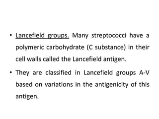
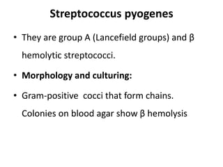
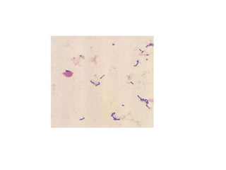
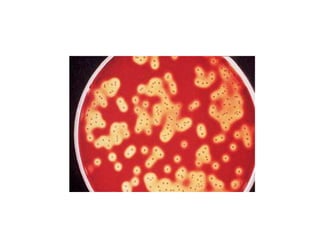
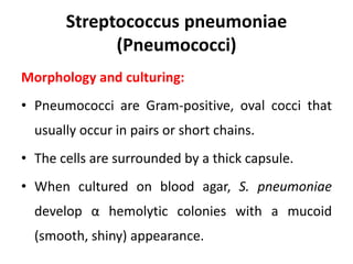
Streptococcus
• Streptococci are Gram-positive, nonmotile,
catalase-negative, facultatively anaerobic cocci
that occur in chains or pairs.
• They are classified based on their hemolytic
capacity (α, β and γ hemolysis) and the
antigenicity of a carbohydrate occurring in
their cell walls (Lancefield antigen A, B, C, D,
…….).
Classification
• α, β and γhemolysis.
• α hemolysis. Colonies on blood agar are surrounded by a green zone.
This“greening” is caused by H₂O₂, which converts hemoglobin into
methemoglobin.
• β hemolysis. Colonies on blood agar are surrounded by a large, yellowish
hemolytic zone in which no more intact erythrocytes are present and the
hemoglobin is lysed.
• γ hemolysis. This term indicates the absence of macroscopically visible
hemolytic zones.
• Lancefield groups. Many streptococci have a
polymeric carbohydrate (C substance) in their
cell walls called the Lancefield antigen.
• They are classified in Lancefield groups A-V
based on variations in the antigenicity of this
antigen.
Streptococcus pyogenes
• They are group A (Lancefield groups) and β
hemolytic streptococci.
• Morphology and culturing:
• Gram-positive cocci that form chains.
Colonies on blood agar show β hemolysis
Fine structures
• The murein layer of the cell wall is followed by
- The serogroup a carbohydrate layer, which consists
of C substance and is covalently bound to the murein.
-Long, twisted protein threads that extend outward are
anchored in the cell wall murein: the M protein.
Streptococci are classified in serovars with characteristic M
protein chemistry.
Extracellular toxins and enzymes:
• Streptolysin O, streptolysin S:
• Destroy the membranes of erythrocytes and other cells.
Streptolysin O acts as an antigen. Past infections can be
detected by measuring the antibodies to this toxin
(antistreptolysin titer).
• Pyrogenic streptococcal exotoxins
• Responsible for fever, scarlet fever, sepsis and septic shock.
• N.B. The pyrogenic exotoxins are superantigens and therefore
induce production of large amounts of cytokines.
• Streptolysin O, streptolysin S:
• Destroy the membranes of erythrocytes and other cells. Streptolysin O
acts as an antigen. Past infections can be detected by measuring the
antibodies to this toxin (antistreptolysin titer).
• Pyrogenic streptococcal exotoxins
• Responsible for fever, scarlet fever, sepsis, and septic shock.
• N.B. The pyrogenic exotoxins are superantigens and therefore induce
production of large amounts of cytokines.
• Streptokinase:
• Dissolves fibrin; facilitates spread of streptococci in tissues.
• Hyaluronidase:
• Breaks down hyaluronic acid a substance that
cements tissues together.
• DNases:
• Breakdown of DNA.
Pathogenesis and clinical pictures:
• Invasive infections: The pathogens enter through
traumas or microtraumas in the skin or mucosa and
cause invasive local or generalized infections.
• (They rarely cause severe septic infection and
necrotizing fasciitis).
• Post streptococcal diseases:
• Glomerulonephritis and acute rheumatic fever.
Diagnosis:
• - By detection of the pathogen by means of
microscopy and culturing.
• - Group A antigen can be detected using
particles coated with antibodies that
precipitate agglutination.
• Transmission is by direct contact (smear
infection) or droplets.
• The incubation period is one to three days.
The incidence of carriers among
• children is 10–20%.
Streptococcus agalactiae
• Group B and β hemolytic streptococci; occasionally
cause infections of the skin and connective tissues,
• urinary tract infections, pneumonia, and peritonitis in
immunocompromised individuals.
• It is one of the common causes of sepsis and meningitis
in neonates.
• Potential predisposing factors include birth
complications, premature birth, and a lack of antibodies
to the capsule in mother and neonate.
Streptococcus pneumoniae
(Pneumococci)
Morphology and culturing:
• Pneumococci are Gram-positive, oval cocci that
usually occur in pairs or short chains.
• The cells are surrounded by a thick capsule.
• When cultured on blood agar, S. pneumoniae
develop α hemolytic colonies with a mucoid
(smooth, shiny) appearance.
Antigen structure:
• Pneumococci are classified in 90 different
serotypes based on the fine chemical structure of
the capsule polysaccharides acting as antigens.
• This capsule antigen can be identified using
specific antisera in a reaction known as capsular
swelling.
Pathogenesis and clinical pictures.
• The capsule protects the pathogens from
phagocytosis and is the most important
determinant of pneumococcal virulence.
• Uncapsulated variants are not capable of causing
disease. Other virulence factors include IgA1
protease.
• The natural habitat of pneumococci is the mucosa
of the upper respiratory tract. About 40–70% of
healthy adults are carriers.
• Pneumococcal infections usually arise from
this normal flora (endogenous infections).
• Predisposing factors include cardiopulmonary
diseases and previous infections (e.g.,
influenza).
• The most important pneumococcal infections
are pneumonia. Other infections include otitis
media, sinusitis, meningitis, and corneal ulcer.
Diagnosis.
• The laboratory diagnosis includes detection of
the pathogen in appropriate test samples by
means of microscopy and culturing.
• Pneumococci can be differentiated from other
α-hemolytic streptococci based on their
sensitivity to optochin (optochin sensitive while
other stryptococci are optochin unsensitive).
Oral Streptococci
• Most of the oral streptococci often known as
the viridans group have no group antigen.
• They usually cause α-hemolysis or γ-
hemolysis.
• Oral streptococci are responsible for 50–70%
of all cases of bacterial endocarditis.
• The origins of endocarditis lie in invasion of the
vascular system through lesions in the oral mucosa.
• A transitory bacteremia results. The heart valves
are colonized and a biofilm is formed by the
organism.
• Predisposing factors include congenital heart
defects, acute rheumatic fever, cardiac surgery.
• Laboratory diagnosis of endocarditis involves
isolation of the pathogen from blood cultures.
• S. mutans, S. sanguis, and S. mitis are responsible
for dental caries
• These streptococci can attach to the proteins
covering the tooth where they then convert
sucrose into polysaccharides (mutan, dextran,
levan…).
• These sticky substances (in which the original
bacterial layers along with secondary bacterial
colonizers are embedded) form dental plaque.
Enterococcus (Enterococci)
• Enterococci are a widespread bacterial genus
normally found in the intestines of humans and
other animals.
• They are nonmotile, catalase-negative, and
characterized by group antigen D.
• They are able to proliferate at 45°C, in the presence
of 6.5% NaCl and at pH 9, qualities that differentiate
them from streptococci.
• E. faecalis, and E. faecium are the most
important species of this genus. They are used
as indicators for fecal pollution of water.
• They cause opportunistic infection specially
nosocomial (hospital acquired infection).
• They are highly resistant to antibiotics.

Streptococcus - 2.pptx

  • 1.
  • 2.
    • Streptococci areGram-positive, nonmotile, catalase-negative, facultatively anaerobic cocci that occur in chains or pairs. • They are classified based on their hemolytic capacity (α, β and γ hemolysis) and the antigenicity of a carbohydrate occurring in their cell walls (Lancefield antigen A, B, C, D, …….).
  • 3.
    Classification • α, βand γhemolysis. • α hemolysis. Colonies on blood agar are surrounded by a green zone. This“greening” is caused by H₂O₂, which converts hemoglobin into methemoglobin. • β hemolysis. Colonies on blood agar are surrounded by a large, yellowish hemolytic zone in which no more intact erythrocytes are present and the hemoglobin is lysed. • γ hemolysis. This term indicates the absence of macroscopically visible hemolytic zones.
  • 4.
    • Lancefield groups.Many streptococci have a polymeric carbohydrate (C substance) in their cell walls called the Lancefield antigen. • They are classified in Lancefield groups A-V based on variations in the antigenicity of this antigen.
  • 5.
    Streptococcus pyogenes • Theyare group A (Lancefield groups) and β hemolytic streptococci. • Morphology and culturing: • Gram-positive cocci that form chains. Colonies on blood agar show β hemolysis
  • 8.
    Fine structures • Themurein layer of the cell wall is followed by - The serogroup a carbohydrate layer, which consists of C substance and is covalently bound to the murein. -Long, twisted protein threads that extend outward are anchored in the cell wall murein: the M protein. Streptococci are classified in serovars with characteristic M protein chemistry.
  • 9.
    Extracellular toxins andenzymes: • Streptolysin O, streptolysin S: • Destroy the membranes of erythrocytes and other cells. Streptolysin O acts as an antigen. Past infections can be detected by measuring the antibodies to this toxin (antistreptolysin titer). • Pyrogenic streptococcal exotoxins • Responsible for fever, scarlet fever, sepsis and septic shock. • N.B. The pyrogenic exotoxins are superantigens and therefore induce production of large amounts of cytokines.
  • 10.
    • Streptolysin O,streptolysin S: • Destroy the membranes of erythrocytes and other cells. Streptolysin O acts as an antigen. Past infections can be detected by measuring the antibodies to this toxin (antistreptolysin titer). • Pyrogenic streptococcal exotoxins • Responsible for fever, scarlet fever, sepsis, and septic shock. • N.B. The pyrogenic exotoxins are superantigens and therefore induce production of large amounts of cytokines. • Streptokinase: • Dissolves fibrin; facilitates spread of streptococci in tissues.
  • 11.
    • Hyaluronidase: • Breaksdown hyaluronic acid a substance that cements tissues together. • DNases: • Breakdown of DNA.
  • 12.
    Pathogenesis and clinicalpictures: • Invasive infections: The pathogens enter through traumas or microtraumas in the skin or mucosa and cause invasive local or generalized infections. • (They rarely cause severe septic infection and necrotizing fasciitis). • Post streptococcal diseases: • Glomerulonephritis and acute rheumatic fever.
  • 13.
    Diagnosis: • - Bydetection of the pathogen by means of microscopy and culturing. • - Group A antigen can be detected using particles coated with antibodies that precipitate agglutination.
  • 14.
    • Transmission isby direct contact (smear infection) or droplets. • The incubation period is one to three days. The incidence of carriers among • children is 10–20%.
  • 16.
    Streptococcus agalactiae • GroupB and β hemolytic streptococci; occasionally cause infections of the skin and connective tissues, • urinary tract infections, pneumonia, and peritonitis in immunocompromised individuals. • It is one of the common causes of sepsis and meningitis in neonates. • Potential predisposing factors include birth complications, premature birth, and a lack of antibodies to the capsule in mother and neonate.
  • 17.
    Streptococcus pneumoniae (Pneumococci) Morphology andculturing: • Pneumococci are Gram-positive, oval cocci that usually occur in pairs or short chains. • The cells are surrounded by a thick capsule. • When cultured on blood agar, S. pneumoniae develop α hemolytic colonies with a mucoid (smooth, shiny) appearance.
  • 18.
    Antigen structure: • Pneumococciare classified in 90 different serotypes based on the fine chemical structure of the capsule polysaccharides acting as antigens. • This capsule antigen can be identified using specific antisera in a reaction known as capsular swelling.
  • 19.
    Pathogenesis and clinicalpictures. • The capsule protects the pathogens from phagocytosis and is the most important determinant of pneumococcal virulence. • Uncapsulated variants are not capable of causing disease. Other virulence factors include IgA1 protease. • The natural habitat of pneumococci is the mucosa of the upper respiratory tract. About 40–70% of healthy adults are carriers.
  • 20.
    • Pneumococcal infectionsusually arise from this normal flora (endogenous infections). • Predisposing factors include cardiopulmonary diseases and previous infections (e.g., influenza). • The most important pneumococcal infections are pneumonia. Other infections include otitis media, sinusitis, meningitis, and corneal ulcer.
  • 21.
    Diagnosis. • The laboratorydiagnosis includes detection of the pathogen in appropriate test samples by means of microscopy and culturing. • Pneumococci can be differentiated from other α-hemolytic streptococci based on their sensitivity to optochin (optochin sensitive while other stryptococci are optochin unsensitive).
  • 22.
    Oral Streptococci • Mostof the oral streptococci often known as the viridans group have no group antigen. • They usually cause α-hemolysis or γ- hemolysis. • Oral streptococci are responsible for 50–70% of all cases of bacterial endocarditis.
  • 23.
    • The originsof endocarditis lie in invasion of the vascular system through lesions in the oral mucosa. • A transitory bacteremia results. The heart valves are colonized and a biofilm is formed by the organism. • Predisposing factors include congenital heart defects, acute rheumatic fever, cardiac surgery. • Laboratory diagnosis of endocarditis involves isolation of the pathogen from blood cultures.
  • 24.
    • S. mutans,S. sanguis, and S. mitis are responsible for dental caries • These streptococci can attach to the proteins covering the tooth where they then convert sucrose into polysaccharides (mutan, dextran, levan…). • These sticky substances (in which the original bacterial layers along with secondary bacterial colonizers are embedded) form dental plaque.
  • 25.
    Enterococcus (Enterococci) • Enterococciare a widespread bacterial genus normally found in the intestines of humans and other animals. • They are nonmotile, catalase-negative, and characterized by group antigen D. • They are able to proliferate at 45°C, in the presence of 6.5% NaCl and at pH 9, qualities that differentiate them from streptococci.
  • 26.
    • E. faecalis,and E. faecium are the most important species of this genus. They are used as indicators for fecal pollution of water. • They cause opportunistic infection specially nosocomial (hospital acquired infection). • They are highly resistant to antibiotics.