The document discusses the stomatognathic system, emphasizing its components, such as the temporomandibular joint and masticatory muscles, and their roles in functions like mastication, deglutition, respiration, and speech. It explores the principles of functional osteology and myology, detailing the historical understanding of bone architecture and muscle physiology. Key concepts include bone remodelling principles and the impact of functional forces on bone structure and muscle function.


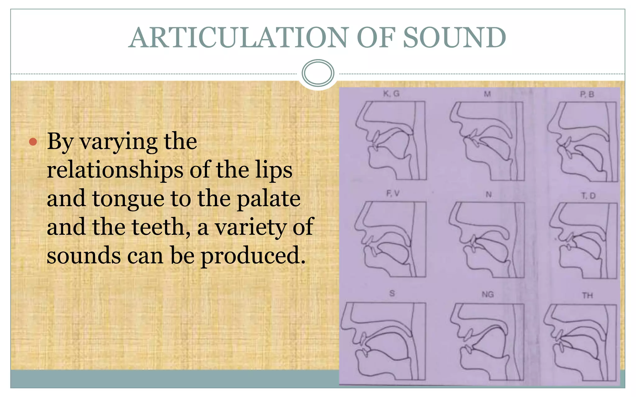
![SPEECH DIFFICULTIES RELATED TO
MALOCCLUSION
Speech Sounds Problem Related Malocclusion
/s/, /z/ (sibilants) Lisp Anterior Open Bite, Large
Gap between Incisors
/t/, /d/ (lingua-alveolar
stops)
Difficulty in production Irregular Incisors,
especially lingual position
of maxillary incisors
/f/, /v/ (labiodental
fricatives)
Distortion Skeletal Class III
th, sh, ch (linguodental
fricatives) [voiced or
voiceless]
Distortion Anterior Open Bite](https://image.slidesharecdn.com/stomatognathicsystem-190715133759/75/Stomatognathic-system-124-2048.jpg)


![Apporach to lung biopsy [Auto-saved].pptx latest](https://cdn.slidesharecdn.com/ss_thumbnails/apporachtolungbiopsyauto-saved-251211225655-93258539-thumbnail.jpg?width=640&height=640&fit=bounds)


