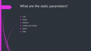
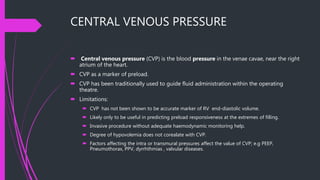
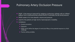
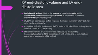
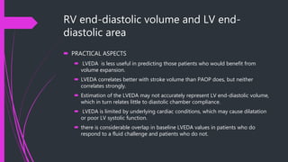
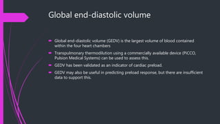
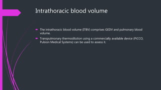
STATIC PARAMETERS OF
HAEMODYNAMIC
MONITORING
DR. ARSALAN ZAKI
DR. S. A. FATIMA
Goals
 Definitions
 what are the static parameters of haemodynamic monitoring?
 Why are these parameters important?
 How these parameters really influence our fluid therapy/management ?
 What are the pros and cons of different static parameters?
 What is next?
Definitions
 Cardiac output is the volume of blood ejected from each of the ventricles of the
heart per minute, and is therefore the product of stroke volume and heart rate. The
unit of cardiac output is L.min-1.
 Cardiac index is the cardiac output of a patient referenced to their body surface
area and has units of L.min-1.m-2.
 Stroke volume is the volume of blood ejected by each contraction of the
ventricle and is determined by the preload, afterload and contractility. The stroke
volume is usually 60-80ml for an average sized adult.
 Preload describes the tension developed in the ventricular wall at end-diastole
(i.e. at maximal filling just prior to contraction). This tension is difficult to measure
and end-diastolic pressure is taken as a surrogate (or estimate) measurement. It is
mainly determined by venous return and gives an indication of the filling pressure
of the ventricle.
Definitions
 Contractility refers to the amount of work the heart can generate, at given levels
of preload and afterload, and is estimated by the maximum rate at which the
ventricle can generate a change of pressure over time. Inotropy is used to explain
an increase in the work done by the heart that is independent of heart rate, preload
and afterload.
 Afterload is the tension that needs to be generated in the ventricular wall in
order to eject blood into the arterial system during systole. This is largely
determined by the resistance of the arterial system – the systemic vascular
resistance (SVR). It is calculated by:
 SVR = Mean arterial pressure (mmHg) – Central venous pressure (mmHg) x 80
/Cardiac output (L.min-1)
Definitions
 Mean arterial pressure (MAP) is the average arterial blood pressure
throughout the cardiac cycle. As 2/3 of the cardiac cycle is spent in diastole, and
1/3 in systole, MAP may be calculated using the formula:
 MAP = Diastolic BP + 1/3(Systolic BP - Diastolic BP)
 Ejection fraction is the fraction of total blood in a ventricle that is ejected per
beat. It applies to both the left and right ventricles. It gives an index of contractility.
Normal value is in the region of 55-65%.
What are the static parameters?
 CVP
 PAOP
 RVEDVI
 LVEDA and LVEDAI
 GEDV
 ITBV
Why are these parameters important?
 Haemodynamic monitoring is essential if fluid therapy is to be accurately titrated.
 Not all patients will respond to a fluid challenge. Therefore, it is useful to predict
fluid responsiveness to identify those patients in whom fluid therapy will be of
benefit.
 Adequate fluid administration is of extreme importance.
 Inadequate fluid-------inadequate tissue blood flow/perfusion.
 Excessive fluid-----------edema, increased cardiac demand, pulmonary edema and
respiratory failure and many other sequelae.
How these parameters help?
Responders and non-responders
 The expected haemodynamic response is an increase in stroke volume and
therefore cardiac output.
 the subsequent increase in stroke volume also depends on ventricular function.
 Accurate assessment of preload responsiveness.
 From the Frank –Starling law of the heart ,an increase in preload will significantly
increase stroke volume only if both ventricles are on the ascending portion of the
curve. If one or both ventricles lie on the flat portion, then the patient will be
regarded as a non-responder; that is, cardiac output will not increase significantly
in response to volume expansion.
FRANK STARLING LAW
CENTRAL VENOUS PRESSURE
 Central venous pressure (CVP) is the blood pressure in the venae cavae, near the right
atrium of the heart.
 CVP as a marker of preload.
 CVP has been traditionally used to guide fluid administration within the operating
theatre.
 Limitations:
 CVP has not been shown to be accurate marker of RV end-diastolic volume.
 Likely only to be useful in predicting preload responsiveness at the extremes of filling.
 Invasive procedure without adequate haemodynamic monitoring help.
 Degree of hypovolemia does not corealate with CVP.
 Factors affecting the intra or transmural pressures affect the value of CVP; e.g PEEP,
Pneumothorax, PPV, dyrrhthmias , valvular diseases.
Pulmonary Artery Occlusion Pressure
 PAOP is the pressure measured by wedging a pulmonary catheter with an inflated
balloon into a small pulmonary arterial branch. It estimates the left atrial pressure.
 PAOP marker of LV end-diastolic volume and pressure.
 Used for the patients at high risk for haemodynamic instability.
 Limitations:
 Invasive procedure.
 Expensive.
 PAOP cannot define the degree of ventricular filling or the potential response to a fluid
challenge.
 Practice setting variables.
RV end-diastolic volume and LV end-
diastolic area
 End-diastolic volume (EDV) is the volume of blood in the right and/or
left ventricle at end load or filling in (diastole) or the amount of blood in
the ventricles just before systole.
 (RVEDV) can be measured by fast-response thermistor pulmonary artery catheter
or by cardiac scintigraphy.
 A response to fluid is likely with an RVEDV index of ,90 ml m22 and unlikely with an
index of .138 ml m22 , respectively.
 Static measurement of LV end-diastolic area (LVEDA), measured by
transoesophageal echo (TOE), correlates well with LVEDV, and as such has been
examined as a parameter of LV preload.
RV end-diastolic volume and LV end-
diastolic area
 PRACTICAL ASPECTS
 LVEDA is less useful in predicting those patients who would benefit from
volume expansion.
 LVEDA correlates better with stroke volume than PAOP does, but neither
correlates strongly.
 Estimation of the LVEDA may not accurately represent LV end-diastolic volume,
which in turn relates little to diastolic chamber compliance.
 LVEDA is limited by underlying cardiac conditions, which may cause dilatation
or poor LV systolic function.
 there is considerable overlap in baseline LVEDA values in patients who do
respond to a fluid challenge and patients who do not.
Global end-diastolic volume
 Global end-diastolic volume (GEDV) is the largest volume of blood contained
within the four heart chambers
 Transpulmonary thermodilution using a commercially available device (PiCCO,
Pulsion Medical Systems) can be used to assess this.
 GEDV has been validated as an indicator of cardiac preload.
 GEDV may also be useful in predicting preload response, but there are insufficient
data to support this.
Intrathoracic blood volume
 The intrathoracic blood volume (ITBV) comprises GEDV and pulmonary blood
volume.
 Transpulmonary thermodilution using a commercially available device (PiCCO,
Pulsion Medical Systems) can be used to assess it.
What next?
 Dynamic parameters of the haemodynamic monitoring
Static parameters of_haemodynamic_monitoring

Static parameters of_haemodynamic_monitoring

  • 1.
  • 2.
    Goals  Definitions  whatare the static parameters of haemodynamic monitoring?  Why are these parameters important?  How these parameters really influence our fluid therapy/management ?  What are the pros and cons of different static parameters?  What is next?
  • 3.
    Definitions  Cardiac outputis the volume of blood ejected from each of the ventricles of the heart per minute, and is therefore the product of stroke volume and heart rate. The unit of cardiac output is L.min-1.  Cardiac index is the cardiac output of a patient referenced to their body surface area and has units of L.min-1.m-2.  Stroke volume is the volume of blood ejected by each contraction of the ventricle and is determined by the preload, afterload and contractility. The stroke volume is usually 60-80ml for an average sized adult.  Preload describes the tension developed in the ventricular wall at end-diastole (i.e. at maximal filling just prior to contraction). This tension is difficult to measure and end-diastolic pressure is taken as a surrogate (or estimate) measurement. It is mainly determined by venous return and gives an indication of the filling pressure of the ventricle.
  • 4.
    Definitions  Contractility refersto the amount of work the heart can generate, at given levels of preload and afterload, and is estimated by the maximum rate at which the ventricle can generate a change of pressure over time. Inotropy is used to explain an increase in the work done by the heart that is independent of heart rate, preload and afterload.  Afterload is the tension that needs to be generated in the ventricular wall in order to eject blood into the arterial system during systole. This is largely determined by the resistance of the arterial system – the systemic vascular resistance (SVR). It is calculated by:  SVR = Mean arterial pressure (mmHg) – Central venous pressure (mmHg) x 80 /Cardiac output (L.min-1)
  • 5.
    Definitions  Mean arterialpressure (MAP) is the average arterial blood pressure throughout the cardiac cycle. As 2/3 of the cardiac cycle is spent in diastole, and 1/3 in systole, MAP may be calculated using the formula:  MAP = Diastolic BP + 1/3(Systolic BP - Diastolic BP)  Ejection fraction is the fraction of total blood in a ventricle that is ejected per beat. It applies to both the left and right ventricles. It gives an index of contractility. Normal value is in the region of 55-65%.
  • 6.
    What are thestatic parameters?  CVP  PAOP  RVEDVI  LVEDA and LVEDAI  GEDV  ITBV
  • 7.
    Why are theseparameters important?  Haemodynamic monitoring is essential if fluid therapy is to be accurately titrated.  Not all patients will respond to a fluid challenge. Therefore, it is useful to predict fluid responsiveness to identify those patients in whom fluid therapy will be of benefit.  Adequate fluid administration is of extreme importance.  Inadequate fluid-------inadequate tissue blood flow/perfusion.  Excessive fluid-----------edema, increased cardiac demand, pulmonary edema and respiratory failure and many other sequelae.
  • 8.
    How these parametershelp? Responders and non-responders  The expected haemodynamic response is an increase in stroke volume and therefore cardiac output.  the subsequent increase in stroke volume also depends on ventricular function.  Accurate assessment of preload responsiveness.  From the Frank –Starling law of the heart ,an increase in preload will significantly increase stroke volume only if both ventricles are on the ascending portion of the curve. If one or both ventricles lie on the flat portion, then the patient will be regarded as a non-responder; that is, cardiac output will not increase significantly in response to volume expansion.
  • 9.
  • 10.
    CENTRAL VENOUS PRESSURE Central venous pressure (CVP) is the blood pressure in the venae cavae, near the right atrium of the heart.  CVP as a marker of preload.  CVP has been traditionally used to guide fluid administration within the operating theatre.  Limitations:  CVP has not been shown to be accurate marker of RV end-diastolic volume.  Likely only to be useful in predicting preload responsiveness at the extremes of filling.  Invasive procedure without adequate haemodynamic monitoring help.  Degree of hypovolemia does not corealate with CVP.  Factors affecting the intra or transmural pressures affect the value of CVP; e.g PEEP, Pneumothorax, PPV, dyrrhthmias , valvular diseases.
  • 11.
    Pulmonary Artery OcclusionPressure  PAOP is the pressure measured by wedging a pulmonary catheter with an inflated balloon into a small pulmonary arterial branch. It estimates the left atrial pressure.  PAOP marker of LV end-diastolic volume and pressure.  Used for the patients at high risk for haemodynamic instability.  Limitations:  Invasive procedure.  Expensive.  PAOP cannot define the degree of ventricular filling or the potential response to a fluid challenge.  Practice setting variables.
  • 12.
    RV end-diastolic volumeand LV end- diastolic area  End-diastolic volume (EDV) is the volume of blood in the right and/or left ventricle at end load or filling in (diastole) or the amount of blood in the ventricles just before systole.  (RVEDV) can be measured by fast-response thermistor pulmonary artery catheter or by cardiac scintigraphy.  A response to fluid is likely with an RVEDV index of ,90 ml m22 and unlikely with an index of .138 ml m22 , respectively.  Static measurement of LV end-diastolic area (LVEDA), measured by transoesophageal echo (TOE), correlates well with LVEDV, and as such has been examined as a parameter of LV preload.
  • 13.
    RV end-diastolic volumeand LV end- diastolic area  PRACTICAL ASPECTS  LVEDA is less useful in predicting those patients who would benefit from volume expansion.  LVEDA correlates better with stroke volume than PAOP does, but neither correlates strongly.  Estimation of the LVEDA may not accurately represent LV end-diastolic volume, which in turn relates little to diastolic chamber compliance.  LVEDA is limited by underlying cardiac conditions, which may cause dilatation or poor LV systolic function.  there is considerable overlap in baseline LVEDA values in patients who do respond to a fluid challenge and patients who do not.
  • 14.
    Global end-diastolic volume Global end-diastolic volume (GEDV) is the largest volume of blood contained within the four heart chambers  Transpulmonary thermodilution using a commercially available device (PiCCO, Pulsion Medical Systems) can be used to assess this.  GEDV has been validated as an indicator of cardiac preload.  GEDV may also be useful in predicting preload response, but there are insufficient data to support this.
  • 15.
    Intrathoracic blood volume The intrathoracic blood volume (ITBV) comprises GEDV and pulmonary blood volume.  Transpulmonary thermodilution using a commercially available device (PiCCO, Pulsion Medical Systems) can be used to assess it.
  • 16.
    What next?  Dynamicparameters of the haemodynamic monitoring