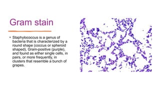
STAPHYLOCOCCUS
Sae Food Poisoning
STAPHYLOCOCCUS
• Domain- Bacteria
• Kingdom- Eubacteria
• Phylum- Firmicutes
• Class- Bacilli
• Order- Bacillales
• Family- Staphylococcaceae
• Genus- Staphylocuccus
• The genus name Staphylococcus is derived from
Greek terms (staphyle and kokkos) that mean "a
bunch of grapes,
• Staphylococci are organisms of universal distribution located in water, air, soil, wastewater,
food, as well as surfaces and all articles that come into contact with humans and animals,
where the nose has been established as its main reservoir, along with the throat, skin and
intestinal tract.
• Non spore-forming, non motile, non flagellated, facultative anaerobic, gram positive bacteria.
• It is catalase and coagulase positive
• Cells are spherical single and often form grape-like clusters
• survive desiccation and tolerate high levels of salt
• sensitive to lysozyme & lysostaphin, which specifically cleaves the pentaglycin bridges of
Staphylococcus spp.
• The organisms are able to grow in a wide range of temperatures (7°C to 48°C with an optimum
of 30°C to 37°C), pH (4.2 to 9.3, with an optimum of 7.0 to 7.5); and sodium chloride
concentrations (up to 15% NaCl).
• indicators of personnel hygiene
• It is commonly detected in foods of animal origin
• The cooked and processed food are more susceptible to Staphaylococcus aureus because of
the occurrence of low number of other competing organisums.
Gram stain
• Staphylococcus is a genus of
bacteria that is characterized by a
round shape (coccus or spheroid
shaped), Gram-positive (purple),
and found as either single cells, in
pairs, or more frequently, in
clusters that resemble a bunch of
grapes.
Staphylococcus aureus in Mannitol Salt Agar S. aureus on Baird-Parker agar plate
Staphylococcal food poisoning
• Staphylococcus aureus infections cause significant morbidity and mortality both in
the developing and developed nations . It accounts for up to one third of foodborne
gastrointestinal illnesses
• The presence of Staphylococcus aureus in fish and products can mainly indicate
contamination from the skin, mouth and nostrils of infected handlers or healthy
carriers, during stages of capture, transport, storage, processing and preparation
processes.
• Source of staphylococci to foods are mainly from human and domestic animals.
• Food handlers are main source of food contamination with S. aureus. Besides,
equipment and food contact surfaces also contribute for contamination with this
organism.
• It is an indicator of hygienic practices in a fish processing unit
• This bacterium produces an exotoxin called enterotoxin which
causes gastroenteritis or inflammation of intestinal tract.
• Staphylococcal enterotoxins are exoproteins of low molecular weight
approximately 27-31 kDa.
• Staphylococcal enterotoxins are highly stable, resist most proteolytic enzymes,
such as pepsin, or trypsin, and thus keep their activity in the digestive tract after
ingestion.
• Entertoxins are heat resistant and not inactivated at pasteurisation temperature.
Normal cooking of food will not destroy toxin present in food.
• High salt tolerance and ability to grow at low water activity (0.86) helps this
organism to have competitive advantage over other organisms especially in
smoked fish and salted fish.
Disease
and
symptoms
• Individuals differ in their susceptibility to food
poisoning and incubation period is more than 4
hrs.
• It cause gastrointestinal illnesses
• Symptoms of food poisoning is characterized
by salivation, nausea, vomiting, abdominal
cramps, diarrhea, blood and mucus in stools,
headache etc.
• Mortality is very low and infected persons
recover except in extreme cases.
In South America, countries such as Peru,
the ministerial resolution for chilled or
frozen raw food maximum allowable limit
is 103
CFU/g.
In Central America for frozen or
refrigerated fish the permitted limit is 10²
CFU/g
Diagnosis
The diagnosis of Staphylococcus aureus is based performing tests with colonies.
Coagulase, haemolysins, and thermostable deoxyribonuclease tests are all
commonly performed
Real-time PCR and quantitative PCR
Immunological methods based on monoclonal antibodies (e.g., ELISA, ELFA, and
Reverse Passive Latex Agglutination) for determining the enterotoxigenicity of
Staphylococcus aureus isolated from foods.
Staphylococci - blood agar culture
• Blood agar is both differential and enriched
medium.
• On blood agar, S. aureus usually displays a light to
golden yellow pigment
• On blood agar, S. aureus is usually beta-hemolytic
• individual colonies on agar are round, convex, and
1-4 mm in diameter with a sharp border
• On blood agar plates, colonies of Staphylococcus
aureus are frequently surrounded by zones of clear
beta-hemolysis. The golden appearance of colonies
of some strains is the etymological root of the
bacteria's name; aureus meaning "golden" in Latin.
Catalase test
• The catalase test is primarily used to distinguish among Gram-positive cocci.
• Members of the genus Staphylococcus are catalase-positive, and members of the
genera Streptococcus and Enterococcus are catalase-negative.
1. Transfer a small amount of bacterial colony to a surface of clean, dry glass
slide using a loop or sterile wooden stick
2. Place a drop of 3% H2O2 onto the slide and mix.
3. A positive result is the rapid evolution of oxygen (within 5-10 sec.) as
evidenced by bubbling (Fig. 5).
4. A negative result is no bubbles or only a few scattered bubbles
Mannitol salt agar culture
• Mannitol salt agar (MSA) is both a selective and differential media used for the isolation
of Staphylococci from mixed cultures.
• 7,5% NaCl – selects for species of Staphylococcus. This concentration of salt is too high
for most other bacteria to withstand and, therefore, inhibits their growth.
• Mannitol – alcohol of the carbohydrate mannose. Mannitol fermentation produces acid
end products which turn the medium yellow. Yellow indicates mannitol positive and no
color change indicates mannitol negative.
• On MSA, only pathogenic Staphylococcus aureus produces small colonies surrounded
by yellow zones.
• The reason for this color change is that S. aureus have the ability to ferment the
mannitol, producing an acid, which changes the indicator color from red to yellow. The
growth of other types of bacteria is usually inhibited.
• This growth differentiates S. aureus from S. epidermidis, which forms colonies with red
zones.
Coagulase test
• The coagulase test is used to distinguish between pathogenic and nonpathogenic members of the
genus Staphylococcus.
• Most strains of S. aureus produce one or two types of coagulase; free coagulase and bound
coagulase.
Detection of bound coagulase - Slide Test
1. Divide the slide into two sections with grease pencil. One should be labeled as „test” and the other
as „control“.
2. Place a small drop of distilled water on each area.
3. Emulsify one or two colonies of Staphylococcus on blood agar plate on each drop to make a smooth
suspension.
4. The test suspension is treated with a drop of citrated plasma and mixed well with a needle.
5. Do not put anything in the other drop that serves as control. The control suspension serves to rule
out false positivity due to auto agglutination.
6. Clumping of cocci within 5-10 seconds is taken as positive
• Some strains of S.aureus may not produce bound coagulase, and such strains must be identified by
tube coagulase test
Detection of free coagulase - Tube Coagulase Test
1. Three test tubes are taken and labeled “test”, “negative
control” and “positive control”.
2. Each tube is filled with 1 ml of 1 in 10 diluted rabbit plasma.
3. To the tube labeled test, 0.2 ml of overnight broth culture of
test bacteria is added.
4. To the tube labeled positive control, 0.2 ml of overnight
broth culture of known S. aureus is added.
5. To the tube labeled negative control, 0.2ml of sterile broth
is added.
6. All the tubes are incubated at 37°C.
7. Positive result is indicated by gelling of the plasma, which
remains in place even after inverting the tube.
8. If the test remains negative until four hours at 37°C, the
tube is kept at room temperature for overnight incubation.
Control
• Avoid the use of raw materials which may be contaminated with high numbers of S. aureus
• Ensure that food handlers are aware of the importance of good hygiene practices,
particularly hand washing and the need to report skin infections so that appropriate
precautions can be taken.
• Hand washing, wearing gloves during food preparation, and storing foods at the proper
temperature to inhibit pathogen growth and minimize toxin production, as heating food after
toxin has formed will not be an effective control measure.
• serving hot meals right after cooking
• thoroughly reheating of cooked foods
• rapid refrigeration of cooked foods
• proper hand washing before and after food preparation
• Avoid food service workers with skin infections in food establishments
• use clean utensils
• Implementation of a food safety management system based on the principles of HACCP.
Thank you

STAPHYLOCOCCUS .pptx

  • 1.
  • 2.
    STAPHYLOCOCCUS • Domain- Bacteria •Kingdom- Eubacteria • Phylum- Firmicutes • Class- Bacilli • Order- Bacillales • Family- Staphylococcaceae • Genus- Staphylocuccus • The genus name Staphylococcus is derived from Greek terms (staphyle and kokkos) that mean "a bunch of grapes,
  • 3.
    • Staphylococci areorganisms of universal distribution located in water, air, soil, wastewater, food, as well as surfaces and all articles that come into contact with humans and animals, where the nose has been established as its main reservoir, along with the throat, skin and intestinal tract. • Non spore-forming, non motile, non flagellated, facultative anaerobic, gram positive bacteria. • It is catalase and coagulase positive • Cells are spherical single and often form grape-like clusters • survive desiccation and tolerate high levels of salt • sensitive to lysozyme & lysostaphin, which specifically cleaves the pentaglycin bridges of Staphylococcus spp. • The organisms are able to grow in a wide range of temperatures (7°C to 48°C with an optimum of 30°C to 37°C), pH (4.2 to 9.3, with an optimum of 7.0 to 7.5); and sodium chloride concentrations (up to 15% NaCl). • indicators of personnel hygiene • It is commonly detected in foods of animal origin • The cooked and processed food are more susceptible to Staphaylococcus aureus because of the occurrence of low number of other competing organisums.
  • 4.
    Gram stain • Staphylococcusis a genus of bacteria that is characterized by a round shape (coccus or spheroid shaped), Gram-positive (purple), and found as either single cells, in pairs, or more frequently, in clusters that resemble a bunch of grapes.
  • 5.
    Staphylococcus aureus inMannitol Salt Agar S. aureus on Baird-Parker agar plate
  • 6.
    Staphylococcal food poisoning •Staphylococcus aureus infections cause significant morbidity and mortality both in the developing and developed nations . It accounts for up to one third of foodborne gastrointestinal illnesses • The presence of Staphylococcus aureus in fish and products can mainly indicate contamination from the skin, mouth and nostrils of infected handlers or healthy carriers, during stages of capture, transport, storage, processing and preparation processes. • Source of staphylococci to foods are mainly from human and domestic animals. • Food handlers are main source of food contamination with S. aureus. Besides, equipment and food contact surfaces also contribute for contamination with this organism. • It is an indicator of hygienic practices in a fish processing unit
  • 7.
    • This bacteriumproduces an exotoxin called enterotoxin which causes gastroenteritis or inflammation of intestinal tract. • Staphylococcal enterotoxins are exoproteins of low molecular weight approximately 27-31 kDa. • Staphylococcal enterotoxins are highly stable, resist most proteolytic enzymes, such as pepsin, or trypsin, and thus keep their activity in the digestive tract after ingestion. • Entertoxins are heat resistant and not inactivated at pasteurisation temperature. Normal cooking of food will not destroy toxin present in food. • High salt tolerance and ability to grow at low water activity (0.86) helps this organism to have competitive advantage over other organisms especially in smoked fish and salted fish.
  • 8.
    Disease and symptoms • Individuals differin their susceptibility to food poisoning and incubation period is more than 4 hrs. • It cause gastrointestinal illnesses • Symptoms of food poisoning is characterized by salivation, nausea, vomiting, abdominal cramps, diarrhea, blood and mucus in stools, headache etc. • Mortality is very low and infected persons recover except in extreme cases.
  • 9.
    In South America,countries such as Peru, the ministerial resolution for chilled or frozen raw food maximum allowable limit is 103 CFU/g. In Central America for frozen or refrigerated fish the permitted limit is 10² CFU/g
  • 10.
    Diagnosis The diagnosis ofStaphylococcus aureus is based performing tests with colonies. Coagulase, haemolysins, and thermostable deoxyribonuclease tests are all commonly performed Real-time PCR and quantitative PCR Immunological methods based on monoclonal antibodies (e.g., ELISA, ELFA, and Reverse Passive Latex Agglutination) for determining the enterotoxigenicity of Staphylococcus aureus isolated from foods.
  • 13.
    Staphylococci - bloodagar culture • Blood agar is both differential and enriched medium. • On blood agar, S. aureus usually displays a light to golden yellow pigment • On blood agar, S. aureus is usually beta-hemolytic • individual colonies on agar are round, convex, and 1-4 mm in diameter with a sharp border • On blood agar plates, colonies of Staphylococcus aureus are frequently surrounded by zones of clear beta-hemolysis. The golden appearance of colonies of some strains is the etymological root of the bacteria's name; aureus meaning "golden" in Latin.
  • 14.
    Catalase test • Thecatalase test is primarily used to distinguish among Gram-positive cocci. • Members of the genus Staphylococcus are catalase-positive, and members of the genera Streptococcus and Enterococcus are catalase-negative. 1. Transfer a small amount of bacterial colony to a surface of clean, dry glass slide using a loop or sterile wooden stick 2. Place a drop of 3% H2O2 onto the slide and mix. 3. A positive result is the rapid evolution of oxygen (within 5-10 sec.) as evidenced by bubbling (Fig. 5). 4. A negative result is no bubbles or only a few scattered bubbles
  • 15.
    Mannitol salt agarculture • Mannitol salt agar (MSA) is both a selective and differential media used for the isolation of Staphylococci from mixed cultures. • 7,5% NaCl – selects for species of Staphylococcus. This concentration of salt is too high for most other bacteria to withstand and, therefore, inhibits their growth. • Mannitol – alcohol of the carbohydrate mannose. Mannitol fermentation produces acid end products which turn the medium yellow. Yellow indicates mannitol positive and no color change indicates mannitol negative. • On MSA, only pathogenic Staphylococcus aureus produces small colonies surrounded by yellow zones. • The reason for this color change is that S. aureus have the ability to ferment the mannitol, producing an acid, which changes the indicator color from red to yellow. The growth of other types of bacteria is usually inhibited. • This growth differentiates S. aureus from S. epidermidis, which forms colonies with red zones.
  • 17.
    Coagulase test • Thecoagulase test is used to distinguish between pathogenic and nonpathogenic members of the genus Staphylococcus. • Most strains of S. aureus produce one or two types of coagulase; free coagulase and bound coagulase. Detection of bound coagulase - Slide Test 1. Divide the slide into two sections with grease pencil. One should be labeled as „test” and the other as „control“. 2. Place a small drop of distilled water on each area. 3. Emulsify one or two colonies of Staphylococcus on blood agar plate on each drop to make a smooth suspension. 4. The test suspension is treated with a drop of citrated plasma and mixed well with a needle. 5. Do not put anything in the other drop that serves as control. The control suspension serves to rule out false positivity due to auto agglutination. 6. Clumping of cocci within 5-10 seconds is taken as positive • Some strains of S.aureus may not produce bound coagulase, and such strains must be identified by tube coagulase test
  • 18.
    Detection of freecoagulase - Tube Coagulase Test 1. Three test tubes are taken and labeled “test”, “negative control” and “positive control”. 2. Each tube is filled with 1 ml of 1 in 10 diluted rabbit plasma. 3. To the tube labeled test, 0.2 ml of overnight broth culture of test bacteria is added. 4. To the tube labeled positive control, 0.2 ml of overnight broth culture of known S. aureus is added. 5. To the tube labeled negative control, 0.2ml of sterile broth is added. 6. All the tubes are incubated at 37°C. 7. Positive result is indicated by gelling of the plasma, which remains in place even after inverting the tube. 8. If the test remains negative until four hours at 37°C, the tube is kept at room temperature for overnight incubation.
  • 19.
    Control • Avoid theuse of raw materials which may be contaminated with high numbers of S. aureus • Ensure that food handlers are aware of the importance of good hygiene practices, particularly hand washing and the need to report skin infections so that appropriate precautions can be taken. • Hand washing, wearing gloves during food preparation, and storing foods at the proper temperature to inhibit pathogen growth and minimize toxin production, as heating food after toxin has formed will not be an effective control measure. • serving hot meals right after cooking • thoroughly reheating of cooked foods • rapid refrigeration of cooked foods • proper hand washing before and after food preparation • Avoid food service workers with skin infections in food establishments • use clean utensils • Implementation of a food safety management system based on the principles of HACCP.
  • 20.

Editor's Notes

  • #5 . Colonies appear as small, black, convex round colonies with clear zone.
  • #17 Coagulases are enzymes that clot blood plasma by a mechanism that is similar to normal clotting. The coagulase test identifies whether an organism produces this exoenzyme. This enzyme clots the plasma component of blood. The only significant disease causing bacteria of humans that produce coagulase enzyme are Staphylococcus aureus. Thus this enzyme is a good indicator of the pathogenic potential of S. aureus.