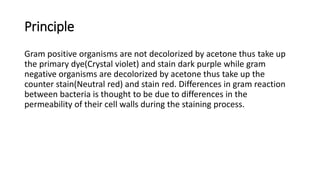
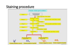
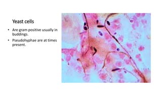
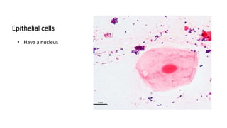
SPUTUM GRAM STAIN
PRESENTED BY RICKY OTEMA
LABORATORY ASSISTANT SEBBI HOSPITAL
Principle
Gram positive organisms are not decolorized by acetone thus take up
the primary dye(Crystal violet) and stain dark purple while gram
negative organisms are decolorized by acetone thus take up the
counter stain(Neutral red) and stain red. Differences in gram reaction
between bacteria is thought to be due to differences in the
permeability of their cell walls during the staining process.
Staining procedure
Sources of errors
• Over decolorization of the smear.
• Use of too old iodine solution i.e yellow instead of brown.
• Excessive heat fixation of the smear.
• Too thick smear which may lead to under decolorization of gram negative organisms
thus appearing as gram positive organisms.
Corrective measures
• Make relatively a good smear, not too thick or too thin measuring 15-20mm in diameter.
• Use of fresh or unexpired reagents.
• Proper labelling of stains and reagents.
• Following exactly a staining technique particularly decolorizing time.
• When heat fixing, gentle heat should be used.
• Smear should be protected from dust, flies, ants and direct sunlight when drying.
Morphology of pus cells, epithelial cells and bacteria
under x10 objective.
Sputum quality score (Bartlett criteria)
Cells criteria Score
Neutrophils(PMNs) >25
10-25
<10
+2
+1
0
Macroscopy Mucoid, mucopurulent,
purulent or blood stained
+
Squamous epithelial cells >25
10-25
<10
-2
-1
0
Interpretation of Q-Score
The “+” Q-Score indicates good sputum quality. A “0” or “-” Q-Score suggests low
sputum quality, excessive oropharyngeal contamination.
Examine the smear under 100x objective and quantify
the organisms as follows;
Criteria Score
<1
1-10
11-25
>25
+
+2
+3
+4
Expected gram organisms in sputum and their
morphology.
Gram positive
• Streptococcus pneumoniae
• Streptococcus pyogenes
• Staphylococcus aureus
Gram negative
• Haemophilus influenza
• Moraxella catarrhalis
• Pseudomonas aeruginosa
• Proteus species
• Yersinia pestis
Fungi
• Cryptococcus neoformans
• Candida albicans
• Nocardia species
• Aspergillus species
• Pneumocystis jiroveci
• Blastomyces dermatitidis
• Histoplasma capsulatum
Streptococcus
pneumoniae
• Are gram positive,lancet
shaped cocci.(elongated
cocci)
• Usually are seen as
pairs(diplococcic) but may
also occur singly and in
short chains.
• May occur intracellularly or
extracellularly
Streptococcus
pyogenes
• Are gram positive,spherical,
ovoid, or round shaped.
• They occur in pairs or
chains of varying lengths.
Staphylococcus aureus
• Are gram positive spherical
cocci.
• They are arranged in grape-
like clusters.
• Measure approximately 1.0
um in diameter.
• A few strains possess
capsules.
Haemophilus
influenzae
• Are slender, short, gram-
negative rods or
coccobacilli.
• Encapsulated form are
coccobacilli.
• Non-capsulated are
pleomorphic.
Klebsiella pneumoniae
• Gram negative rod-shaped,
singly and in pairs or short
chains.
Moraxella catarrhalis
• Are gram-negative
diplococcic(kidney-bean
shaped).
Pseudomonas
aeruginosa
• Are thin gram-negative
rods.
Yeast cells
• Are gram positive usually in
buddings.
• Pseudohyphae are at times
present.
Epithelial cells
• Have a nucleus
Reporting of Gram organisms
• Gram positive bacteria………………………….Dark purple
• Yeast cells……………………………………………..Dark purple
• Gram negative bacteria…………………………Pale to dark red
• Nuclei of pus cells…………………………………Red
• Epithelial cells……………………………………….Pale red
The report should include the following information;
• Numbers of bacteria present
• Gram reaction of the bacteria
• Morphology of the bacteria
• Presence and number of pus cells
• Presence of yeast cells and epithelial cells.
Quality Assurance / Quality control
• Training and competency assessment of all appropriate staff.
• Always check new batches of stain and reagents for correct staining
reactions using known gram negative and gram positive smears.
• Subsequent periodic refresher training and observation by senior
laboratory staff.
• Continuous monitoring and review of patient records.
• Review of every gram smear by a second senior technologist/scientist.
End

SPUTUM GRAM STAIN.pptx

  • 1.
    SPUTUM GRAM STAIN PRESENTEDBY RICKY OTEMA LABORATORY ASSISTANT SEBBI HOSPITAL
  • 2.
    Principle Gram positive organismsare not decolorized by acetone thus take up the primary dye(Crystal violet) and stain dark purple while gram negative organisms are decolorized by acetone thus take up the counter stain(Neutral red) and stain red. Differences in gram reaction between bacteria is thought to be due to differences in the permeability of their cell walls during the staining process.
  • 3.
  • 4.
    Sources of errors •Over decolorization of the smear. • Use of too old iodine solution i.e yellow instead of brown. • Excessive heat fixation of the smear. • Too thick smear which may lead to under decolorization of gram negative organisms thus appearing as gram positive organisms. Corrective measures • Make relatively a good smear, not too thick or too thin measuring 15-20mm in diameter. • Use of fresh or unexpired reagents. • Proper labelling of stains and reagents. • Following exactly a staining technique particularly decolorizing time. • When heat fixing, gentle heat should be used. • Smear should be protected from dust, flies, ants and direct sunlight when drying.
  • 5.
    Morphology of puscells, epithelial cells and bacteria under x10 objective.
  • 6.
    Sputum quality score(Bartlett criteria) Cells criteria Score Neutrophils(PMNs) >25 10-25 <10 +2 +1 0 Macroscopy Mucoid, mucopurulent, purulent or blood stained + Squamous epithelial cells >25 10-25 <10 -2 -1 0 Interpretation of Q-Score The “+” Q-Score indicates good sputum quality. A “0” or “-” Q-Score suggests low sputum quality, excessive oropharyngeal contamination.
  • 7.
    Examine the smearunder 100x objective and quantify the organisms as follows; Criteria Score <1 1-10 11-25 >25 + +2 +3 +4
  • 8.
    Expected gram organismsin sputum and their morphology. Gram positive • Streptococcus pneumoniae • Streptococcus pyogenes • Staphylococcus aureus Gram negative • Haemophilus influenza • Moraxella catarrhalis • Pseudomonas aeruginosa • Proteus species • Yersinia pestis Fungi • Cryptococcus neoformans • Candida albicans • Nocardia species • Aspergillus species • Pneumocystis jiroveci • Blastomyces dermatitidis • Histoplasma capsulatum
  • 9.
    Streptococcus pneumoniae • Are grampositive,lancet shaped cocci.(elongated cocci) • Usually are seen as pairs(diplococcic) but may also occur singly and in short chains. • May occur intracellularly or extracellularly
  • 10.
    Streptococcus pyogenes • Are grampositive,spherical, ovoid, or round shaped. • They occur in pairs or chains of varying lengths.
  • 11.
    Staphylococcus aureus • Aregram positive spherical cocci. • They are arranged in grape- like clusters. • Measure approximately 1.0 um in diameter. • A few strains possess capsules.
  • 12.
    Haemophilus influenzae • Are slender,short, gram- negative rods or coccobacilli. • Encapsulated form are coccobacilli. • Non-capsulated are pleomorphic.
  • 13.
    Klebsiella pneumoniae • Gramnegative rod-shaped, singly and in pairs or short chains.
  • 14.
    Moraxella catarrhalis • Aregram-negative diplococcic(kidney-bean shaped).
  • 15.
  • 16.
    Yeast cells • Aregram positive usually in buddings. • Pseudohyphae are at times present.
  • 17.
  • 18.
    Reporting of Gramorganisms • Gram positive bacteria………………………….Dark purple • Yeast cells……………………………………………..Dark purple • Gram negative bacteria…………………………Pale to dark red • Nuclei of pus cells…………………………………Red • Epithelial cells……………………………………….Pale red The report should include the following information; • Numbers of bacteria present • Gram reaction of the bacteria • Morphology of the bacteria • Presence and number of pus cells • Presence of yeast cells and epithelial cells.
  • 19.
    Quality Assurance /Quality control • Training and competency assessment of all appropriate staff. • Always check new batches of stain and reagents for correct staining reactions using known gram negative and gram positive smears. • Subsequent periodic refresher training and observation by senior laboratory staff. • Continuous monitoring and review of patient records. • Review of every gram smear by a second senior technologist/scientist. End