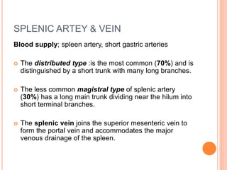
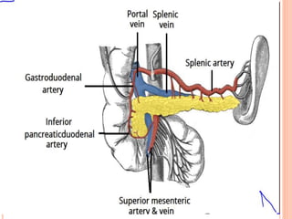
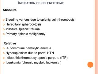
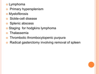
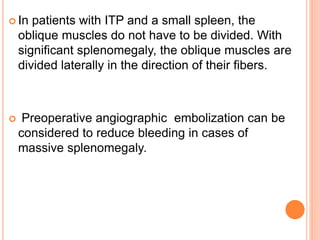
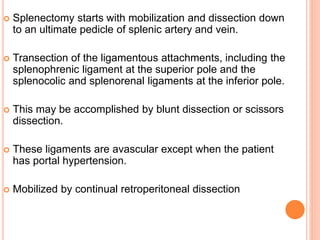
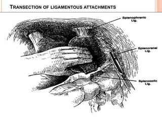
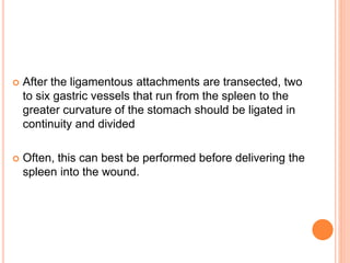
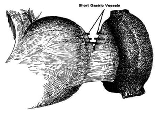
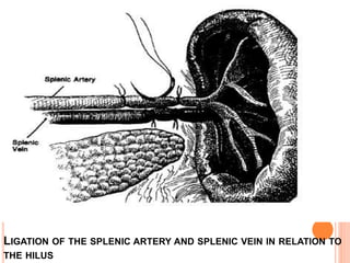
This document provides information on hypersplenism and its surgical management. It begins with definitions of hypersplenism and classifications of primary and secondary hypersplenism. The indications for splenectomy include bleeding varices, hereditary spherocytosis, massive splenic trauma, and others. The surgical technique involves mobilizing the spleen through ligation of attachments and dissection of the hilum before removing the spleen and ligating vessels. Precise ligation of the splenic artery and vein is important to safely remove an enlarged spleen.
1. The spleen develops from mesenchymal cells in the dorsal mesogastrium during the fifth week of gestation. It is located in the left upper quadrant of the abdomen and has multiple functions including filtration, host defense, storage, and cytopoiesis.
2. Common indications for splenectomy include trauma, hereditary spherocytosis, idiopathic thrombocytopenic purpura, and various blood disorders. Splenic injuries are graded based on their severity and treatment may involve observation, splenic repair or resection, or splenectomy depending on the grade and stability of the patient.
3. Splenic abscesses are rare but can develop due to hematogenous spread,
Bilirubin is formed from the breakdown of heme in hemoglobin. It is transported to the liver bound to albumin, where it enters hepatocytes. The liver conjugates bilirubin with glucuronic acid via the enzyme bilirubin UDP-glucuronosyltransferase to increase its solubility. The conjugated bilirubin is excreted in the bile or undergoes enterohepatic circulation and is eventually excreted in the urine. Certain drugs can displace bilirubin from albumin, allowing it to enter the brain and cause neural damage in infants due to their immature bilirubin metabolism.
Right iliac fossa mass is a common clinical presentation and has a range of differentials that need to be excluded.
In this presentation will discuss RIF masses in briefly.
contact me / dr.3shaq@gmail.com
“Love is like the human appendix. You take it for granted while it's there, but when it's suddenly gone you're forced to endure horrible pain that can only be alleviated through drugs.”
― Reverend Jen,
The document discusses carcinoma penis, including its epidemiology, risk factors, pathology, staging, investigations, and treatment options. Premalignant lesions like erythroplasia of Queyrat and balanitis xerotica obliterans are described. Treatment depends on the stage and includes circumcision for small tumors, local excision, glansectomy, Mohs micrographic surgery, and laser surgery to preserve the organ while wide local excision or penectomy may be needed for more advanced cases.
Primary retroperitoneal tumors are rare neoplasms that arise in the retroperitoneum and pelvis. Liposarcoma is the most common type of primary retroperitoneal tumor, while lymphoma is the most common retroperitoneal malignancy overall. These tumors often grow extensively before causing symptoms. Diagnostic imaging includes CT or MRI to evaluate the tumor characteristics and relationship to surrounding structures. Surgical resection with negative margins is the standard treatment for localized primary retroperitoneal sarcomas, while chemotherapy or radiation may be used in certain settings. Prognosis depends on tumor grade, stage, and ability to achieve a complete resection.
Extra Levator Abdomino Perineal Resection Dr Harsh Shah
This document discusses extralevator abdominoperineal excision (ELAPE) for rectal cancer. It describes how ELAPE aims to improve on conventional abdominoperineal resection (APR) by removing a larger volume of tissue, including the levator muscles, to obtain clear margins. Meta-analyses have found ELAPE results in lower rates of circumferential resection margin involvement, local recurrence, and involved nodes compared to APR. However, ELAPE also increases morbidity risks and requires perineal wound reconstruction. While ELAPE shows potential oncological benefits, more high-quality studies are still needed to prove its superiority over selective approaches.
LAPAROSCOPIC INGUINAL HERNIA REPAIR- OPERATIVE SURGERY
#surgicaleducator #operativesurgery #laparascopicinguinalherniarepair #usmle #babysurgeon #surgicaltutor
Dear viewers,
• Greetings from “Surgical Educator”
• Because of the popular demand by viewers of the YouTube channel “Surgical Educator”, I have decided to create and upload videos on common surgeries.
• I have already uploaded videos on open and Laparoscopic Appendicectomy, Thyroidectomy, Modified Radical Mastectomy and open and
Laparoscopic Cholecystectomy and Open inguinal hernia repair
• In this video today, I have discussed Laparoscopic Inguinal Hernia Repair- both TAPP and TEP approaches.
• However, these videos are not real surgeries but the theoretical aspect of operative surgery like going through an atlas of operative surgery.
• Along with these videos, I recommend you to watch real operative surgery videos as well and I will give a link for each surgery in the end of the video as end-cards, which I think will be very useful.
• This will give a very good opportunity for the surgical trainees to mentally rehearse various surgical steps in a sequential manner prior to actual surgery. You can watch the video in the following links:
• surgicaleducator.blogspot.com
• youtube.com/c/surgicaleducator
• Thank you for watching the videos.
The spleen is an organ located in the upper left abdomen. It filters blood and fights infections. A splenectomy is the surgical removal of the spleen. It is usually performed laparoscopically to avoid complications of open surgery. During the procedure, the surgeon uses cameras and surgical tools inserted through small incisions to carefully dissect and divide attachments of the spleen. This allows the spleen to be removed while preserving surrounding structures like the pancreas and stomach. A splenectomy may be recommended for conditions like immune thrombocytopenia or certain blood disorders.
1. Choledochal cysts are abnormal dilations of the bile ducts that are more common in Asia and women.
2. They are classified into 5 types based on location and extent of dilation.
3. Presentation varies from jaundice and abdominal mass in children to pain and cholangitis in older patients.
4. Investigation involves ultrasound, CT, MRCP and cholangiography to determine type and rule out complications.
5. Treatment is complete excision of the cysts and biliary tree with Roux-en-Y hepaticojejunostomy, except for type III which can be managed endoscopically.
Mesenteric cysts are rare fluid-filled masses that form between the two layers of the small bowel mesentery. They have an incidence of 1 in 100,000 hospital admissions, with about 1/3 diagnosed in patients under 15 years old. The most common type is the chylolymphatic cyst, which occurs due to congenitally misplaced lymphatic tissue. Ultrasound and CT scans are used to identify the fluid-filled cystic structures. Surgical treatment involves enucleation of chylolymphatic cysts or excision with intestinal resection. Prognosis is generally favorable with low recurrence rates following complete excision.
gastric resection, reconstruction and post gastrectomy syndromessanyal1981
discussion regarding history of gastrectomy, types of gastrectomy, billroth I, billroth II and roux en y gastrojejunostomy........discussion of post gastrectomy syndromes
This document describes various anal and perianal diseases. It discusses the surgical anatomy of the anal canal and lists common diseases such as hemorrhoids, anal fissures, fistulas, abscesses, and various cancers. It describes the pathology, clinical features, investigations, differential diagnoses, and treatments for each condition. A key point is Goodsall's rule, which states that fistulas with an external opening in the anterior anal region typically have a direct track, while posterior openings often curve with a midline internal opening.
The document discusses tumors of the small and large intestines. It classifies intestinal tumors and provides details on various benign and malignant tumor types. The most common tumors are epithelial tumors, with colorectal cancer representing 70% of all gastrointestinal malignancies. Adenomas are precursors to most colorectal cancers. Risk factors include inflammatory bowel disease, familial polyposis, and diet. Prognosis and treatment depend on tumor stage and characteristics.
Circulation of liver & Portosystemic collateralsPratap Tiwari
The document summarizes the circulation of the liver and portosystemic collateral veins. It discusses:
- The dual blood supply to the liver from the hepatic artery (25-30% of flow) and portal vein (70-75% of flow).
- The portal vein is formed by the superior mesenteric vein and splenic vein. It divides within the liver into right and left branches.
- Portosystemic collateral veins develop to bypass portal hypertension and include veins around the falciform ligament, umbilical veins, and abdominal wall veins.
- The presence of dilated umbilical or abdominal wall veins indicates high pressure within the left branch of the portal vein and
An abdominal mass can have various causes and require different treatments depending on the underlying condition. Examination of the patient and medical tests are needed to identify the location and cause of the mass. Common symptoms include abdominal pain, changes in appetite or bowel habits, weight changes, and the appearance of a mass. Serious symptoms may indicate life-threatening conditions like rapid mass growth or expansion accompanied by severe pain. Treatment options range from observation to surgery and may involve medications, drainage/removal of the mass, removal of part of an organ, or removal of the entire organ along with chemotherapy or radiation.
The document describes the anatomy and physiology of the biliary tree. It details the structures of the gallbladder, bile ducts, and their variations. Bile aids in digesting lipids and eliminating waste. The liver produces bile which is stored in the gallbladder and released in response to cholecystokinin after eating to help break down fats in the small intestine.
Colon and Rectum Surgical Anatomy and PhysiologyFaz Halim
The document summarizes the surgical anatomy and physiology of the colon and rectum. It discusses the arterial supply to different parts of the colon from the terminal ileum to the sigmoid colon. It also describes the venous supply and how infections can spread. The physiology section covers fermentation in the colon and absorption of water, sodium, potassium and short chain fatty acids. For the rectum, it outlines the arterial supply from branches of the internal pudendal and internal iliac arteries. Investigation methods like radiology, endoscopy, colonoscopy and CT scans are mentioned.
The spleen is responsible for filtering blood and mounting immune responses. Indications for splenectomy include trauma, idiopathic thrombocytopenic purpura refractory to steroids, and hematological conditions causing abnormal red blood cell morphology. Splenectomy may be performed open or laparoscopically and indications include trauma, hematological diseases, neoplasms, and spontaneous rupture. Complications include infection, bleeding, and thrombocytosis.
SURGICAL EXPLORATION OF THE COMMON BILE DUCT.pptxmasoom parwez
Surgical exploration of the common bile duct involves removing gallstones discovered during cholecystectomy. Key steps include:
1. Performing intraoperative cholangiography to identify stones
2. Making an incision in the bile duct and extracting stones using forceps, balloons, or baskets
3. Placing a T-tube for drainage and performing a follow up cholangiogram to ensure clearance
Post-operatively, patients are monitored for complications like bleeding or leakage and the T-tube is typically removed after 2 weeks if follow up imaging is normal. Surgical exploration effectively treats gallstones and provides pain relief for most patients.
This document discusses Vitello-intestinal duct anomalies including Meckel's diverticulum. Meckel's diverticulum is the most common congenital anomaly of the small intestine, occurring in about 2% of the population. It can cause hemorrhage, intestinal obstruction, or diverticulitis. Investigations like USG, CT, and 99m Tc scans can help detect Meckel's diverticulum. Treatment involves resection of symptomatic diverticula with adjacent ileum, while asymptomatic cases may be resected or left alone.
The document provides detailed anatomical information about the anal canal. It describes the length and divisions of the anal canal. It discusses the muscles including the internal and external sphincters. It covers the blood supply, lymphatic drainage and innervation of the anal canal. It also describes anal gland anatomy and various congenital anomalies that can occur in the anal region.
This document discusses the anatomy, physiology, diagnostic studies, diseases, and surgical procedures related to the gallbladder and extrahepatic biliary system. Key points include:
- The gallbladder is a pear-shaped sac located in the liver that stores and concentrates bile. Bile is produced by the liver and aids in fat digestion.
- Gallstones are a common problem, forming when bile becomes supersaturated, and can cause conditions like cholecystitis. Surgical interventions include cholecystectomy.
- Other issues discussed include bile duct injuries, tumors, and abnormalities like sclerosing cholangitis. A variety of imaging studies and endoscopic procedures are used for diagnosis and treatment.
This document discusses hypersplenism and its surgical management. It begins with an introduction to the spleen's history and the term "hypersplenism." It then covers the anatomy, histology, functions, and causes of hypersplenism. The main treatment approaches discussed are medical management, partial splenic embolization, and splenectomy, with details provided on the surgical techniques for open and laparoscopic splenectomy. Indications for splenectomy include bleeding varices, hereditary spherocytosis, trauma, malignancy, and various hematological disorders.
This document provides information about splenomegaly (enlarged spleen). It begins by discussing the spleen's anatomy and location. The spleen's functions include immune filtration and pitting of blood cells. Splenomegaly is defined based on spleen size or weight. Causes of splenomegaly include inflammatory, infectious, and infiltrative diseases that increase spleen demand or alter blood flow. Evaluation involves history, exam, labs, imaging and biopsies. Splenectomy is indicated for trauma, malignancy, or refractory diseases and can be open or laparoscopic. Complications include infection due to loss of immune function.
This document discusses anaesthesia considerations for EHPVO (extrahepatic portal venous obstruction) and meso-Rex shunt surgery. EHPVO is a non-cirrhotic cause of portal hypertension most common in children, while IPH (idiopathic portal hypertension) typically affects adults. Key differences are noted. Meso-Rex shunt restores hepatic blood flow more physiologically than non-physiological shunts. Anaesthesia must consider issues like malnutrition, anemia, ascites, and potential for bleeding or thrombosis. Careful monitoring is needed due to fluid shifts and potential liver or cardiac dysfunction.
This document provides an overview of splenomegaly, including the anatomy and functions of the spleen, causes of splenomegaly, examination techniques, classification of splenomegaly by size, potential symptoms, initial lab and imaging workup, and step-wise approach to evaluating a patient with splenomegaly. Common causes discussed include infections such as viral hepatitis, infiltrative diseases such as Gaucher's disease, malignancies such as lymphoma, and congestive states related to conditions like cirrhosis.
The document discusses several indications for splenectomy including immune thrombocytopenic purpura (ITP), hereditary spherocytosis, hemoglobinopathies, malignancy, splenic abscess, cysts, and vein thrombosis. For ITP, splenectomy is considered if thrombocytopenia is refractory to steroids, relapse occurs after treatment, or platelet levels remain low during pregnancy. It has a 65% success rate for improving thrombocytopenia. Hereditary spherocytosis and hemoglobinopathies can cause hemolytic anemia treated with splenectomy. Splenectomy may also be used for staging or treatment of certain lymphomas and metastases to the spleen. It is often recommended for
This document discusses several liver conditions including portal vein thrombosis (PVT), peliosis hepatis, and their associated features. It notes that PVT can be acute or chronic, and describes the clinical manifestations, imaging findings, and treatment approaches for each. Peliosis hepatis is characterized by multiple blood-filled cavities in the liver and has been associated with medications, transplantation, and certain infections. The pathogenesis of peliosis hepatis remains unknown.
Anatomy and physiology of spleen and laparoscopic management of splenic diso...Dr Sajad Nazir
This ppt. Is about the surgical anatomy, physiology,functions of spleen and laparoscopic management of splenic disorder.
Most of the anatomy is depicted pictorial and with suitable diagrams and is recommended for postgraduates only.
Most of Laparoscopic splenectomy has been made understand by diagrams only after consulting different standard surgical books.
The spleen is normally located in the left upper quadrant of the abdomen. This case presents a 20-year-old female with abdominal pain who was found to have a torsed wandering spleen at the center of her abdomen. Wandering spleen is a condition where the spleen lacks normal ligamentous support, causing it to be mobile within the abdomen. At surgery, her enlarged spleen was found to have torsed along its vascular pedicle, cutting off its blood supply. A splenectomy was performed to remove the non-viable spleen. Histopathology confirmed splenic infarction due to the torsion.
brief lecture notes for 5th sem MBBS, on portal hypertension and varices. Introduction to portal hypertension and esophageal and gastric varices and management of variceal bleeding.
Hyperviscosity syndrome (HVS) is a condition caused by an increase in the viscosity of blood, making it thicker and less able to flow easily. This can lead to organ dysfunction. HVS occurs when there is an abnormal increase in certain blood components like red blood cells, white blood cells, platelets or proteins. Common causes include polycythemia, leukemia, myeloma and other blood disorders. Patients may experience symptoms like bleeding, visual changes, neurological issues, respiratory problems and renal impairment. Diagnosis involves blood tests, imaging and assessing the underlying condition causing the increased viscosity. Treatment focuses on treating the underlying cause, stabilizing the patient, and procedures like plasmapheresis, hydration and phlebot
This document discusses various motility disorders and conditions that affect the esophagus. It begins by describing different types of motility disorders like achalasia and diverticula. It then focuses on achalasia, describing its pathogenesis, clinical presentation, diagnostic tests like esophagram and manometry, and various treatment methods. The document also discusses other topics like esophageal diverticula, benign and malignant neoplasms, perforations, injuries, and acid reflux conditions like Barrett's esophagus. Esophageal manometry is described as the most accurate way to assess motility disorders, and high-resolution manometry is mentioned as an improved technique.
The document provides information on the anatomy, microscopic anatomy, blood supply, nerve supply, and common conditions of the prostate gland such as benign prostatic hyperplasia. It discusses the procedure of transurethral resection of the prostate in detail, including preoperative considerations, choices of anesthesia, intraoperative monitoring, complications such as TURP syndrome, and their prevention and management. TURP syndrome is caused by excessive absorption of irrigating fluids and can lead to hyponatremia, hypervolemia, and other electrolyte abnormalities.
This document discusses splenic trauma, including its anatomy, presentation, management, and complications. The spleen lies in the left upper abdominal quadrant and is supplied by the splenic artery. Patients may present with left upper abdominal or shoulder pain following blunt trauma. Management depends on hemodynamic stability and injury grade, and may involve non-operative treatment or surgery such as splenorrhaphy or splenectomy. Complications include recurrent bleeding, infection, and thrombocytosis.
This document discusses portal hypertension and anesthetic concerns for lienorenal shunt surgery. It begins by defining portal hypertension as an increase in pressure gradient between the portal vein and hepatic veins/inferior vena cava. Common causes include increased resistance to hepatic blood flow from cirrhosis and increased splanchnic blood flow from splanchnic vasodilation. Major consequences include ascites, portosystemic shunts/varices, splenomegaly, and hepatic encephalopathy. Management of acute variceal bleeding and procedures like TIPS and surgery are discussed. Anesthetic considerations include aspiration prophylaxis, hemodynamic monitoring, and managing complications of variceal bleeding and procedures.
The document summarizes anatomy, functions, evaluation, indications for splenectomy, preoperative considerations, techniques for open and laparoscopic splenectomy, complications, and prevention of overwhelming post-splenectomy infection. Key points include that the spleen filters blood and fights infection, splenectomy is commonly performed for trauma or hematologic disorders, vaccinations and antibiotics can help prevent post-operative infection, and complications include bleeding, infection, and thrombosis.
This document provides information about hydrocephalus, including:
- It is a common pediatric disorder caused by an increase in CSF volume within the ventricles of the brain, putting pressure on the brain.
- Hydrocephalus can be obstructive or non-obstructive, depending on where the blockage of CSF flow occurs. Common causes include congenital abnormalities, infections, tumors, or hemorrhages.
- Clinical features in infants include an enlarged head circumference, bulging fontanelles, vomiting, and irritability. Older children may experience headaches, nausea, and vision problems.
- Treatment involves addressing the underlying cause if possible, or surgically placing a shunt to drain CSF out
With 14.4 million obese children in India, childhood obesity is a growing problem. Key risk factors include lack of physical activity due to increased screen time, overprotective parenting, excessive caloric intake from snacks and fast food, and aggressive marketing of unhealthy foods. Obese children often develop serious health conditions like diabetes, high blood pressure, and sleep apnea. Preventing childhood obesity requires efforts like educating parents, promoting community support, ensuring good nutrition early on, and limiting screen time in favor of physical activity. Countries have implemented strategies such as food labeling, marketing restrictions, and subsidizing healthy options. A comprehensive approach is needed to address this issue.
This document provides an overview of urinary tract infections (UTIs) in children from a surgeon's perspective. Some key points:
- UTIs are common in infants and children, especially girls under 5 years old. Boys are more commonly affected in the first year of life.
- Evaluation of a child with UTI includes a physical exam, urine culture, and consideration of imaging like ultrasound based on factors like age, symptoms, recurrence.
- Common causes of UTIs include anatomical abnormalities like vesicoureteral reflux, posterior urethral valves, or ureteroceles.
- Treatment involves antibiotics tailored to culture results. Children with recurrent UTIs or anatomical issues may
Primordial germ cells migrate during fetal development and can become arrested, resulting in extragonadal germ cell tumors like sacrococcygeal teratomas. Sacrococcygeal teratomas are the most common extragonadal germ cell tumors in neonates, occurring more frequently in females. They may be partially or completely external (Altman types I and II) or primarily internal with extension into the pelvis or abdomen (types III and IV). Complete surgical excision including coccygectomy is the primary treatment, with chemotherapy for malignant histology, and alpha-fetoprotein monitoring post-surgery to detect recurrence.
This document discusses testicular cancer, including:
- Risk factors include history of undescended testes, contralateral testicular tumor, or Klinefelter syndrome.
- Tumors are classified as germ cell tumors (most common), interstitial cell tumors, lymphoma, or other rare tumors.
- Seminoma and non-seminomatous germ cell tumors (NSGCT) are the main types of germ cell tumors.
- Diagnostic workup includes scrotal ultrasound, serum tumor markers, chest imaging and lymph node assessment to determine clinical stage according to the TNM system.
This document provides information on nuclear medicine and radiopharmaceuticals. It defines key terms like radionuclides, radiopharmaceuticals, and units of radiation measurement. It describes imaging modalities like scintigraphy, SPECT, PET and the equipment used. Various radiotracers are discussed along with their clinical uses for imaging organs like bone, thyroid, liver and brain. Positron emission and annihilation are also summarized.
1. Hirschsprung disease is a developmental disorder caused by the absence of ganglion cells in the myenteric and submucosal plexuses of the distal intestine.
2. It presents in neonates as intestinal obstruction due to functional obstruction at the level of aganglionosis.
3. Diagnosis involves clinical features like failure to pass meconium, imaging studies to identify transition zone, and rectal biopsy to show absence of ganglion cells.
4. Surgical treatment involves pull-through procedures like Swenson, Duhamel or Soave to resect aganglionic segment and join normal bowel above.
Resection & anastomosis of boweL its complications PRANAYA PPTPRANAYA PANIGRAHI
This document discusses intestinal resection and anastomosis. It defines anastomosis as establishing communication between two portions of intestine after removal of diseased bowel. Factors that influence healing, techniques for performing anastomoses (hand sewn vs. stapling), and common complications are described. Maintaining adequate blood supply, tension-free closure, and paying attention to technical details are emphasized for achieving successful anastomotic healing.
A 3-day-old female child presented with abdominal distension and inability to pass meconium. Examination revealed a normal anal opening but resistance to passage of a feeding tube beyond 2 cm. An invertogram showed rectal atresia. Endoscopy visualized a distal rectal membrane, which was incised. A fistulogram then revealed a fistula between the upper anal canal and the labia. Key points discussed include techniques for evaluating anorectal malformations like invertograms and classifications of ARM like the Wingspread and PENA systems. Relationship between sacral development and pelvic floor muscle function is also summarized.
This document discusses congenital anomalies of the esophagus. It begins by classifying anomalies and discussing their embryology. The most common anomaly is esophageal atresia with distal tracheoesophageal fistula, occurring in 84% of cases. Prenatal diagnosis and clinical findings are outlined. Treatment involves repairing the fistula and reconnecting the esophagus. Complications can include anastomotic leaks, strictures, and gastroesophageal reflux disease. The document provides details on surgical techniques, pre- and post-operative management of various anomaly subtypes.
Integrating Ayurveda into Parkinson’s Management: A Holistic ApproachAyurveda ForAll
Explore the benefits of combining Ayurveda with conventional Parkinson's treatments. Learn how a holistic approach can manage symptoms, enhance well-being, and balance body energies. Discover the steps to safely integrate Ayurvedic practices into your Parkinson’s care plan, including expert guidance on diet, herbal remedies, and lifestyle modifications.
Muktapishti is a traditional Ayurvedic preparation made from Shoditha Mukta (Purified Pearl), is believed to help regulate thyroid function and reduce symptoms of hyperthyroidism due to its cooling and balancing properties. Clinical evidence on its efficacy remains limited, necessitating further research to validate its therapeutic benefits.
Osteoporosis - Definition , Evaluation and Management .pdfJim Jacob Roy
Osteoporosis is an increasing cause of morbidity among the elderly.
In this document , a brief outline of osteoporosis is given , including the risk factors of osteoporosis fractures , the indications for testing bone mineral density and the management of osteoporosis
8 Surprising Reasons To Meditate 40 Minutes A Day That Can Change Your Life.pptxHolistified Wellness
We’re talking about Vedic Meditation, a form of meditation that has been around for at least 5,000 years. Back then, the people who lived in the Indus Valley, now known as India and Pakistan, practised meditation as a fundamental part of daily life. This knowledge that has given us yoga and Ayurveda, was known as Veda, hence the name Vedic. And though there are some written records, the practice has been passed down verbally from generation to generation.
share - Lions, tigers, AI and health misinformation, oh my!.pptxTina Purnat
• Pitfalls and pivots needed to use AI effectively in public health
• Evidence-based strategies to address health misinformation effectively
• Building trust with communities online and offline
• Equipping health professionals to address questions, concerns and health misinformation
• Assessing risk and mitigating harm from adverse health narratives in communities, health workforce and health system
Cell Therapy Expansion and Challenges in Autoimmune DiseaseHealth Advances
There is increasing confidence that cell therapies will soon play a role in the treatment of autoimmune disorders, but the extent of this impact remains to be seen. Early readouts on autologous CAR-Ts in lupus are encouraging, but manufacturing and cost limitations are likely to restrict access to highly refractory patients. Allogeneic CAR-Ts have the potential to broaden access to earlier lines of treatment due to their inherent cost benefits, however they will need to demonstrate comparable or improved efficacy to established modalities.
In addition to infrastructure and capacity constraints, CAR-Ts face a very different risk-benefit dynamic in autoimmune compared to oncology, highlighting the need for tolerable therapies with low adverse event risk. CAR-NK and Treg-based therapies are also being developed in certain autoimmune disorders and may demonstrate favorable safety profiles. Several novel non-cell therapies such as bispecific antibodies, nanobodies, and RNAi drugs, may also offer future alternative competitive solutions with variable value propositions.
Widespread adoption of cell therapies will not only require strong efficacy and safety data, but also adapted pricing and access strategies. At oncology-based price points, CAR-Ts are unlikely to achieve broad market access in autoimmune disorders, with eligible patient populations that are potentially orders of magnitude greater than the number of currently addressable cancer patients. Developers have made strides towards reducing cell therapy COGS while improving manufacturing efficiency, but payors will inevitably restrict access until more sustainable pricing is achieved.
Despite these headwinds, industry leaders and investors remain confident that cell therapies are poised to address significant unmet need in patients suffering from autoimmune disorders. However, the extent of this impact on the treatment landscape remains to be seen, as the industry rapidly approaches an inflection point.
ABDOMINAL TRAUMA in pediatrics part one.drhasanrajab
Abdominal trauma in pediatrics refers to injuries or damage to the abdominal organs in children. It can occur due to various causes such as falls, motor vehicle accidents, sports-related injuries, and physical abuse. Children are more vulnerable to abdominal trauma due to their unique anatomical and physiological characteristics. Signs and symptoms include abdominal pain, tenderness, distension, vomiting, and signs of shock. Diagnosis involves physical examination, imaging studies, and laboratory tests. Management depends on the severity and may involve conservative treatment or surgical intervention. Prevention is crucial in reducing the incidence of abdominal trauma in children.
Adhd Medication Shortage Uk - trinexpharmacy.comreignlana06
The UK is currently facing a Adhd Medication Shortage Uk, which has left many patients and their families grappling with uncertainty and frustration. ADHD, or Attention Deficit Hyperactivity Disorder, is a chronic condition that requires consistent medication to manage effectively. This shortage has highlighted the critical role these medications play in the daily lives of those affected by ADHD. Contact : +1 (747) 209 – 3649 E-mail : sales@trinexpharmacy.com
- Video recording of this lecture in English language: https://youtu.be/kqbnxVAZs-0
- Video recording of this lecture in Arabic language: https://youtu.be/SINlygW1Mpc
- Link to download the book free: https://nephrotube.blogspot.com/p/nephrotube-nephrology-books.html
- Link to NephroTube website: www.NephroTube.com
- Link to NephroTube social media accounts: https://nephrotube.blogspot.com/p/join-nephrotube-on-social-media.html
NVBDCP.pptx Nation vector borne disease control programSapna Thakur
NVBDCP was launched in 2003-2004 . Vector-Borne Disease: Disease that results from an infection transmitted to humans and other animals by blood-feeding arthropods, such as mosquitoes, ticks, and fleas. Examples of vector-borne diseases include Dengue fever, West Nile Virus, Lyme disease, and malaria.
2. INTRODUCTION
The spleen was regarded by Galen as “an organ of
mystery,”by Aristotle as unnecessary, and by Pliny
as an organ that might hinder the speed of runners
The term ‘hypersplenism’ first appeared in the
thesis of Anatole Chauffard in 1907 and
subsequently in the study of Morawitz and
Denecked
3. EMBRYOLOGY
Spleen is the largest reticuloendothelial organ in the
body
From the primitive mesoderm of dorsal mesogastrium
Evident in the fifth week of gestation in an 8 mm
embryo
The most common variation of splenic embryology is
the accessory spleen . Present in up to 14-29 % of
the population
4. ANATOMY
The spleen is located in the left upperquadrant.
Lies between the 9th -11th rib and weighs about
150(75-250g). Measures about13x 7 x 45cm in
dimention.
Attachement
Laterally- lienorenal ligament
Anteriorly-gastrosplenic ligament (contains short
gastric arteries)
Superiorly-splenophrenic ligament
Inferiorly-splenocolic ligament
5.
6. RELATIONS
Anterior-fundus of
the stomach
Medially- tail of
the pancreas
Inferiorly- splenic
flexure
Superiorly-
diaphragm
Posteriorly- upper
part of the kidney
7.
8.
9. SPLENIC ARTEY & VEIN
Blood supply; spleen artery, short gastric arteries
The distributed type :is the most common (70%) and is
distinguished by a short trunk with many long branches.
The less common magistral type of splenic artery
(30%) has a long main trunk dividing near the hilum into
short terminal branches.
The splenic vein joins the superior mesenteric vein to
form the portal vein and accommodates the major
venous drainage of the spleen.
10.
11. NORMAL PHYSIOLOGIC ROLES
Red pulp(90%)- Cords and sinuses Phagocytosis
White pulp- Periarticular lymphatic sheets
Immunoglobulins.
12. Reservior for platelets,monocytes,FVIII etc.
Haematopoiesis in fetus
Repairs and destruction of RBC’s by culling & pitting.
Immune function: IgM ,properidin,tuftsin are produced
by spleen.
prevention of infection By capsulated org.(H.influ
etc)role in phagocytosis.
13. APPROACH TO THE PATIENT
The most common symptoms produced by
diseases involving the spleen are pain and a
heavy sensation in the LUQ
14. Inspection may reveal fullness in the LUQ.
Palpation.
Percussion- Nixon, Castell, Percussion of Traube's
semilunar space.
Auscultation- reveal a venous hum or friction rub.
15. HYPERSPLENISM
DEFINITION-
Hypersplenism is a clinical syndrome characterized by:
SPLENOMEGALY, although this may be only moderate
PANCYTOPENIA or a reduction in the number of one or more
types of blood cells, neutropenia is less common than anemia
and thrombocytopenia
HYPERPLASIA of the precursor cells in the marrow or a so
called maturation arrest
Decreased RBCsurvival
Decreased platelet survival.
16. In Hypersplenism, its normal function
accelerates, and begins automatically to
remove cells that may still be normal in
function.
Sometimes, the spleen will temporarily
sequestrate 90% of the body platelets and
45% of the red cells.
17. Hypersplenism can be classified into three categories by its
etiological causes as follows.( Yunfu et al., 2016)
Primary hypersplenism
Cause is not clear.
1. Primary splenic hyperplasia
2. Non-tropical idiopathic splenomegaly
3. Primary splenic granulocytopenia
4. Primary splenic pancytopenia
5. Splenic anemia or thrombocytopenia
18. Secondary hypersplenism
Cause is clear
A. Infections - viral hepatitis, brucellosis, subacute or chronic
diseases, infectious mononucleosis syndrome and malaria.
B. Alcohol use such as long-term or excessive drinking
C. Portal hypertension (PH) such as liver cirrhosis of various
causes including Post-hepatitic Cirrhosis, Alcoholic
Cirrhosis, Biliary Cirrhosis, Fatty Liver Cirrhosis, Post-
hepatitic Autoimmune Cirrhosis, Schistosomiasis-induced
Cirrhosis, & Drug-induced Cirrhosis, as well as
Hemosiderosis And Portal Vein Thrombosis.
D. Granulomatous inflammation - Systemic Lupus
Erythematosus, Rheumatoid Arthritis, Chronic Syphilis,
Chronic Tuberculosis, Felty's Syndrome, & Sarcoidosis.
19. Malignancies - Splenic lymphosarcoma, leukemia, and
cancer metastasis.
Chronic hemolytic diseases such as hereditary
spherocytosis, autoimmune hemolytic anemia, and
thalassemia.
Lipidosis such as Gaucher's disease, and Niemann-Pick
disease.
Myeloproliferative disorders- Polycythemia Vera, Chronic
Myeloid Leukemia, Myelofibrosis
20. OCCULT HYPERSPLENISM
Sometimes due to benign bone marrow hyperplasia and
sufficient bone marrow compensation, peripheral
cytopenias may not occur.
In this case, hypersplenism becomes occult with no
symptoms.
However, once the bone marrow hematopoietic function is
suppressed by factors such as infection or drugs,
monolineage or multilineage peripheral cytopenia occurs,
accompanied by clinical symptoms, which is not classified
as occult hypersplenism.
22. ON CT OR POST-RESECTION WEIGHT
SPLENIC LENGTH (CM) SPLENIC WEIGHT
(GM)
Normal spleen Up to 13 <300
Mild splenomegaly >13–15 300–500
Moderate splenomegaly 16–20 500–1000
Massive splenomegaly >20 >1000
gm with etiological dia
gnosis
23. HACKETT’S GRADING SYSTEM FOR PALPABLE
SPLENOMEGALY
MILD-palpable <3cms below LCM
MODERATE-4-7 below LCM
SEVERE- >7cms below LCM
24. INVESTIGATION
Ultrasound -
The spleen is considered to be normal in size if its length is <13
cm or its thickness is ≤5 cm
CT Scanning-
In general, the spleen can be considered enlarged if its
craniocaudal length is more than 10cm on conventional
CT scans.
Spleen that extends below the lower third pole of the kidney
is also indicative of splenomegaly
25. LiverSpleen Colloid Scanning-
A splenic length of greater than 14 cm is considered enlarged on
liverspleen scan .
Erythrocytes are labeled with chromium51, mercury197 ,rubidium-
81
Splenectomy and Splenic Biopsy
26. LABORATORY STUDIES
Complete blood cell count (CBC) with differential.
Liver function testing
Hepatitis B and C testing
Lactate dehydrogenase (LDH)
Erythrocyte sediumentation rate (ESR)
Evaluation of peripheral blood smear for RBC morphology & sig
ns of myeloproliferative disorders underlying
bone marrow disorders
Prothrombin time (INR) and activated partial
thromboplastin time (aPTT)
28. Blood grouping and Cross matching
Platelets should not be administered preoperatively in
patient with ITP
In myeloproliferative disorders administer low-dose
heparin and aspirin on the day before surgery upto 5
days postoperatively.
Orogastric tube is used during the operation
Preoperative embolization(massive spleen)
Perioperative steroids are usually given if a patient has
had prolonged steroid treatment
29. INDICATION OF SPLENECTOMY
Absolute
Bleeding varices due to splenic vein thrombosis
Hereditary spherocytosis
Massive splenic trauma
Primary splenic malignancy
Relative
Autoimmune hemolytic anemia
Hypersplenism due to portal HTN
Idiopathic thrombocytopenic purpura (ITP)
Leukemia (chronic myeloid leukemia )
31. SURGICAL TECHNIQUE
Splenectomy related to blunt abdominal trauma, staging
of Hodgkin disease an upper midline incision to facilitate
dissection of the lower peri-aortic and iliac nodes.
In hematologic disorder, a left oblique subcostal incision
beginning to the right of the midline and proceeding
obliquely outward and downward approximately two
finger breadths below the costal margin give excellent
exposure.
32. In patients with ITP and a small spleen, the
oblique muscles do not have to be divided. With
significant splenomegaly, the oblique muscles are
divided laterally in the direction of their fibers.
Preoperative angiographic embolization can be
considered to reduce bleeding in cases of
massive splenomegaly.
33. Splenectomy starts with mobilization and dissection down
to an ultimate pedicle of splenic artery and vein.
Transection of the ligamentous attachments, including the
splenophrenic ligament at the superior pole and the
splenocolic and splenorenal ligaments at the inferior pole.
This may be accomplished by blunt dissection or scissors
dissection.
These ligaments are avascular except when the patient
has portal hypertension.
Mobilized by continual retroperitoneal dissection
35. After the ligamentous attachments are transected, two
to six gastric vessels that run from the spleen to the
greater curvature of the stomach should be ligated in
continuity and divided
Often, this can best be performed before delivering the
spleen into the wound.
36.
37. Spleen mobilized and elevated into the wound following division of
ligament attachments and posterior dissection
38. After these maneuvers are completed, the spleen can be
delivered into the wound by blunt dissection of the posterior
attachments.
Care should be taken not to divide the posterior
attachments too far medially to avoid entering the splenic
vein.
One should also avoid axial rotation of the spleen because
this may lead to disruption of the splenic artery or vein.
39. Dissection is carried out at the hilus as close to the spleen
as possible to avoid injury to the pancreas.
Individual ligation of the splenic artery or arterial branches
and the splenic vein or venous branches is generally
preferable.
40. Splenic artery ligation is managed by double ligation and
suture ligature, whereas the splenic vein can be doubly
ligated and divided.
In the case of a markedly enlarged spleen, occasionally one
must place a vascular clamp on the splenic vein and close
the lumen with continuous vascular suture.
41. LIGATION OF THE SPLENIC ARTERY AND SPLENIC VEIN IN RELATION TO
THE HILUS
42. Three major areas to be inspected for bleeding:-
(a) the inferior surface of the diaphragm.
(b) the greater curvature of the stomach and the region of
the short gastric vessels.
(c) the region of the hilus.
(d)short gastric vessels that have been divided.
43. An integral part of splenectomy for
hematologic disease is a thorough
exploration to detect any accessory
spleens.
44. PREOPERATIVE SPLENIC ARTERY EMBOLIZATION
(SPIGOS ET AL, 1979, )
Applied in the treatment of PH, hypersplenism, and
bleeding esophagogastric varices.
Increases platelet and leukocyte counts.
Reduces splenic size, improves pancytopenia, and
stimulates the immune system.
RISKS OF SPLENIC ARTERY EMBOLIZATION (SAE)
Post-embolization syndrome: pain, fever, ileus, pleural
effusion
Pancreatitis
Splenic abscess or rupture
Peritonitis
45. SAE can be used preoperative intervention to
reduce vascularity and size of massive spleen in
preparation for a laparoscopic approach.
Embolization is achieved using microcoils and/ or
Gelfoam.
46. LAPAROSCOPIC SPLENECTOMY
Laparoscopic techniques have improved and most
patients today are considered for elective
laparoscopic splenectomy.
The complicating factors are a large spleen (>500
g), suspected perisplenitis (most common in
patients with previous infectious diseases of the
spleen or portal hypertension) and previous gastric
surgery.
47. ITP patients and staging laparotomy is suited ideally for
laparoscopic approaches as well.
Position- right side down
Ports-
1. midline and 4 cm below the spleen tip,
2. near the tip of the 11th rib along the posterior axillary line, and a
third is a
3. half way between the other two, along the anterior axillary line.
Occasionally, a fourth port may be required.
scissors with cautery or preferably the harmonic Scalpel can be
used to take down the lateral peritoneal attachments and can be
used to ligate short gastric vessels.
ligate and divide the short gastric vessels then ligate the splenic
artery and vein.
48.
49. Specimen delivery - morselization of the spleen in a bag or port
site can be enlarged to facilitate removal
If the spleen is too large, a small Pfannenstiel incision and
removing the spleen through a suprapubic area may be more
cosmetically satisfactory.
Laparoscopic splenectomy in children has increased with
frequency and can be done for hematologic disease, hereditary
spherocytosis.
Accessory spleens can be localized and removed
laparoscopically by following the pattern of the most common
location.
50. HAND-ASSISTED SPLENECTOMY
Hand-assisted laparoscopic surgery (HALS ---
As an alternative to the LS approach with same positioning
Spleen greater than 22 cm in craniocaudal length or 19 cm in
width may benefit.
Merit
Marked reduction in average operative time.
This technique allows for a tactile feedback and atraumatic
manipulation of the enlarged spleen.
Demerit
Require a small incision (7–8 cm) for hand insertion and
specimen extraction.
51. SINGLE-INCISION LAPAROSCOPIC SURGERY
(SILS)
One small transabdominal incision
Theoretical benefits of less pain and better cosmetics.
incision -periumbilical and is used as the specimen
extraction site.
Technical challenging for solid organs- since all
instruments are closely aligned together.
Limited degrees of movement
52. ROBOTIC SPLENECTOMY
Unique three-dimensional visualization of the surgical field.
Facilitates movement with higher precision than standard
laparoscopy.
Robotic splenectomy is very similar to standard laparoscopy,
although not as cost effective.
No clear benefit of robotic versus laparoscopic splenectomy.
53. Splenoptosis (wandering spleen) refers to a rare
condition in which the spleen hangs by a long pedicle
from the mesentery and may present itself as an
asymptomatic mass or with symptoms of intermittent or
acute abdominal pain due to torsion.
Treatment involves splenectomy in cases of ischemia but
splenopexy should be considered in other cases
54. POST OPERATIVE MANAGEMENT
Remove NG tube and the suction drain when
drainage is minimal (usually 24 - 48hours)
Commence oral when bowel activity resumes.
Long term oral penicillin 250mg daily.
Pneumococcal vaccine 2weeks post op.
Anti-malaria prophylaxis.
55. COMPLICATIONS
Early
Acute gastric dilatation
Fundal ischemia- hematemesis, perforation
Pancreatic fistula
Portal vein thrombosis
Reactionary hemorrhage from splenic vessel
The most common site of bleeding is the short gastric
vessels - 4% to 16% of patients
Late
Infection; pneumococcal, viral, OPSI thrombocytosis
56. OVERWHELMING POSTSPLENECTOMY
INFECTION (OPSI)
Post Splenectomy leads to reduced IgM, tuftin, properdin
and other antibodies, phagocytosis of encapsulated bacteria
is defective.
Post-splenectomised patient is more prone for
Pneumococcal septicaemia (commonest), N. meningitides,
H. infl uenzae, Babesia microti infections.
Incidence is 4%.
Common in first two years after splenectomy.
57. Clinical Features-
Prodromal phase—fever, chills, sore throat.
Hypotension, shock.
DIC.
Respiratory distress, coma, death.
Mortality for fully developed OPSI—50-70%.
Prevention
Pneumococcal vaccine should be given to all splenectomised
patients.
Polyvalent pneumo-vac is given 2-3 weeks prior to surgery and
soon after recovery from surgery and it is repeated once in 5
years (Given to patients older than 2 years).
Other vaccines - meningococcal vaccine (only to those who
travel with high-risk), H. influenzae ‘B’ vaccine (to all whatever
the age, once in 10 years).
In malaria endemic areas, anti-malarial prophylaxis is given for
patients after splenectomy.
58. Treatment of OPSI
Antibiotics like Cefoperazone, Ceftazidime, Amikacin
Ventilatory support—ICU care.
Blood transfusion.
Immunoglobulin transfusion.
Nutrition (TPN) and maintaining of urine output.
59. PREVENTION OF OPSI
Life long prophylaxis using benzathine penicillin 12-24 lac
units—controversial in adults.
Pneumococcal vaccine given 2-3 weeks prior to
splenectomy— 70% protection.
H. influenzae-B type vaccine.
Meningococcal vaccine is given only to high-risk groups,
as its effects are short term. So it is not routinely given
60. CONCLUSION
Management and treatment should therefore be
administered taking into account the specific etiology and be
individualized for each patient.
Available treatment options include non-surgical and
surgical methods.
Surgical outcome following splenectomy is usually
satisfactory
Continuous basic and clinical studies will advance our
understanding of the underlying mechanisms of the
development of hypersplenism, and provide better
management strategies for the treatment of patients with
hypersplenism.