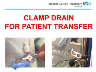
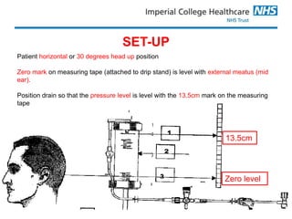
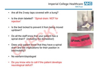
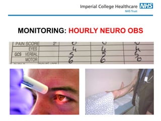
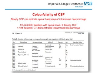
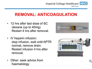
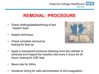
SPINAL DRAINS
POST-OPERATIVE MANAGEMENT
IN RECOVERY/ITU/HDU
Rachael Lear
Advanced Vascular Nurse Practitioner
CLAMP DRAIN
FOR PATIENT TRANSFER
13.5cm
Zero level
SET-UP
Patient horizontal or 30 degrees head up position
Zero mark on measuring tape (attached to drip stand) is level with external meatus (mid
ear).
Position drain so that the pressure level is level with the 13.5cm mark on the measuring
tape
13.5cm
Zero level
Why is the set up important?
Normal CSF pressure is 10mmHg
13.5 cmH20 = 10mmHg
If the drain is kept at 13.5cm above the spinal cord, CSF will drain into the
chamber if the pressure rises above normal.
The Golden Rule
DO NOT MOVE THE PATIENT OR THE
CHAMBER WITHOUT CLAMPING THE DRAIN
Chamber too
high
Reduced CSF
drainage
Increased
CSF pressure
Paraplegia
The Golden Rule
DO NOT MOVE THE PATIENT OR THE
CHAMBER WITHOUT CLAMPING THE DRAIN
Chamber too low
Excess of CSF drainage
Reduced intracranial
pressure
‘Coning’ of
brain/intracranial bleeding
Subdural
haematoma
caused by tearing
of the dural
bridging vein
attributed to
excessive CSF
drainage
Murakami et al. 2004
• Are all the 3-way taps covered with a bung?
• Is the drain labeled? “Spinal drain: NOT for
injection”
• Is the bed locked to prevent it from being moved
up/down?
• Do all the staff know that your patient has a
spinal drain? (including the domestics!)
• Does your patient know that they have a spinal
drain and the implications for their position in
bed/mobility?
• No warfarin/clopidogrel
• Do you know who to call if the patient develops
neurological deficit?
MONITORING: PATIENT PARAMETERS
MAP >85-90 mmHg (inotropes)
Haemoglobin >10 g/dl
PO2 7.3 – 10.6 kPa
O2 saturation >95%
Spinal cord
perfusion
pressure
=
MAP - CSFP
MONITORING: HOURLY NEURO OBS
MONITORING: VOLUME & COLOUR OF CSF
• Measure & record the volume & colour CSF drained hourly.
• If >10mls/hour or 5-10mls for 2 consecutive hours, inform the
vascular registrar immediately.
• If CSF has not drained for a period of two consecutive
hours, nursing staff should check patency of spinal catheter.
TO CHECK CATHETER PATENCY
STRICT ASEPTIC TECHNIQUE
TO CHECK CATHETER PATENCY
Aspirate
0.5mls max
Clamp
closed to
chamber
Three-way
tap open to
the patient
Colour/clarity of CSF
Bloody CSF can indicate spinal haematoma/ intracranial haemorrhage
5% (24/486) patients with spinal drain  bloody CSF
17/24 patients: CT demonstrated intracranial haemorrhage
• ITU: sedation hold every morning to check neuro status & motor
function
• Insertion site check every 12 hours for evidence of infection/CSF
leak
• Drain may be clamped briefly (<5 minutes) for care activities
• Prophylactic LMWH can be given daily
• NO INTRATHECAL INJECTIONS via spinal drain
• CPAP increases CSF pressure & may lead to paraplegia:
commencing CPAP is a consultant decision
MANAGEMENT: other important points
REMOVAL: ANTICOAGULATION
• 12 hrs after last dose of SC
clexane (up to 40mg)
Restart 4 hrs after removal.
• IV heparin infusion:
stop infusion, wait until APTR
normal, remove drain.
Restart infusion 4 hrs after
removal.
• Other: seek advice from
haematology
REMOVAL: PROCEDURE
• Check clotting/platelets/timing of last
heparin dose
• Aseptic technique
• Check complete removal by
looking for blue tip
• Apply a transparent occlusive dressing once the catheter is
removed and Inspect the insertion site every 2 hours for 24
hours, looking for CSF leak
• Neuro obs for 24hrs
• Handover timing for safe administration of anti-coagulation
Spinal drain post-op management 2

Spinal drain post-op management 2

  • 1.
    SPINAL DRAINS POST-OPERATIVE MANAGEMENT INRECOVERY/ITU/HDU Rachael Lear Advanced Vascular Nurse Practitioner
  • 2.
  • 3.
    13.5cm Zero level SET-UP Patient horizontalor 30 degrees head up position Zero mark on measuring tape (attached to drip stand) is level with external meatus (mid ear). Position drain so that the pressure level is level with the 13.5cm mark on the measuring tape
  • 4.
    13.5cm Zero level Why isthe set up important? Normal CSF pressure is 10mmHg 13.5 cmH20 = 10mmHg If the drain is kept at 13.5cm above the spinal cord, CSF will drain into the chamber if the pressure rises above normal.
  • 5.
    The Golden Rule DONOT MOVE THE PATIENT OR THE CHAMBER WITHOUT CLAMPING THE DRAIN Chamber too high Reduced CSF drainage Increased CSF pressure Paraplegia
  • 6.
    The Golden Rule DONOT MOVE THE PATIENT OR THE CHAMBER WITHOUT CLAMPING THE DRAIN Chamber too low Excess of CSF drainage Reduced intracranial pressure ‘Coning’ of brain/intracranial bleeding Subdural haematoma caused by tearing of the dural bridging vein attributed to excessive CSF drainage Murakami et al. 2004
  • 8.
    • Are allthe 3-way taps covered with a bung? • Is the drain labeled? “Spinal drain: NOT for injection” • Is the bed locked to prevent it from being moved up/down? • Do all the staff know that your patient has a spinal drain? (including the domestics!) • Does your patient know that they have a spinal drain and the implications for their position in bed/mobility? • No warfarin/clopidogrel • Do you know who to call if the patient develops neurological deficit?
  • 9.
    MONITORING: PATIENT PARAMETERS MAP>85-90 mmHg (inotropes) Haemoglobin >10 g/dl PO2 7.3 – 10.6 kPa O2 saturation >95% Spinal cord perfusion pressure = MAP - CSFP
  • 10.
  • 11.
    MONITORING: VOLUME &COLOUR OF CSF • Measure & record the volume & colour CSF drained hourly. • If >10mls/hour or 5-10mls for 2 consecutive hours, inform the vascular registrar immediately. • If CSF has not drained for a period of two consecutive hours, nursing staff should check patency of spinal catheter.
  • 12.
    TO CHECK CATHETERPATENCY STRICT ASEPTIC TECHNIQUE
  • 13.
    TO CHECK CATHETERPATENCY Aspirate 0.5mls max Clamp closed to chamber Three-way tap open to the patient
  • 14.
    Colour/clarity of CSF BloodyCSF can indicate spinal haematoma/ intracranial haemorrhage 5% (24/486) patients with spinal drain  bloody CSF 17/24 patients: CT demonstrated intracranial haemorrhage
  • 15.
    • ITU: sedationhold every morning to check neuro status & motor function • Insertion site check every 12 hours for evidence of infection/CSF leak • Drain may be clamped briefly (<5 minutes) for care activities • Prophylactic LMWH can be given daily • NO INTRATHECAL INJECTIONS via spinal drain • CPAP increases CSF pressure & may lead to paraplegia: commencing CPAP is a consultant decision MANAGEMENT: other important points
  • 16.
    REMOVAL: ANTICOAGULATION • 12hrs after last dose of SC clexane (up to 40mg) Restart 4 hrs after removal. • IV heparin infusion: stop infusion, wait until APTR normal, remove drain. Restart infusion 4 hrs after removal. • Other: seek advice from haematology
  • 17.
    REMOVAL: PROCEDURE • Checkclotting/platelets/timing of last heparin dose • Aseptic technique • Check complete removal by looking for blue tip • Apply a transparent occlusive dressing once the catheter is removed and Inspect the insertion site every 2 hours for 24 hours, looking for CSF leak • Neuro obs for 24hrs • Handover timing for safe administration of anti-coagulation

Editor's Notes

  • #8 So it is essential that you get the set up right. This is what it looks like in reality. O at the level of the mid ear. Pressure level at 13.5cm.
  • #19 I