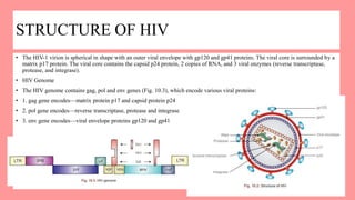
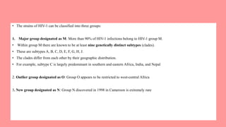
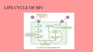
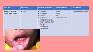
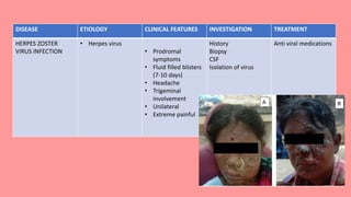
HUMAN IMMUNODEFICIENCY
VIRUS INFECTION / ACQUIRED
IMMUNE DEFICIENCY
SYNDROME
DR PRIYANKA
ORAL MEDICINE AND RADIOLOGY
INTRODUCTION
โ€ข Infection with the RNA retroviruses known as human immunodeficiency viruses (HIV)
โ€ข HIV infection which eventually damages T lymphocytes, especially those that are CD4+ , thus causing HIV disease and ultimately
the acquired immune deficiency syndrome (AIDS).
โ€ข In healthy persons aged 5years and older, with normally functioning immune systems, the CD4+ T-cell counts usually range from
500โ€“ 1500 cells/mL.
โ€ข CDC, in collaboration with Council of State and Territorial Epidemiologists (CSTE), has expanded the AIDS surveillance case
definition to include all HIV-infected persons with CD4+ T cell counts of less than 200 cells/ฮผl
โ€ข These immunocytes are crucial to host defences against fungi, viruses, mycobacteria and parasites
EPIDEMIOLOGY
โ€ข The total number of people living with the human immunodeficiency virus (HIV) reached its highest level: an estimated 40.3
million people are now living with HIV. Close to 5 (4.9) million people were newly infected with the virus in 2005
โ€ข Approximately 35.2 million persons were living with HIV at the end of 2012, with an estimated 0.8% of reproductive age adults
globally living
โ€ข Approximately 33 million persons are living with HIV at end of 2022
โ€ข Approximately 2 millions persons are dying per year
โ€ข Approximately 2.5 millions are getting infections every year
โ€ข 70% are from Africa, 20 % are from Asia and 10% rest
HISTORICAL BACKGROUND
โ€ข Dr. Luc Montagnier (1983) of Pasteur Institute, Paris, first identified and named it Lymphadenopathy Associated Virus.
โ€ข Dr. Roberto Gallo identified it later at NIH Bethesda, Maryland, U.S.A in 1984 and named it as Human T cell Lymphotropic Virus.
STRUCTURE OF HIV
โ€ข The HIV-1 virion is spherical in shape with an outer viral envelope with gp120 and gp41 proteins. The viral core is surrounded by a
matrix p17 protein. The viral core contains the capsid p24 protein, 2 copies of RNA, and 3 viral enzymes (reverse transcriptase,
protease, and integrase).
โ€ข HIV Genome
โ€ข The HIV genome contains gag, pol and env genes (Fig. 10.3), which encode various viral proteins:
โ€ข 1. gag gene encodesโ€”matrix protein p17 and capsid protein p24
โ€ข 2. pol gene encodesโ€”reverse transcriptase, protease and integrase
โ€ข 3. env gene encodesโ€”viral envelope proteins gp120 and gp41
Classification of HIV
โ€ข HIV is a highly variable virus, which mutates very readily, which means there are many different strains of HIV, even within the
body of a single infected person.
โ€ข Based on genetic similarities, the numerous virus strains may be classified into types, groups, and subtypes.
โ€ข There are 2 genetically different types of HIV: HIV-1 and HIV-2.
โ€ข Both types are transmitted by sexual contact, through blood, and from mother to child, and they cause AIDS.
โ€ข However, it seems that HIV-2 is less easily transmitted, and the period between initial infection and illness is longer in the case of
HIV-2.
โ€ข The relatively uncommon HIV-2 type is mostly confined to West Africa and is rarely found elsewhere.
โ€ข Worldwide, the predominant virus is HIV-1, and in general when people refer to HIV without specifying the type of virus they
will be referring to HIV-1.
โ€ข The strains of HIV-1 can be classified into three groups:
1. Major group designated as M: More than 90% of HIV-1 infections belong to HIV-1 group M.
โ€ข Within group M there are known to be at least nine genetically distinct subtypes (clades).
โ€ข These are subtypes A, B, C, D, E, F, G, H, J.
โ€ข The clades differ from each other by their geographic distribution.
โ€ข For example, subtype C is largely predominant in southern and eastern Africa, India, and Nepal
2. Outlier group designated as O: Group O appears to be restricted to west-central Africa
3. New group designated as N: Group N discovered in 1998 in Cameroon is extremely rare
Transmission of HIV
1. Sexual (homosexuals/heterosexuals)
2. Parenteral (intravenous drug abusers/ hemophiliacs)
3. Mother to infant (vertical transmission)
โ€ข a. Transplacental
โ€ข b. During delivery
โ€ข c. HIV infected milk
HIV Is Not Transmitted Through
1. Saliva
2. Kissing
3. Mosquito bite
4. Touching
5. Sharing toilets
โ€ข Saliva:
Saliva contains a protein called โ€œsecretary leukocyte proteinase inhibitorโ€ which inactivates the HIV.
Antibodies (IgA) are present in the saliva against HIV.
โ€ข Mosquitoes:
the virus is present at very low levels in the blood of HIV positive individuals.
In order for a mosquito to transmit HIV from an infected person to an uninfected, host:
โ€ข 1. The levels of HIV in the circulating blood should be high
โ€ข 2. They should survive in the mosquito
LIFE CYCLE OF HIV
Course of HIV Infection
โ€ข Early โ€œAcuteโ€ Phase
โ€ข The early โ€œacuteโ€ phase represents the initial response of the immune system to HIV infection.
โ€ข Clinically, this self-limiting illness develops in 50 to 70% of adults 3 to 6 weeks after infection and is characterized by flu-like
symptoms, which include fever, malaise, generalized lymphadenopathy, pharyngitis, headache, myalgia, arthralgia, skin
rash, and sometimes aseptic meningitis.
โ€ข During this phase, HIV initially infects CD4+ T cells and macrophages directly or is carried to regional lymph nodes by
Langerhans cells where these cells are present.
โ€ข Viral replication in the regional lymph nodes leads to viremia (high levels of viral particles in the blood) and widespread seeding
of HIV in the lymphoid tissues (lymph nodes, spleen, and tonsils) resulting in reduction of CD4+ T cell count.
โ€ข The viremia is controlled by the development of antiHIV antibodies (usually within 3 to 12 weeks). As viremia dies away,
CD4+ T cell count returns to normal number.
โ€ข Middle โ€œChronicโ€ Phase
โ€ข The immune system is stable.
โ€ข Patients are usually asymptomatic or develop persistent lymphadenopathy, and many patients have minor opportunistic
infections such as oral candidosis or shingles.
โ€ข The virus replicates in the CD4+ T cells and macrophages present in the lymphoid tissues for several years and gradually
destroy the CD4+ T cells.
โ€ข However, when the CD4+ T cells that are destroyed cannot be replenished, the CD4+ T cell count decreases and the patient enters
the final crisis phase.
โ€ข The macrophages are not destroyed by the virus replication and they transport the virus to various tissues, particularly the brain.
โ€ข Final โ€œCrisisโ€ Phase
โ€ข This phase is characterized by the appearance of opportunistic infections and specific conditions as outlined by CDC.
โ€ข Approximately 90% of patients with untreated AIDS die from opportunistic infections.
โ€ข Patients with AIDS often have generalized lymphadenopathy, severe weight loss, fatigue, chronic diarrhea, chronic fever, and
night sweats.
โ€ข Laboratory tests reveal increased viral load, altered CD4/CD8 ratio, and decrease in CD4+ T cell count.
โ€ข The number of CD4+ T cells decrease from a normal range of 1500 to 200 cells/mm3.
โ€ข The normal CD4+ T cell/CD8+ T cell ratio is about 2.
โ€ข In AIDS patients, it is < 0. 5 and therefore the ratio is an important measure of the disease progression.
โ€ข Thus, as the disease progresses the number of CD4+ T cells decrease and CD8+ T cells increase.
โ€ข The marked reduction of CD4+ T cells explains the lack of immune response and is most likely related to the increase in malignant
disease associated with AIDS.
Causes for Decreased CD4+T-lymphocyte Count
1. Because of HIV infection.
2. Killing of infected CD4+ T cells by CD8+ T cells
3. Formation of syncytia (giant cells) by fusion of infected CD4+ T cells and uninfected CD4+ T cells
4. Uninfected CD4+ T cell may bind to free gp120 resulting in apoptosis
5. Depletion of CD4+ T cell precursor cells, either by direct infection of thymus cells or by infection of cells that secrete cytokines
essential for CD4+ T cell differentiation
Consequences of CD4+ T-cell (โ€œMaster Cellโ€)
Destruction
1. Decreased antigen presentation by macrophages
2. Decreased antibody production by plasma cells in response to antigen
3. Decreased killing of tumor cells by Natural Killer (NK) cells
4. Decreased CD8+ T cell cytotoxicity
5. Decreased response to antigen and decreased cytokine production by CD4+ T cells
ORAL MANIFESTATIONS
โ€ข WHO collaborating center and EC clearing house (London) revised the classification of oral lesions associated with HIV infection
on September 1992 which was reported by Williams DM (1993).
โ€ข Group I: Lesions strongly associated with HIV infection
โ€ข Group II: Lesions commonly associated with HIV infection
โ€ข Group III: Lesions uncommonly associated with HIV infection
Group I: Lesions Strongly Associated with HIV Infection
โ€ข Candidiasisโ€”Erythematous, Pseudomembranous
โ€ข Hairy leukoplakia
โ€ข Kaposiโ€™s sarcoma
โ€ข Non-Hodgkinโ€™s lymphoma
โ€ข Periodontal diseasesโ€”Linear Gingival Erythema, Necrotizing Gingivitis/Periodontitis
DISEASE ETIOLOGY CLINICAL FEATURE INVESTIGATION TREATMENT
CANDIDIASIS โ€ข candida
โ€ข hyposalivation
โ€ข smoking
โ€ข broad-spectrum
antimicrobials
โ€ข Corticosteroids
โ€ข Malnutrition
โ€ข age
โ€ข Middle age
โ€ข Tongue and palate
โ€ข Curdy white
appearance
โ€ข Scrapable
Cytosmear
PCR
Antifungal
HAIRY TONGUE โ€ข Smoking
โ€ข Soft diet
โ€ข Hyposalivation
โ€ข Antimicrobial
โ€ข EBV
โ€ข Posterior tongue
โ€ข Vary color from yellow
to black
โ€ข Affect filiform
papillae(elongated)
cytosmear โ€ข increasing their oral hygiene
โ€ข using a tongue scraper
โ€ข brushing the tongue before
retiring at night, with a hard
toothbrush and cold water
โ€ข using sodium bicarbonate
mouthwashes
DISEASE ETIOLOGY CLINICAL FEATURES INVESTIGATION TREATMENT
KAPOSI SARCOMA HPV โ€ข asymptomatic red,
blue or purple
macules
โ€ข progresses to
papules, nodules or
ulcers and may
become painful
โ€ข is most common
around the face
(especially on the
nose) and mouth
โ€ข occurs especially at
the hard/soft
palate junction or
the anterior
maxillary gingivae
Biopsy intralesional injections
of vinblastine, or
systemic
chemotherapy
DISEASE ETIOLOGY CLINICAL FEATURES INVESTIGATION TREATMENT
NON HODGKINโ€™S
LYMPHOMA
โ€ข EBV
โ€ข HTLV-1
โ€ข HCV
โ€ข KSHV
โ€ข PESTICIDES,
SMOKING
โ€ข Old age
โ€ข M>F
โ€ข Painless
enlargement of
one or more
lymph node
โ€ข Unexplained
weight loss
โ€ข Fever
โ€ข Severe itching
Biopsy
PET/CT
Radiaiotherapy
Chemotherapy
Immunotherapy
Linear gingival
erythema
candida โ€ข erythematous
distinctive linear
band (1-3 mm
wide) at the free
gingival margin
โ€ข gentle probing
produces bleeding
CBC โ€ข intensive daily oral
hygiene,
โ€ข oral rinsing with
chlorhexidine
gluconate 0.20%
solution,
โ€ข scaling and root
planing (if
necessary) with 10%
betadine solution
irrigation, and
โ€ข careful follow-up
and maintenance
`wwwww
Nectrotizing
Ulcerative
Periodontitis (NUP)
Microbes
Poor oral hygiene
Smoking
Malnutrition
pain, generalized
gingival erythema
bleeding,
interproximal gingival
necrosis,
Punched out crater,
rapidly progressive
destruction of the
periodontal
attachment and bone
Psuedomembranous
slough
Ultrasonic scaling โ€“
supragingival
Metronidazole
250mg
Amoxycillin 500mg
Group II: Lesions Commonly Associated with HIV Infection
โ€ข Bacterial infectionsโ€”Mycobacterium avium, Mycobacterium tuberculosis
โ€ข Melanotic hyperpigmentation
โ€ข Nectrotizing ulcerative stomatitis
โ€ข Salivary gland diseaseโ€”Dry mouth (decreased salivary flow) or unilateral and bilateral swelling of the salivary glands
โ€ข Thrombocytopenic purpura
โ€ข Ulceration ( not otherwise specified)
โ€ข Viral infectionsโ€”Herpes simplex virus, Human papilloma virus, varicella zoster virus (herpes zoster- varicella)
DISEASE ETIOLOGY CLINICAL FEATURES INVESTIGATION TREATMENT
Tuberculosis โ€ข Mycobacterium
tuberculosis
โ€ข Current cough
โ€ข weight loss
โ€ข Fever
โ€ข Night sweats
โ€ข Tongue
โ€ข Undermined edge
โ€ข Irregular punched
out
โ€ข painful
History
Biopsy
Chest CT scan
Sputum examination
Tuberculin skin test
Anti tubercular
medications
DISEASE ETIOLOGY CLINICAL FEATURES INVESTIGATION
MELANOTIC
HYPERPIGMENTATION
direct result of HIV
infection
medication
โ€ข Brown macule
โ€ข Non tender
โ€ข Non scrapable
โ€ข Irregular shape
โ€ข Buccal mucosa
โ€ข tongue
Biopsy
DISEASE ETIOLOGY CLINICAL FEATURES INVESTIGATION
Salivary gland disease Bacteria
Viral
HIV
โ€ข Painful
โ€ข Enlargement of
parotid gland
โ€ข Not movable
โ€ข Xerostomia
โ€ข Sicca symptoms
History
CT scan
FNAC
DISEASE ETIOLOGY CLINICAL FEATURES INVESTIGATION TREATMENT
HERPES SIMPLEX
VIRUS INFECTION
โ€ข HSV โ€ข vesicles
โ€ข Multiples
โ€ข Painful
โ€ข Regional
lymphadenopathy
โ€ข More prolong than
hiv-ve
โ€ข Prodromal
symptoms
History
Biopsy
CSF
Isolation of virus
Anti viral medications
DISEASE ETIOLOGY CLINICAL FEATURES INVESTIGATION TREATMENT
HERPES ZOSTER
VIRUS INFECTION
โ€ข Herpes virus
โ€ข Prodromal
symptoms
โ€ข Fluid filled blisters
(7-10 days)
โ€ข Headache
โ€ข Trigeminal
involvement
โ€ข Unilateral
โ€ข Extreme painful
History
Biopsy
CSF
Isolation of virus
Anti viral medications
Group III: Lesions Uncommonly Associated with HIV Infection
โ€ข Bacterial infectionsโ€”Actinomyces israelii, Escherichia coli, Klebsiella pneumonia
โ€ข Cat scratch disease
โ€ข Drug reactions
โ€ข Epithelioid (bacillary) angiomatosis
โ€ข Fungal infections other than candidiasis
โ€ข Neurological disturbances
โ€ข Recurrent aphthous ulceration (RAU)
โ€ข Viral infectionsโ€”Cytomegalovirus, Molluscum contagiosum
DISEASE ETIOLOGY CLINICAL FEATURES INVESTIGATION TREATMENT
RECURRENT
APTHOUS
ULCERATION
โ€ข genetic factors,
hematologic or
immunologic
abnormalities,
and local factors,
such as trauma
and smoking.
โ€ข Burning
โ€ข small white
papule forms,
ulcerates, and
gradually enlarges
over the next 48โ€“
72 hours
History
CBC
Isolation of virus
benzocaine or
lidocaine
high-potency topical
steroid preparation
LABORATORY DIAGNOSIS
โ€ข There are two main approaches for the diagnosis of HIV infection, the direct and the indirect.
โ€ข Classical method of isolation for HIV is time consuming, expensive, requires special containment laboratory and highly
trained personnel.
โ€ข Molecular biology technique such as polymerase chain reaction (PCR) is expensive and should be done by highly trained
personnel but it is less expensive than classical virus isolation and less time consuming.
โ€ข PCR finds application under special circumstances such as the presence of early stages of infection.
โ€ข The indirect methods are based upon the observation that infected persons make antibody eventually.
โ€ข In most cases, the IgG class of antibody can be detected on the serum 6โ€“8 weeks after infection.
Nonspecific Tests
โ€ข Hematological picture may show
1. Lymphocytopenia (below 2000/ cu. mm)
2. Decrease in CD4 count
3. Low T4 : T8 cell ratioโ€” T helper/T suppressor cells.
4. Increase in IgG and IgA
5. Decreased delayed hypersensitivity on skin testing and deceased natural killer cell activity.
โ€ข HIV infected person remain seronegative for about 6-12 weeks during โ€œwindow periodโ€ when initial viral replication takes place.
โ€ข Antibody test at this stage does not reveal the true status as it takes some time for formation of antibodies.
Specific Tests
โ€ข HIV ANTIBODY TESTS
โ€ข Screening testโ€”ELISA (Enzyme linked immunosorbent assay) is most widely used, inexpensive, good screening test where the
HIV infected serum shows positive to antibodies. It has sensitivity of about 99.5 percent
โ€ข Confirmatory testsโ€”Western Blot is a very useful confirmatory test, where screening test shows positive
โ€ข IFA (Indirect Immunofluorescent Assay)โ€”This test can detect IgM HIV antibodies very early.
โ€ข RIPA (Radio-immunoprecipitation Assay)โ€”This is more sensitive and specific than the western blot test. But it is more time
consuming and expensive test than the western blot.
ELISA- ENZYME LINKED
IMMUNOSORBENT ASSAY
โ€ข most widely used, inexpensive,
โ€ข good screening test
โ€ข performed on a patients serum or blood,
โ€ข it identifies antiHIV antibodies.
โ€ข highly sensitive but not always specific.
โ€ข This means that other illnesses besides HIV can cause a positive test. (autoimmune diseases, certain viral infections, syphilis, and
hematological malignancies.)
โ€ข Pregnancy may also cause a false-positive ELISA.
โ€ข As maternal antibodies are present in an infantโ€™s blood for up to 18 months after birth,
โ€ข an infected patient may have a negative ELISA during a โ€œwindow periodโ€
WINDOW PERIOD
โ€ข The window period is the time between initial infection with HIV and the development of enough antiHIV antibodies to be
detected through testing.
โ€ข In general, 3 weeks to 3 months (12 weeks) is required after infection.
โ€ข A recently infected individual, therefore, may not test positive for HIV but could still transmit HIV to other
โ€ข Understanding the window period is important for recommendations regarding for risk prevention and further testing as
mentioned below:
โ€ข 1. Persons should be counseled to take precautions to prevent transmission of HIV during the window period
โ€ข 2. A recently exposed person should be advised to return for antiHIV antibody testing within 3 weeks after the exposure incident
โ€ข 3. It is important to ensure that persons receiving pre-HIV test counseling and postHIV test counseling understand the window
period concept.
WESTERN BLOT
โ€ข A western blot is a polyacrylamide gel electrophoresis that detects bands of proteins specific to antiHIV antibodies.
โ€ข If no bands are seen, the western blot is negative.
โ€ข If most or all of the protein bands are seen, the western blot is positive.
โ€ข In the event that an inconclusive test result is found, the test should be repeated on the same serum sample and then repeated
again in two weeks.
โ€ข If the inconclusive pattern persists, the western blot needs to be repeated periodically for the next six months.
โ€ข If the pattern persists after six months, the person is most likely not infected with HIV.
DNA/RNA PCR
โ€ข Finally, polymerase chain reactions (PCR) for the DNA or RNA of the HIV virus can be performed.
โ€ข These tests are highly sensitive and specific for HIV infection.
โ€ข They are often used if the results of other diagnostic tests are unclear. DNA PCR is most commonly utilized to diagnose HIV
infection in children under the age of 18 months.
MANAGEMENT
โ€ข Use of HAART
โ€ข Highly Active Anti-retroviral Therapy (HAART)โ€”use a combination of the anti-retroviral drugs like Efavirenz, Nelfinavir, and
nucleoside reverse transcriptase inhibitors has considerable caused relief for the HIV patients in the west.
โ€ข This reduces the emergence of drug resistant mutants as compared with the single drug therapy.
โ€ข CLASSIFICATION OF ANTI RETROVIRAL
โ€ข 1. Nucleoside reverse transcriptase inhibitors
โ€ข 2. Non-nucleoside reverse transcriptase inhibitors
โ€ข 3. Protease inhibitors
โ€ข 4. Fusion inhibitors. - Enfuvirtide (T-20)
What should a Dentist do?
โ€ข 1. All HIV infected individuals develop oral alterations during the course of HIV disease.
โ€ข 2. Take a good history as a part of evaluation of oral mucosal disorders.
โ€ข 3. Ask for preliminary HIV screening tests in dental patients who have tuberculosis, unexplained cervical lymphadenopathy or
any nonhealing oral ulcers that is not carcinoma.
โ€ข 4. The dental care provider is the most appropriate health care professional who can treat pain and discomfort associated with
different oral manifestations in HIV infected patients.
โ€ข 5. To minimize complications after dental procedures while treating HIV infected individuals, dentists should be aware of
increased bleeding tendencies, postoperative infections, drug interactions and adverse reactions.
โ€ข 6. Dentist must be a member of the multi-disciplinary team of health care professionals who can administer HAART and other
modalities to the patient.
โ€ข 7. Dental professionals should educate other health care colleagues about screening or oral abnormalities in HIV infected
patients.
โ€ข 8. Take active part in promoting awareness of AIDS, the role of prostitution and sex workers in spread of AIDS and the importance
of clean and protected sex.
Post-exposure Prophylaxis (PEP)
โ€ข Healthcare personnel are at risk for occupational exposure to blood borne pathogens, including human immunodeficiency virus or
HIV.
โ€ข Exposures occur through needle sticks or cuts from sharp instruments contaminated with an infected patients blood.
โ€ข Several clinical studies have demonstrated that HIV transmission following occupational exposure can be significantly reduced by
administration of antiretroviral agents or ARV.
โ€ข Therefore, postexposure prophylaxis or PEP is an integral component of a complete program to prevent infection after an
occupational exposure to blood.
PEP Indications
โ€ข Implementing PEP
โ€ข PEP should be initiated as soon as possible, ideally within 2 hours and generally no later than 36 hours post-exposure.
โ€ข PEP provides a โ€œwindow of opportunityโ€ in limiting and preventing viral replication following exposure to blood.
โ€ข The prescribing provider should ensure that the health care worker or HCW has access to the full course of ARV drugs.
โ€ข PEP is indicated under the following circumstances:
โ€ข 1. Break in the skin by a sharp object that is contaminated with blood
โ€ข 2. Bite from a HIV infected patient with visible bleeding in the mouth that causes bleeding in the health care worker
โ€ข 3. Splash of blood to a mucosal surface (mouth, nose, or eyes)
โ€ข 4. A nonintact skin exposure to blood (e.g., dermatitis).
Infection Status of the Source
โ€ข The infection status of the source according to CDC is classified as:
1. Class 1: Asymptomatic HIV infection or patient has a known viral load of < 1500 RNA copies/milliliter
2. Class 2: Symptomatic HIV infection, full blown AIDS or patient with known viral load of > 1500 RNA copies/milliliter
3. Unknown HIV Status: The HIV status of the patient is unknown
โ€ข In most cases, if the HIV status is unknown, no PEP is recommended.
โ€ข However, for source with HIV risk factors or in settings where they are likely to be HIV-positive, a basic two regimen using two
nucleoside reverse transcriptase inhibitors is recommended.
1. Class 1: Zidovudine (ZDV) + lamivudine (3TC); lamivudine (3TC) + stavudine (d4T); stavudine (d4T) + didanosine (ddL).
2. Class 2: Zidovudine + lamivudine + indinavir or nelfinavir or efavirenz; lamivudine + stavudine + indinavir or nelfinavir or
efavirenz.
3. Unknown HIV status: Zidovudine + lamivudine; lamivudine + stavudine; stavudine + didanosine.
HUMAN IMMUNODEFICIENCY VIRUS INFECTION.pptx

HUMAN IMMUNODEFICIENCY VIRUS INFECTION.pptx

  • 1.
    HUMAN IMMUNODEFICIENCY VIRUS INFECTION/ ACQUIRED IMMUNE DEFICIENCY SYNDROME DR PRIYANKA ORAL MEDICINE AND RADIOLOGY
  • 2.
    INTRODUCTION โ€ข Infection withthe RNA retroviruses known as human immunodeficiency viruses (HIV) โ€ข HIV infection which eventually damages T lymphocytes, especially those that are CD4+ , thus causing HIV disease and ultimately the acquired immune deficiency syndrome (AIDS). โ€ข In healthy persons aged 5years and older, with normally functioning immune systems, the CD4+ T-cell counts usually range from 500โ€“ 1500 cells/mL. โ€ข CDC, in collaboration with Council of State and Territorial Epidemiologists (CSTE), has expanded the AIDS surveillance case definition to include all HIV-infected persons with CD4+ T cell counts of less than 200 cells/ฮผl โ€ข These immunocytes are crucial to host defences against fungi, viruses, mycobacteria and parasites
  • 3.
    EPIDEMIOLOGY โ€ข The totalnumber of people living with the human immunodeficiency virus (HIV) reached its highest level: an estimated 40.3 million people are now living with HIV. Close to 5 (4.9) million people were newly infected with the virus in 2005 โ€ข Approximately 35.2 million persons were living with HIV at the end of 2012, with an estimated 0.8% of reproductive age adults globally living โ€ข Approximately 33 million persons are living with HIV at end of 2022 โ€ข Approximately 2 millions persons are dying per year โ€ข Approximately 2.5 millions are getting infections every year โ€ข 70% are from Africa, 20 % are from Asia and 10% rest
  • 4.
    HISTORICAL BACKGROUND โ€ข Dr.Luc Montagnier (1983) of Pasteur Institute, Paris, first identified and named it Lymphadenopathy Associated Virus. โ€ข Dr. Roberto Gallo identified it later at NIH Bethesda, Maryland, U.S.A in 1984 and named it as Human T cell Lymphotropic Virus.
  • 5.
    STRUCTURE OF HIV โ€ขThe HIV-1 virion is spherical in shape with an outer viral envelope with gp120 and gp41 proteins. The viral core is surrounded by a matrix p17 protein. The viral core contains the capsid p24 protein, 2 copies of RNA, and 3 viral enzymes (reverse transcriptase, protease, and integrase). โ€ข HIV Genome โ€ข The HIV genome contains gag, pol and env genes (Fig. 10.3), which encode various viral proteins: โ€ข 1. gag gene encodesโ€”matrix protein p17 and capsid protein p24 โ€ข 2. pol gene encodesโ€”reverse transcriptase, protease and integrase โ€ข 3. env gene encodesโ€”viral envelope proteins gp120 and gp41
  • 6.
    Classification of HIV โ€ขHIV is a highly variable virus, which mutates very readily, which means there are many different strains of HIV, even within the body of a single infected person. โ€ข Based on genetic similarities, the numerous virus strains may be classified into types, groups, and subtypes. โ€ข There are 2 genetically different types of HIV: HIV-1 and HIV-2. โ€ข Both types are transmitted by sexual contact, through blood, and from mother to child, and they cause AIDS. โ€ข However, it seems that HIV-2 is less easily transmitted, and the period between initial infection and illness is longer in the case of HIV-2. โ€ข The relatively uncommon HIV-2 type is mostly confined to West Africa and is rarely found elsewhere. โ€ข Worldwide, the predominant virus is HIV-1, and in general when people refer to HIV without specifying the type of virus they will be referring to HIV-1.
  • 7.
    โ€ข The strainsof HIV-1 can be classified into three groups: 1. Major group designated as M: More than 90% of HIV-1 infections belong to HIV-1 group M. โ€ข Within group M there are known to be at least nine genetically distinct subtypes (clades). โ€ข These are subtypes A, B, C, D, E, F, G, H, J. โ€ข The clades differ from each other by their geographic distribution. โ€ข For example, subtype C is largely predominant in southern and eastern Africa, India, and Nepal 2. Outlier group designated as O: Group O appears to be restricted to west-central Africa 3. New group designated as N: Group N discovered in 1998 in Cameroon is extremely rare
  • 8.
    Transmission of HIV 1.Sexual (homosexuals/heterosexuals) 2. Parenteral (intravenous drug abusers/ hemophiliacs) 3. Mother to infant (vertical transmission) โ€ข a. Transplacental โ€ข b. During delivery โ€ข c. HIV infected milk HIV Is Not Transmitted Through 1. Saliva 2. Kissing 3. Mosquito bite 4. Touching 5. Sharing toilets
  • 9.
    โ€ข Saliva: Saliva containsa protein called โ€œsecretary leukocyte proteinase inhibitorโ€ which inactivates the HIV. Antibodies (IgA) are present in the saliva against HIV. โ€ข Mosquitoes: the virus is present at very low levels in the blood of HIV positive individuals. In order for a mosquito to transmit HIV from an infected person to an uninfected, host: โ€ข 1. The levels of HIV in the circulating blood should be high โ€ข 2. They should survive in the mosquito
  • 10.
  • 12.
    Course of HIVInfection โ€ข Early โ€œAcuteโ€ Phase โ€ข The early โ€œacuteโ€ phase represents the initial response of the immune system to HIV infection. โ€ข Clinically, this self-limiting illness develops in 50 to 70% of adults 3 to 6 weeks after infection and is characterized by flu-like symptoms, which include fever, malaise, generalized lymphadenopathy, pharyngitis, headache, myalgia, arthralgia, skin rash, and sometimes aseptic meningitis. โ€ข During this phase, HIV initially infects CD4+ T cells and macrophages directly or is carried to regional lymph nodes by Langerhans cells where these cells are present. โ€ข Viral replication in the regional lymph nodes leads to viremia (high levels of viral particles in the blood) and widespread seeding of HIV in the lymphoid tissues (lymph nodes, spleen, and tonsils) resulting in reduction of CD4+ T cell count. โ€ข The viremia is controlled by the development of antiHIV antibodies (usually within 3 to 12 weeks). As viremia dies away, CD4+ T cell count returns to normal number.
  • 13.
    โ€ข Middle โ€œChronicโ€Phase โ€ข The immune system is stable. โ€ข Patients are usually asymptomatic or develop persistent lymphadenopathy, and many patients have minor opportunistic infections such as oral candidosis or shingles. โ€ข The virus replicates in the CD4+ T cells and macrophages present in the lymphoid tissues for several years and gradually destroy the CD4+ T cells. โ€ข However, when the CD4+ T cells that are destroyed cannot be replenished, the CD4+ T cell count decreases and the patient enters the final crisis phase. โ€ข The macrophages are not destroyed by the virus replication and they transport the virus to various tissues, particularly the brain.
  • 14.
    โ€ข Final โ€œCrisisโ€Phase โ€ข This phase is characterized by the appearance of opportunistic infections and specific conditions as outlined by CDC. โ€ข Approximately 90% of patients with untreated AIDS die from opportunistic infections. โ€ข Patients with AIDS often have generalized lymphadenopathy, severe weight loss, fatigue, chronic diarrhea, chronic fever, and night sweats. โ€ข Laboratory tests reveal increased viral load, altered CD4/CD8 ratio, and decrease in CD4+ T cell count. โ€ข The number of CD4+ T cells decrease from a normal range of 1500 to 200 cells/mm3. โ€ข The normal CD4+ T cell/CD8+ T cell ratio is about 2. โ€ข In AIDS patients, it is < 0. 5 and therefore the ratio is an important measure of the disease progression. โ€ข Thus, as the disease progresses the number of CD4+ T cells decrease and CD8+ T cells increase. โ€ข The marked reduction of CD4+ T cells explains the lack of immune response and is most likely related to the increase in malignant disease associated with AIDS.
  • 15.
    Causes for DecreasedCD4+T-lymphocyte Count 1. Because of HIV infection. 2. Killing of infected CD4+ T cells by CD8+ T cells 3. Formation of syncytia (giant cells) by fusion of infected CD4+ T cells and uninfected CD4+ T cells 4. Uninfected CD4+ T cell may bind to free gp120 resulting in apoptosis 5. Depletion of CD4+ T cell precursor cells, either by direct infection of thymus cells or by infection of cells that secrete cytokines essential for CD4+ T cell differentiation
  • 16.
    Consequences of CD4+T-cell (โ€œMaster Cellโ€) Destruction 1. Decreased antigen presentation by macrophages 2. Decreased antibody production by plasma cells in response to antigen 3. Decreased killing of tumor cells by Natural Killer (NK) cells 4. Decreased CD8+ T cell cytotoxicity 5. Decreased response to antigen and decreased cytokine production by CD4+ T cells
  • 17.
    ORAL MANIFESTATIONS โ€ข WHOcollaborating center and EC clearing house (London) revised the classification of oral lesions associated with HIV infection on September 1992 which was reported by Williams DM (1993). โ€ข Group I: Lesions strongly associated with HIV infection โ€ข Group II: Lesions commonly associated with HIV infection โ€ข Group III: Lesions uncommonly associated with HIV infection Group I: Lesions Strongly Associated with HIV Infection โ€ข Candidiasisโ€”Erythematous, Pseudomembranous โ€ข Hairy leukoplakia โ€ข Kaposiโ€™s sarcoma โ€ข Non-Hodgkinโ€™s lymphoma โ€ข Periodontal diseasesโ€”Linear Gingival Erythema, Necrotizing Gingivitis/Periodontitis
  • 18.
    DISEASE ETIOLOGY CLINICALFEATURE INVESTIGATION TREATMENT CANDIDIASIS โ€ข candida โ€ข hyposalivation โ€ข smoking โ€ข broad-spectrum antimicrobials โ€ข Corticosteroids โ€ข Malnutrition โ€ข age โ€ข Middle age โ€ข Tongue and palate โ€ข Curdy white appearance โ€ข Scrapable Cytosmear PCR Antifungal HAIRY TONGUE โ€ข Smoking โ€ข Soft diet โ€ข Hyposalivation โ€ข Antimicrobial โ€ข EBV โ€ข Posterior tongue โ€ข Vary color from yellow to black โ€ข Affect filiform papillae(elongated) cytosmear โ€ข increasing their oral hygiene โ€ข using a tongue scraper โ€ข brushing the tongue before retiring at night, with a hard toothbrush and cold water โ€ข using sodium bicarbonate mouthwashes
  • 19.
    DISEASE ETIOLOGY CLINICALFEATURES INVESTIGATION TREATMENT KAPOSI SARCOMA HPV โ€ข asymptomatic red, blue or purple macules โ€ข progresses to papules, nodules or ulcers and may become painful โ€ข is most common around the face (especially on the nose) and mouth โ€ข occurs especially at the hard/soft palate junction or the anterior maxillary gingivae Biopsy intralesional injections of vinblastine, or systemic chemotherapy
  • 20.
    DISEASE ETIOLOGY CLINICALFEATURES INVESTIGATION TREATMENT NON HODGKINโ€™S LYMPHOMA โ€ข EBV โ€ข HTLV-1 โ€ข HCV โ€ข KSHV โ€ข PESTICIDES, SMOKING โ€ข Old age โ€ข M>F โ€ข Painless enlargement of one or more lymph node โ€ข Unexplained weight loss โ€ข Fever โ€ข Severe itching Biopsy PET/CT Radiaiotherapy Chemotherapy Immunotherapy
  • 21.
    Linear gingival erythema candida โ€ขerythematous distinctive linear band (1-3 mm wide) at the free gingival margin โ€ข gentle probing produces bleeding CBC โ€ข intensive daily oral hygiene, โ€ข oral rinsing with chlorhexidine gluconate 0.20% solution, โ€ข scaling and root planing (if necessary) with 10% betadine solution irrigation, and โ€ข careful follow-up and maintenance
  • 22.
    `wwwww Nectrotizing Ulcerative Periodontitis (NUP) Microbes Poor oralhygiene Smoking Malnutrition pain, generalized gingival erythema bleeding, interproximal gingival necrosis, Punched out crater, rapidly progressive destruction of the periodontal attachment and bone Psuedomembranous slough Ultrasonic scaling โ€“ supragingival Metronidazole 250mg Amoxycillin 500mg
  • 23.
    Group II: LesionsCommonly Associated with HIV Infection โ€ข Bacterial infectionsโ€”Mycobacterium avium, Mycobacterium tuberculosis โ€ข Melanotic hyperpigmentation โ€ข Nectrotizing ulcerative stomatitis โ€ข Salivary gland diseaseโ€”Dry mouth (decreased salivary flow) or unilateral and bilateral swelling of the salivary glands โ€ข Thrombocytopenic purpura โ€ข Ulceration ( not otherwise specified) โ€ข Viral infectionsโ€”Herpes simplex virus, Human papilloma virus, varicella zoster virus (herpes zoster- varicella)
  • 24.
    DISEASE ETIOLOGY CLINICALFEATURES INVESTIGATION TREATMENT Tuberculosis โ€ข Mycobacterium tuberculosis โ€ข Current cough โ€ข weight loss โ€ข Fever โ€ข Night sweats โ€ข Tongue โ€ข Undermined edge โ€ข Irregular punched out โ€ข painful History Biopsy Chest CT scan Sputum examination Tuberculin skin test Anti tubercular medications
  • 25.
    DISEASE ETIOLOGY CLINICALFEATURES INVESTIGATION MELANOTIC HYPERPIGMENTATION direct result of HIV infection medication โ€ข Brown macule โ€ข Non tender โ€ข Non scrapable โ€ข Irregular shape โ€ข Buccal mucosa โ€ข tongue Biopsy
  • 26.
    DISEASE ETIOLOGY CLINICALFEATURES INVESTIGATION Salivary gland disease Bacteria Viral HIV โ€ข Painful โ€ข Enlargement of parotid gland โ€ข Not movable โ€ข Xerostomia โ€ข Sicca symptoms History CT scan FNAC
  • 27.
    DISEASE ETIOLOGY CLINICALFEATURES INVESTIGATION TREATMENT HERPES SIMPLEX VIRUS INFECTION โ€ข HSV โ€ข vesicles โ€ข Multiples โ€ข Painful โ€ข Regional lymphadenopathy โ€ข More prolong than hiv-ve โ€ข Prodromal symptoms History Biopsy CSF Isolation of virus Anti viral medications
  • 28.
    DISEASE ETIOLOGY CLINICALFEATURES INVESTIGATION TREATMENT HERPES ZOSTER VIRUS INFECTION โ€ข Herpes virus โ€ข Prodromal symptoms โ€ข Fluid filled blisters (7-10 days) โ€ข Headache โ€ข Trigeminal involvement โ€ข Unilateral โ€ข Extreme painful History Biopsy CSF Isolation of virus Anti viral medications
  • 29.
    Group III: LesionsUncommonly Associated with HIV Infection โ€ข Bacterial infectionsโ€”Actinomyces israelii, Escherichia coli, Klebsiella pneumonia โ€ข Cat scratch disease โ€ข Drug reactions โ€ข Epithelioid (bacillary) angiomatosis โ€ข Fungal infections other than candidiasis โ€ข Neurological disturbances โ€ข Recurrent aphthous ulceration (RAU) โ€ข Viral infectionsโ€”Cytomegalovirus, Molluscum contagiosum
  • 30.
    DISEASE ETIOLOGY CLINICALFEATURES INVESTIGATION TREATMENT RECURRENT APTHOUS ULCERATION โ€ข genetic factors, hematologic or immunologic abnormalities, and local factors, such as trauma and smoking. โ€ข Burning โ€ข small white papule forms, ulcerates, and gradually enlarges over the next 48โ€“ 72 hours History CBC Isolation of virus benzocaine or lidocaine high-potency topical steroid preparation
  • 31.
    LABORATORY DIAGNOSIS โ€ข Thereare two main approaches for the diagnosis of HIV infection, the direct and the indirect. โ€ข Classical method of isolation for HIV is time consuming, expensive, requires special containment laboratory and highly trained personnel. โ€ข Molecular biology technique such as polymerase chain reaction (PCR) is expensive and should be done by highly trained personnel but it is less expensive than classical virus isolation and less time consuming. โ€ข PCR finds application under special circumstances such as the presence of early stages of infection. โ€ข The indirect methods are based upon the observation that infected persons make antibody eventually. โ€ข In most cases, the IgG class of antibody can be detected on the serum 6โ€“8 weeks after infection.
  • 32.
    Nonspecific Tests โ€ข Hematologicalpicture may show 1. Lymphocytopenia (below 2000/ cu. mm) 2. Decrease in CD4 count 3. Low T4 : T8 cell ratioโ€” T helper/T suppressor cells. 4. Increase in IgG and IgA 5. Decreased delayed hypersensitivity on skin testing and deceased natural killer cell activity. โ€ข HIV infected person remain seronegative for about 6-12 weeks during โ€œwindow periodโ€ when initial viral replication takes place. โ€ข Antibody test at this stage does not reveal the true status as it takes some time for formation of antibodies.
  • 33.
    Specific Tests โ€ข HIVANTIBODY TESTS โ€ข Screening testโ€”ELISA (Enzyme linked immunosorbent assay) is most widely used, inexpensive, good screening test where the HIV infected serum shows positive to antibodies. It has sensitivity of about 99.5 percent โ€ข Confirmatory testsโ€”Western Blot is a very useful confirmatory test, where screening test shows positive โ€ข IFA (Indirect Immunofluorescent Assay)โ€”This test can detect IgM HIV antibodies very early. โ€ข RIPA (Radio-immunoprecipitation Assay)โ€”This is more sensitive and specific than the western blot test. But it is more time consuming and expensive test than the western blot.
  • 34.
    ELISA- ENZYME LINKED IMMUNOSORBENTASSAY โ€ข most widely used, inexpensive, โ€ข good screening test โ€ข performed on a patients serum or blood, โ€ข it identifies antiHIV antibodies. โ€ข highly sensitive but not always specific. โ€ข This means that other illnesses besides HIV can cause a positive test. (autoimmune diseases, certain viral infections, syphilis, and hematological malignancies.) โ€ข Pregnancy may also cause a false-positive ELISA. โ€ข As maternal antibodies are present in an infantโ€™s blood for up to 18 months after birth, โ€ข an infected patient may have a negative ELISA during a โ€œwindow periodโ€
  • 36.
    WINDOW PERIOD โ€ข Thewindow period is the time between initial infection with HIV and the development of enough antiHIV antibodies to be detected through testing. โ€ข In general, 3 weeks to 3 months (12 weeks) is required after infection. โ€ข A recently infected individual, therefore, may not test positive for HIV but could still transmit HIV to other โ€ข Understanding the window period is important for recommendations regarding for risk prevention and further testing as mentioned below: โ€ข 1. Persons should be counseled to take precautions to prevent transmission of HIV during the window period โ€ข 2. A recently exposed person should be advised to return for antiHIV antibody testing within 3 weeks after the exposure incident โ€ข 3. It is important to ensure that persons receiving pre-HIV test counseling and postHIV test counseling understand the window period concept.
  • 37.
    WESTERN BLOT โ€ข Awestern blot is a polyacrylamide gel electrophoresis that detects bands of proteins specific to antiHIV antibodies. โ€ข If no bands are seen, the western blot is negative. โ€ข If most or all of the protein bands are seen, the western blot is positive. โ€ข In the event that an inconclusive test result is found, the test should be repeated on the same serum sample and then repeated again in two weeks. โ€ข If the inconclusive pattern persists, the western blot needs to be repeated periodically for the next six months. โ€ข If the pattern persists after six months, the person is most likely not infected with HIV.
  • 39.
    DNA/RNA PCR โ€ข Finally,polymerase chain reactions (PCR) for the DNA or RNA of the HIV virus can be performed. โ€ข These tests are highly sensitive and specific for HIV infection. โ€ข They are often used if the results of other diagnostic tests are unclear. DNA PCR is most commonly utilized to diagnose HIV infection in children under the age of 18 months.
  • 40.
    MANAGEMENT โ€ข Use ofHAART โ€ข Highly Active Anti-retroviral Therapy (HAART)โ€”use a combination of the anti-retroviral drugs like Efavirenz, Nelfinavir, and nucleoside reverse transcriptase inhibitors has considerable caused relief for the HIV patients in the west. โ€ข This reduces the emergence of drug resistant mutants as compared with the single drug therapy. โ€ข CLASSIFICATION OF ANTI RETROVIRAL โ€ข 1. Nucleoside reverse transcriptase inhibitors โ€ข 2. Non-nucleoside reverse transcriptase inhibitors โ€ข 3. Protease inhibitors โ€ข 4. Fusion inhibitors. - Enfuvirtide (T-20)
  • 41.
    What should aDentist do? โ€ข 1. All HIV infected individuals develop oral alterations during the course of HIV disease. โ€ข 2. Take a good history as a part of evaluation of oral mucosal disorders. โ€ข 3. Ask for preliminary HIV screening tests in dental patients who have tuberculosis, unexplained cervical lymphadenopathy or any nonhealing oral ulcers that is not carcinoma. โ€ข 4. The dental care provider is the most appropriate health care professional who can treat pain and discomfort associated with different oral manifestations in HIV infected patients. โ€ข 5. To minimize complications after dental procedures while treating HIV infected individuals, dentists should be aware of increased bleeding tendencies, postoperative infections, drug interactions and adverse reactions.
  • 42.
    โ€ข 6. Dentistmust be a member of the multi-disciplinary team of health care professionals who can administer HAART and other modalities to the patient. โ€ข 7. Dental professionals should educate other health care colleagues about screening or oral abnormalities in HIV infected patients. โ€ข 8. Take active part in promoting awareness of AIDS, the role of prostitution and sex workers in spread of AIDS and the importance of clean and protected sex.
  • 43.
    Post-exposure Prophylaxis (PEP) โ€ขHealthcare personnel are at risk for occupational exposure to blood borne pathogens, including human immunodeficiency virus or HIV. โ€ข Exposures occur through needle sticks or cuts from sharp instruments contaminated with an infected patients blood. โ€ข Several clinical studies have demonstrated that HIV transmission following occupational exposure can be significantly reduced by administration of antiretroviral agents or ARV. โ€ข Therefore, postexposure prophylaxis or PEP is an integral component of a complete program to prevent infection after an occupational exposure to blood.
  • 44.
    PEP Indications โ€ข ImplementingPEP โ€ข PEP should be initiated as soon as possible, ideally within 2 hours and generally no later than 36 hours post-exposure. โ€ข PEP provides a โ€œwindow of opportunityโ€ in limiting and preventing viral replication following exposure to blood. โ€ข The prescribing provider should ensure that the health care worker or HCW has access to the full course of ARV drugs. โ€ข PEP is indicated under the following circumstances: โ€ข 1. Break in the skin by a sharp object that is contaminated with blood โ€ข 2. Bite from a HIV infected patient with visible bleeding in the mouth that causes bleeding in the health care worker โ€ข 3. Splash of blood to a mucosal surface (mouth, nose, or eyes) โ€ข 4. A nonintact skin exposure to blood (e.g., dermatitis).
  • 45.
    Infection Status ofthe Source โ€ข The infection status of the source according to CDC is classified as: 1. Class 1: Asymptomatic HIV infection or patient has a known viral load of < 1500 RNA copies/milliliter 2. Class 2: Symptomatic HIV infection, full blown AIDS or patient with known viral load of > 1500 RNA copies/milliliter 3. Unknown HIV Status: The HIV status of the patient is unknown โ€ข In most cases, if the HIV status is unknown, no PEP is recommended. โ€ข However, for source with HIV risk factors or in settings where they are likely to be HIV-positive, a basic two regimen using two nucleoside reverse transcriptase inhibitors is recommended. 1. Class 1: Zidovudine (ZDV) + lamivudine (3TC); lamivudine (3TC) + stavudine (d4T); stavudine (d4T) + didanosine (ddL). 2. Class 2: Zidovudine + lamivudine + indinavir or nelfinavir or efavirenz; lamivudine + stavudine + indinavir or nelfinavir or efavirenz. 3. Unknown HIV status: Zidovudine + lamivudine; lamivudine + stavudine; stavudine + didanosine.