The Working Cell Chapter 5
Cell Activities 
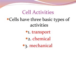
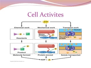
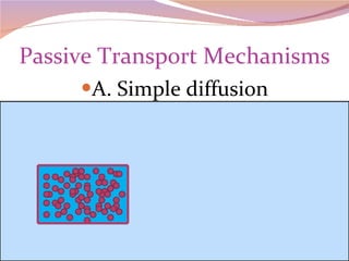
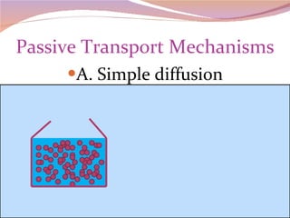
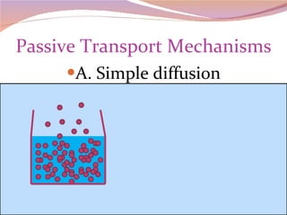
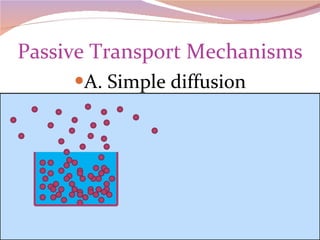
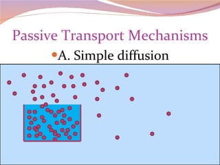
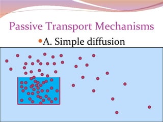
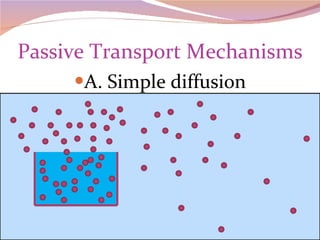
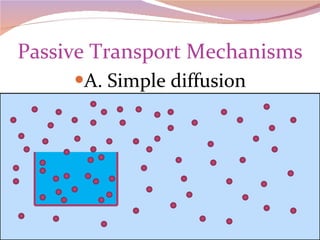
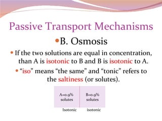
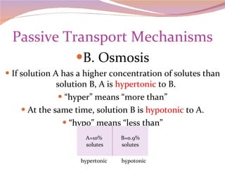
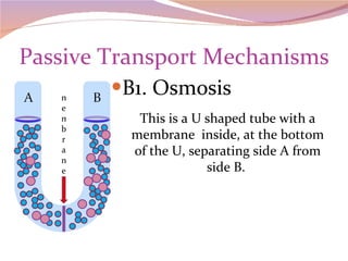
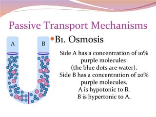
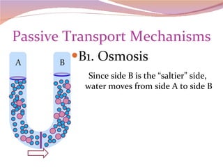
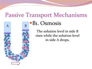
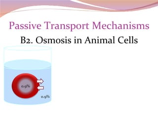
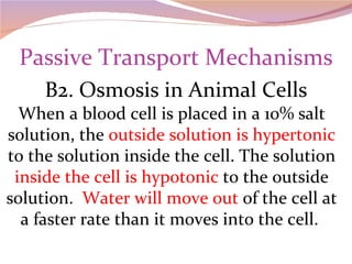
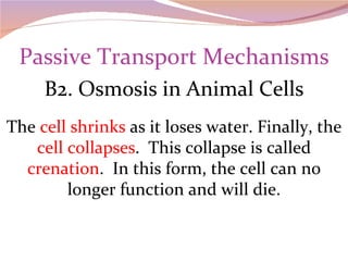
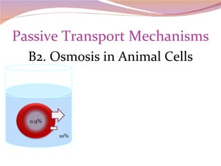
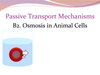
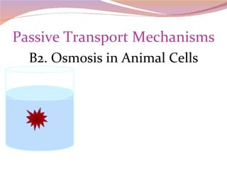
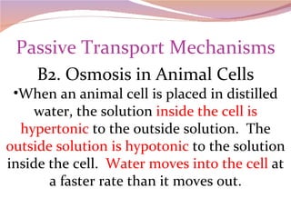
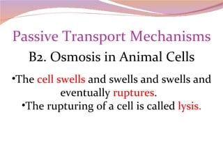
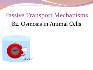
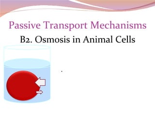
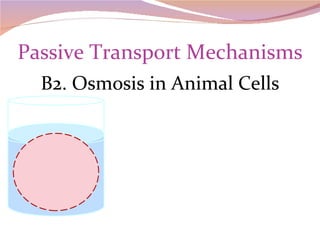
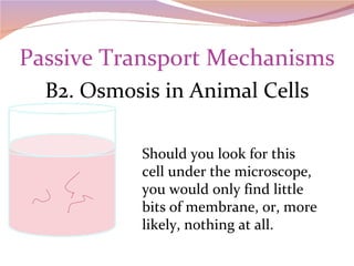
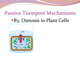
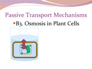
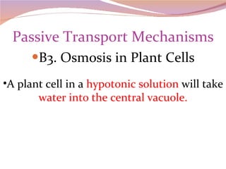
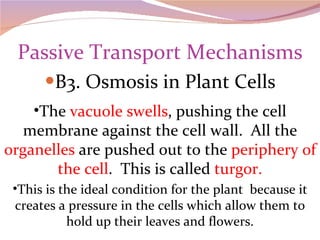
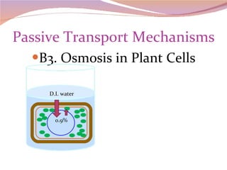
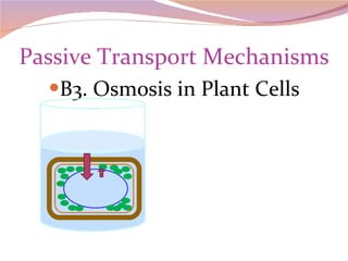
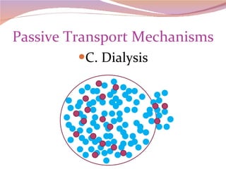
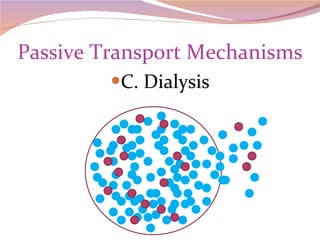
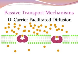
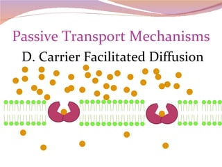
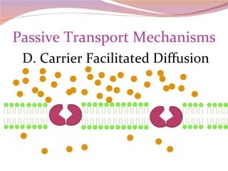
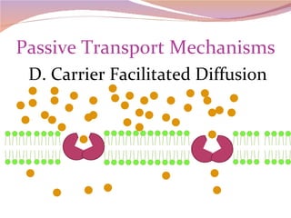
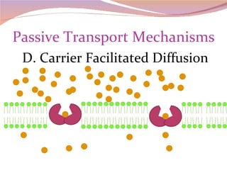
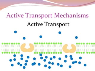
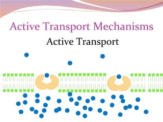
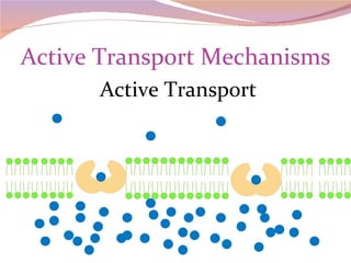
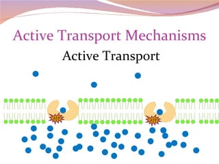
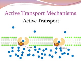
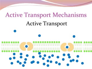
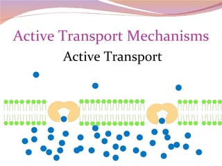
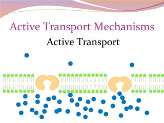
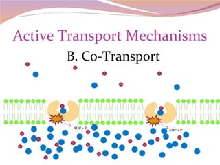
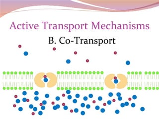
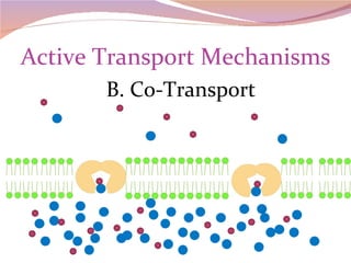
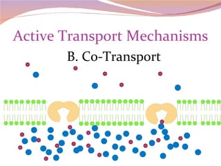
Cells have three basic types of activities 1.  transport 2.  chemical 3.  mechanical
Cell Activites
Transport Activities
  Transport Mechanisms
  Transport Mechanisms I.  Passive transport  Passive transport mechanisms  do not require the cell to input any energy . It is  dependent on two things: 1. The  innate energy  and movement of atoms and molecules 2. A  concentration gradient
  Transport Mechanisms I. Passive transport All atoms and molecules are in a constant state of motion.  Large molecules  move slower than small ones.  Warm molecules  move faster than cold ones.  Gases  are more active than  liquids  which are more active than  solids , but all molecules move.
  Transport Mechanisms I. Passive Transport A concentration gradient occurs as  molecules spread out  into a space. According to Newton’s Laws of motion, an atom or molecule moves in a  straight line  until it collides with another  force that causes it to change direction .
  Transport Mechanisms I. Passive transport  All passive transport mechanisms are really  variations of diffusion . Diffusion  refers to a  movement of molecules  from an area of  greater concentration  to an area of  lesser concentration  along a concentration gradient. If the molecules are in an idealized closed system, they will ultimately spread out and become  equally dispersed throughout the space.
Passive Transport Mechanisms A.  Simple diffusion Simple diffusion is the diffusion of molecules as they move through  open space  or through a  membrane. As with all diffusion, the molecules move from an area of greater concentration to an area of lesser concentration.
  Transport Mechanisms A. Simple Diffusion Initially the molecules are  highly concentrated  in one area. As they move,  they bump into each other  and the confining  walls  of their space.
Passive Transport Mechanisms A. Simple diffusion When we open a perfume bottle in a room, the aromatic molecules begin to move out of the bottle and into the larger space.  This is an example of simple diffusion. Oxygen moves out of the lung and into the blood by simple diffusion. Also, carbon dioxide moves out of the blood and into the lung by simple diffusion.
Passive Transport Mechanisms A. Simple diffusion
Passive Transport Mechanisms B.  Osmosis Osmosis is the  diffusion of  water  through a membrane . The process depends on the  concentration of the molecules dissolved in the water  since these molecules exert osmotic pressure. However, the dissolved molecules  do not pass through the membrane  during the process of osmosis .
Passive Transport Mechanisms B. Osmosis During osmosis, water will move to the side of the membrane with the most concentrated solution ( the saltier side ). Although, when we label a solution, we label it according to the  concentration of the solute  (dissolved material), not the solvent (water). A 10% salt solution is 90% water.  It is important to keep this in mind when discussing osmosis.
Passive Transport Mechanisms B. Osmosis Also, in considering osmosis, it is important to understand that we are comparing the solution  outside the cell  (A) to the solution  inside the cell  (B).  In other words, we compare the solutions on  either side of the membrane.
Passive Transport Mechanisms B. Osmosis If the two solutions are equal in concentration, than A is  isotonic  to B and B is  isotonic  to A. “ iso ” means “the same” and “tonic” refers to the  saltiness  (or solutes). Isotonic  isotonic A=0.9%  solutes B=0.9% solutes
Passive Transport Mechanisms B. Osmosis If solution A has a higher concentration of solutes than solution B, A is  hypertonic  to B. “ hyper” means “more than” At the same time, solution B is  hypotonic  to A. “ hypo” means “less than” hypertonic  hypotonic A=10%  solutes B=0.9% solutes
Passive Transport Mechanisms B1. Osmosis This is a U shaped tube with a membrane  inside, at the bottom of the U, separating side A from side B.  membrane
Passive Transport Mechanisms B1. Osmosis Side A has a concentration of 10% purple molecules  (the blue dots are water). Side B has a concentration of 20% purple molecules. A is hypotonic to B. B is hypertonic to A.
Passive Transport Mechanisms B1. Osmosis Since side B is the “saltier” side,  water moves from side A to side B
Passive Transport Mechanisms B1. Osmosis The solution level in side B rises while the solution level in side A drops.
Passive Transport Mechanisms B. How Osmosis affects cells The solution (cytoplasm) inside most cells has a  concentration of approximately  o.9%. Although this does vary some, for our purposes, we are going to consider this concentration a  constant  for all cells .
Passive Transport Mechanisms B2. Osmosis in  Animal Cells When a blood cell is placed into a solution with a concentration of 0.9% salt, the solutions  inside and outside of the cell are isotonic  to each other.  Water moves into and out of the cell at an equal rate. The cell is not affected by this solution.  It remains  normal and functional .
Passive Transport Mechanisms B2. Osmosis in Animal Cells 0.9% 0.9%
Passive Transport Mechanisms B2. Osmosis in Animal Cells When a blood cell is placed in a 10% salt solution, the  outside solution is hypertonic  to the solution inside the cell. The solution  inside the cell is hypotonic  to the outside solution.  Water will move out  of the cell at a faster rate than it moves into the cell.
Passive Transport Mechanisms B2. Osmosis in Animal Cells The  cell shrinks  as it loses water. Finally, the  cell collapses .  This collapse is called  crenation .  In this form, the cell can no longer function and will die.
Passive Transport Mechanisms B2. Osmosis in Animal Cells 0.9% 10%
Passive Transport Mechanisms B2. Osmosis in Animal Cells
Passive Transport Mechanisms B2. Osmosis in Animal Cells When an animal cell is placed in distilled water, the solution  inside the cell is hypertonic  to the outside solution.  The  outside solution is hypotonic  to the solution inside the cell.  Water moves into the cell  at a faster rate than it moves out.
Passive Transport Mechanisms B2. Osmosis in Animal Cells The  cell swells  and swells and swells and eventually  ruptures .  The rupturing of a cell is called  lysis.
Passive Transport Mechanisms B2. Osmosis in Animal Cells 0.9% D.I. water
Passive Transport Mechanisms B2. Osmosis in Animal Cells .
Passive Transport Mechanisms B2. Osmosis in Animal Cells
Passive Transport Mechanisms B2. Osmosis in Animal Cells Should you look for this cell under the microscope, you would only find little bits of membrane, or, more likely, nothing at all.
Passive Transport Mechanisms B3. Osmosis in  Plant Cells A plant cell in  a hypertonic solution  will  lose water  mainly from its  central vacuole .
Passive Transport Mechanisms B3. Osmosis in Plant Cells The central  vacuole shrinks , the cell  membrane collapses  and all the  organelles  inside the membrane  get crowded together .  This is called  plasmolysis .
Passive Transport Mechanisms B3. Osmosis in Plant Cells In general, the size of the cell does not change because the  cell wall is rigid  and does not collapse.  However, in this condition, the cells have no pressure to hold up the leaves of the plant .  It wilts.
Passive Transport Mechanisms B3. Osmosis in Plant Cells 0.9% 10%
Passive Transport Mechanisms B3. Osmosis in Plant Cells
Passive Transport Mechanisms B3. Osmosis in Plant Cells A plant cell in a  hypotonic solution  will take  water into the central vacuole.
Passive Transport Mechanisms B3. Osmosis in Plant Cells The  vacuole swells , pushing the cell membrane against the cell wall.  All the  organelles  are pushed out to the  periphery of the cell .  This is called  turgor. This is the ideal condition for the plant  because it creates a pressure in the cells which allow them to hold up their leaves and flowers.
Passive Transport Mechanisms B3. Osmosis in Plant Cells The cell does not rupture because the  cell wall is rigid  and creates an opposing pressure that  equalizes  with the osmotic pressure.
Passive Transport Mechanisms B3. Osmosis in Plant Cells 0.9% D.I. water
Passive Transport Mechanisms B3. Osmosis in Plant Cells
Passive Transport Mechanisms C.  Dialysis Dialysis is the  movement of particles  across the membrane. These particles are pulled through the membrane with  water that is diffusing  through.
Passive Transport Mechanisms C. Dialysis
Passive Transport Mechanisms D.  Carrier Facilitated Diffusion Carrier facilitated diffusion relies not only on the  concentration of particles  on either side of the membrane.  Carrier facilitated diffusion requires a  membrane carrier protein  to carry the particles across the membrane. Still, it is a diffusion process and does not require an input of energy from ATP.
Passive Transport Mechanisms D. Carrier Facilitated Diffusion
Active Transport Mechanisms II.  Active Transport Unlike passive transport, active transport does  require an input of energy from the cell . Another important difference between active and passive transport mechanisms is that active transport  can move particles against the concentration gradient , from an area of lesser concentration to an area of greater concentration.
Active Transport Mechanisms II. Active Transport In situations where the cell needs to take in all the particles it can despite concentration, it is important to have this option. As you take in and digest nutrients, the cells that absorb the nutrients may start out with a lesser concentration, but as more and more move into the cell, the concentration becomes greater inside the cell, but the cell still needs to continue taking in more.
Active Transport Mechanisms A.  Typical Active Transport Like carrier facilitated diffusion, typical active transport  requires a membrane carrier  protein to bring the particles across the membrane. However,  ATP is required  to activate the carrier and bring the particle across.
Active Transport Mechanisms Active Transport
Active Transport Mechanisms Active Transport ATP ATP
Active Transport Mechanisms Active Transport ADP + P ADP + P
Active Transport Mechanisms Active Transport
Active Transport Mechanisms B.  Co-Transport Co-transport is similar to typical active transport except, in this case, a  second type of particle attaches itself to the original particle  and both are pulled through the membrane at the same time (piggyback).
Active Transport Mechanisms B. Co-Transport
Active Transport Mechanisms B. Co-Transport ATP ATP
Active Transport Mechanisms B. Co-Transport ADP + P ADP + P
Active Transport Mechanisms B. Co-Transport
Active Transport Mechanisms C.  Exchange pump Another example of an active transport mechanism is the  exchange pump . This process involves moving one type of molecule  to the inside of the cell  while moving a different type of molecule  to the outside of the cell.
Active Transport Mechanisms C. Exchange pump When a nerve or muscle cell becomes electrically charged, sodium ions rush out of the cell and potassium ions rush in (by diffusion). In order for these cells to come back to their resting state, the ions must be returned to their original place. The Na+/K+ pump pulls sodium ions into the cell and potassium ions are pulled out of the cell.
Active Transport Mechanisms C. Exchange pump
Active Transport Mechanisms C. Exchange pump ATP ATP
Active Transport Mechanisms C. Exchange pump ATP ADP + P ADP + P
Active Transport Mechanisms C. Exchange pump
Transport by Vacuole III.  Transport by Vacuole Large molecules and cells  cannot pass through the membrane by passive or active transport. The only way to bring these into the cell is by  forming vacuoles . The cell also transports large molecules  out of the cell by vacuole.  These mechanisms require the cell to expend a  considerable amount of energy .
Transport by Vacuole A.  Endocytosis The process of bringing cells and large molecules into the cell by vacuole is called Endocytosis . There are 3 forms of endocytosis. 1. Phagocytosis  means “cell eating”. 2. Pinocytosis  means “cell drinking”.  3. Receptor (membrane) mediated endocytosis  which uses receptor proteins on the membrane to initiate the reaction.
 
Transport by Vacuole A1. Endocytosis/ Phagocytosis Cells like your white blood cells often take in  bacterial cells  to protect you from infection.  Other cells (especially unicellular organisms or protozoa) take in  small cells as food .  In order to bring in a complete cell, the cell membrane rises up, surrounds and engulfs the cell. Also, phagocytosis may bring in large chunks of materials like  splinter fragments .
Transport by Vacuole A1. Endocytosis/ Phagocytosis As the membrane closes over the material to come in, membrane touches membrane and the  molecules reorganize  so that the inner membrane forms  a vacuole  inside the cell that separates from the membrane.
Transport by Vacuole A1. Endocytosis/Phagocytosis
Transport by Vacuole A2. Endocytosis/ Pinocytosis Taking in fluid requires a different approach. The cell membrane  invaginates  forming an inpocket.  As the pocket forms, it  creates a vacuum  that sucks the extracellular fluid along with large molecules into the pocket. The membrane then closes over the top and  pinches off the vacuole  inside the cell.
Transport by Vacuole A2. Endocytosis/Pinocytosis
Transport by Vacuole A3. Endocytosis/Receptor Mediated Receptor Mediated Endocytosis  is much like pinocytosis, but more specific. Receptors in the membrane  attract and capture specific molecules , although extracellular  fluids do enter the newly forming vacuole as well.
Transport by Vacuole A3. Endocytosis/Receptor Mediated Y Y Y Y Y
Transport by Vacuole B.  Exocytosis The process of  releasing vacuole-encased molecules from the inside of the cell  is called Exocytosis.  The vacuole is formed within the cell and then fuses with the cell membrane as it releases its contents.
Transport by Vacuole B. Exocytosis As the cell forms proteins and other materials for export out of the cell, the  endoplasmic reticulum or the golgi package  these materials in vacuoles. This vacuole moves through the cell until it  reaches the cell membrane .
Transport by Vacuole B. Exocytosis As the vacuole bumps into the cell membrane, membrane touches membrane and the  molecules reorganize . The membrane of the  vacuole is incorporated into the cell membrane  and as the membrane stretches out, the materials inside the vacuole is left on the  outside of the cell .
Transport by Vacuole B. Exocytosis
Enzyme Activities
Energy and the Cell Energy Energy  = the capacity to do work. Kinetic energy  = energy of motion or energy used to do work. Potential energy  = stored energy.
Energy and the Cell Laws of Thermodynamics There are 2 laws of physics that concern  energy transformations . They are called the Laws of Thermodynamics.
Energy and the Cell 1 st  Law  of Thermodynamics = Energy conservation  the amount of energy and matter in the universe is  constant ; It can neither be  created nor destroyed …it can  change form .
Energy and the Cell 2 nd  Law  of Thermodynamics  = the  Law of Entropy (chaos)  The universe is moving towards entropy or  energy in the universe is becoming more chaotic . Energy transfers  or transformations are not  100% efficient .  Some energy is always lost in the form of  heat .
Energy and the Cell Chemical reactions Endergonic   reactions = require energy Exergonic reactions = release energy Exergonic reactions often drive endergonic reactions. This is called  Energy or reaction coupling. ATP  ADP CO 2  + H 2 O  Glucose + O 2
Chemical Reactions The chemical reactions in the cell are collectively called  metabolism or metabolic reactions . There are 2 forms of metabolism 1.  anabolic reactions  – build large molecules from smaller ones.  2.  catabolic reactions  – break down large molecules into smaller ones.
Chemical Reactions Typical reactions  Substrate(s) (reactants)  are converted to  product(s) Or  Or  + anabolic catabolic mixed + + +
Chemical Reactions Enzymes Catalyst  - speeds up the rate of a reaction Biological catalyst  - a catalyst that is safe to use in a living cell. Enzymes are biological catalysts .
Enzymes are  not changed  by  the reaction. Can be used again and again enzyme Substrates  are converted to  product with the help of the enzyme +
Enzymes Enzyme at the start of the reaction Unchanged Enzyme at the end of the reaction
Metabolic Pathway Metabolic pathway = a  series or chain of reactions. Product  of one reaction becomes  substrate  for next + + + A  +  B F  +  G D  +  E C
Metabolic Pathway Each reaction has a  separate enzyme. Enz 1 Enz 2 Enz 3 + + + A  +  B F  +  G D  +  E C
Metabolic Pathway Alternate pathways  are determined by enzymes If Enzyme 3 is present, D will be converted to F and G the original pathway will be completed with these final products.  But if Enzyme 4 is present D will be converted to W and X and the alternate pathway will occur, resulting in the final products Y and Z. Enz 4 Enz 5 Enz 1 Enz 2 Enz 3 + + + A  +  B F  +  G D  +  E C + W +  X Y  +  Z +
Enzymes Enzymes : 1. are  biological catalysts . 2. are usually made at least partly of  protein . 3. are  substrate specific . 4. can be  induced or inhibited .
Enzymes Generalized enzyme structure Apoenzyme   = protein part of the enzyme Coenzyme  (organic) or  cofactor (inorganic) = non-protein part of the enzyme Active Site  = where the substrate fits into the enzyme Allosteric site  = where the enzyme is activated or deactivated enzyme
Enzymes Activation  of an enzyme An enzyme may be activated by placing an  activator molecule  into the  allosteric site .  This process causes the  shape of the active site  to change so that the substrate can fit into the active site.
Enzymes Activation   of an enzyme This is called  positive allosterism . Without the activator molecule,  the active site remains closed off  so that the substrate cannot fit into it.
Enzymes Generalized enzyme structure Inactive enzyme activator substrate
Enzymes Generalized enzyme structure activated enzyme activator substrate
Enzymes Inhibition  of an enzyme An enzyme may be inhibited by placing an  inhibitor molecule  into the  allosteric site .  This process causes the  shape of the active site  to change so that the substrate can no longer fit into the active site.
Enzymes Inhibition of an enzyme This is called  negative allosterism . Without the inhibitor molecule,  the active site remains open  so that the substrate fits into it.
Enzymes Example of negative allosterism
Enzymes E + S  The substrate bumps into the enzyme and aligns with the active site. E-S complex  The substrate fits into the active  site, and the enzyme shifts, stressing the bonds of the substrate. E + P  The products are formed and released. The  enzyme  returns to its original shape and  can be used again . How an Enzyme Works
Enzymes How an Enzyme Works
Enzymes All enzymatic reactions require some energy to get them started.  This energy is called the  energy of activation. Enzymes reduce the energy of activation  so that it takes much less energy to get the reaction going.  Once started, the reaction will continue on its own steam. How an Enzyme Works
Enzymes
Enzymes
Enzymes Factors that affect the rate of an enzymatic reaction 1.  temperature 2.  pH 3.  enzyme concentration 4.  inhibitors
Enzymes 1. Temperature  profile Every enzyme has a temperature profile.  This profile is a  graph showing at what rates the reactions  take place at  various temperatures. The temperature profile generally forms a “ bell-shaped ” curve.
Enzymes 1. Temperature profile The peak of the curve represents the  optimal temperature.   This is the temperature at which the reaction rate is fastest.  As  temperature cools  from optimal,  molecules slow down  and do not encounter each other as often or with as much energy, so the reaction rate slows down until the reaction no longer occurs.  This temperature is called the  minimal temperature .
Enzymes 1. Temperature profile As temperature becomes  warmer  than optimal, enzymes begin to change shape.  This change in shape is called  denaturing the enzyme.  If the temperature gets too warm, the enzyme changes so radically that the substrate can no longer fit into the active site.  The reaction rate is then 0 (no reaction occurs) and the enzyme is irreversibly denatured.  This temperature is called the  maximal temperature.
Enzymes 1. Temperature profile The range of temperatures between the minimal and maximal is called the  functional range .  This is the range of temperatures within which the enzyme works.
Enzymes 1. Temperature profile Minimal temperature Maximal temperature Functional range
Enzymes 1. Temperature profile Every enzyme has a  unique profile  based on the  type of organism  it is found in and/or the  location  in the organism where it does its work.
Enzymes 1. Temperature profile Among mammals, smaller mammals tend to have  higher normal body temperatures  than larger mammals.  As a result, one would expect the  optimal temperature  for a small mammal to be at a  higher temperature  than the optimal for larger mammals.
Enzymes 1. Temperature profile Cold blooded animals do not regulate their body temperatures as closely as mammals, so their functional range may be broader than that for mammals. The enzymes that work in cells all over the body also need a broader functional range. Enzymes that work outside the body (digestive enzymes of fungi or the enzymes that work in the testes) usually have a cooler optimal temperature than enzymes that work inside the body.
Temperature in  o C Temperature Profile for various enzymes
Enzymes 2. pH profile As with temperature, enzymes have a pH profile.  This profile is a graph showing at what rate the reaction takes place a various pHs. This profile also, generally forms a “ bell-shaped ” curve.
Enzymes 2. pH profile The peak of the curve represents the  optimal pH.   This is the pH at which the reaction rate is fastest.  As the pH becomes  more acidic or more basic  than optimal, the enzyme begins to  denature .  When the enzyme no longer functions, it is irreversibly denatured.  The points where this occurs will be  the minimal and maximal pH  and the range between the two is the  functional pH range.
Enzymes 2. pH profile Minimal pH Maximal pH Functional range
Enzymes 2. pH profile Every enzyme has a unique profile based primarily on where in the organism it functions. A blood enzyme has a very narrow range that is slightly basic. Stomach enzymes have a very low pH optimal. Pancreatic enzymes work best at a neutral pH. Catalase, which breaks down hydrogen peroxide, has a broad range of pHs at which it works, since it must work in every cell of the body.
Enzymes 3. Enzyme Concentration The concentration profile for enzymes is quite different than the temperature and pH profiles. Initially, there is a  steep positive correlation  between enzyme concentration and reaction rate, but at a certain point, using more enzyme cannot make the rate go faster and the curve  levels off into a plateau.  There is no minimal or maximal concentration.  The  optimal concentration  occurs just before the plateau .
Enzymes 3. Enzyme concentration
Enzymes 4. Inhibitors The more inhibitor present, the slower the reaction rate. 2 types a. Competitive inhibitors b. Non-competitive inhibitors
Enzymes 4a.  Competitive Inhibitors A competitive inhibitor partially mimics the substrate molecule and  blocks the active site . This inhibition is  temporary  since the inhibitor can move into or out of the active site. The substrate and the inhibitor  compete  with each other for access to the enzyme. Often competitive inhibitors are produced by the body to  slow down the speed  of a reaction.
Enzymes 4b. Non-competitive Inhibitors A Non-Competitive inhibitor blocks the  allosteric site  or removes  co-enzyme or co-factor  from the enzyme. A blocked allosteric is sometimes  temporary  and reversible. A removed co-factor or co-enzyme  destroys the enzyme  and is permanent.
Enzymes Inhibitors
Mechanical Activities
Mechanical Activities  Mechanical activities of the cell involve  movement of some sort . At the cellular level , mechanical activities would include the following: cytoplasmic streaming  amoeboid movement  The beating of cilia and flagella The movement of centrioles, microtubules and chromosomes during cell division
Mechanical Activities  Other cell-level  mechanical activities  include the following: The movement of RNA out of the nucleus. The movement of vacuoles, mitochondria, plastids and other organelles. Endocytosis and exocytosis are transport activities with a mechanical aspect.
Mechanical Activities  Mechanical activities at the  organism level  might include: Muscle contraction allowing gross movement of the body. Peristaltic contractions of the digestive tract The pumping action of the heart The movement of blood through the vessels. The movement of air into and out of the lungs.
Mechanical Activities  Most mechanical activities  require ATP  breakdown and the cells must metabolize foods in order to maintain a  constant supply of the ATP.

Sp2010 chapter 5 old

  • 1.
  • 2.
    Cell Activities Cellshave three basic types of activities 1. transport 2. chemical 3. mechanical
  • 3.
  • 4.
  • 5.
    TransportMechanisms
  • 6.
    TransportMechanisms I. Passive transport Passive transport mechanisms do not require the cell to input any energy . It is dependent on two things: 1. The innate energy and movement of atoms and molecules 2. A concentration gradient
  • 7.
    TransportMechanisms I. Passive transport All atoms and molecules are in a constant state of motion. Large molecules move slower than small ones. Warm molecules move faster than cold ones. Gases are more active than liquids which are more active than solids , but all molecules move.
  • 8.
    TransportMechanisms I. Passive Transport A concentration gradient occurs as molecules spread out into a space. According to Newton’s Laws of motion, an atom or molecule moves in a straight line until it collides with another force that causes it to change direction .
  • 9.
    TransportMechanisms I. Passive transport All passive transport mechanisms are really variations of diffusion . Diffusion refers to a movement of molecules from an area of greater concentration to an area of lesser concentration along a concentration gradient. If the molecules are in an idealized closed system, they will ultimately spread out and become equally dispersed throughout the space.
  • 10.
    Passive Transport MechanismsA. Simple diffusion Simple diffusion is the diffusion of molecules as they move through open space or through a membrane. As with all diffusion, the molecules move from an area of greater concentration to an area of lesser concentration.
  • 11.
    TransportMechanisms A. Simple Diffusion Initially the molecules are highly concentrated in one area. As they move, they bump into each other and the confining walls of their space.
  • 12.
    Passive Transport MechanismsA. Simple diffusion When we open a perfume bottle in a room, the aromatic molecules begin to move out of the bottle and into the larger space. This is an example of simple diffusion. Oxygen moves out of the lung and into the blood by simple diffusion. Also, carbon dioxide moves out of the blood and into the lung by simple diffusion.
  • 13.
    Passive Transport MechanismsA. Simple diffusion
  • 14.
    Passive Transport MechanismsA. Simple diffusion
  • 15.
    Passive Transport MechanismsA. Simple diffusion
  • 16.
    Passive Transport MechanismsA. Simple diffusion
  • 17.
    Passive Transport MechanismsA. Simple diffusion
  • 18.
    Passive Transport MechanismsA. Simple diffusion
  • 19.
    Passive Transport MechanismsA. Simple diffusion
  • 20.
    Passive Transport MechanismsA. Simple diffusion
  • 21.
    Passive Transport MechanismsA. Simple diffusion
  • 22.
    Passive Transport MechanismsB. Osmosis Osmosis is the diffusion of water through a membrane . The process depends on the concentration of the molecules dissolved in the water since these molecules exert osmotic pressure. However, the dissolved molecules do not pass through the membrane during the process of osmosis .
  • 23.
    Passive Transport MechanismsB. Osmosis During osmosis, water will move to the side of the membrane with the most concentrated solution ( the saltier side ). Although, when we label a solution, we label it according to the concentration of the solute (dissolved material), not the solvent (water). A 10% salt solution is 90% water. It is important to keep this in mind when discussing osmosis.
  • 24.
    Passive Transport MechanismsB. Osmosis Also, in considering osmosis, it is important to understand that we are comparing the solution outside the cell (A) to the solution inside the cell (B). In other words, we compare the solutions on either side of the membrane.
  • 25.
    Passive Transport MechanismsB. Osmosis If the two solutions are equal in concentration, than A is isotonic to B and B is isotonic to A. “ iso ” means “the same” and “tonic” refers to the saltiness (or solutes). Isotonic isotonic A=0.9% solutes B=0.9% solutes
  • 26.
    Passive Transport MechanismsB. Osmosis If solution A has a higher concentration of solutes than solution B, A is hypertonic to B. “ hyper” means “more than” At the same time, solution B is hypotonic to A. “ hypo” means “less than” hypertonic hypotonic A=10% solutes B=0.9% solutes
  • 27.
    Passive Transport MechanismsB1. Osmosis This is a U shaped tube with a membrane inside, at the bottom of the U, separating side A from side B. membrane
  • 28.
    Passive Transport MechanismsB1. Osmosis Side A has a concentration of 10% purple molecules (the blue dots are water). Side B has a concentration of 20% purple molecules. A is hypotonic to B. B is hypertonic to A.
  • 29.
    Passive Transport MechanismsB1. Osmosis Since side B is the “saltier” side, water moves from side A to side B
  • 30.
    Passive Transport MechanismsB1. Osmosis The solution level in side B rises while the solution level in side A drops.
  • 31.
    Passive Transport MechanismsB. How Osmosis affects cells The solution (cytoplasm) inside most cells has a concentration of approximately o.9%. Although this does vary some, for our purposes, we are going to consider this concentration a constant for all cells .
  • 32.
    Passive Transport MechanismsB2. Osmosis in Animal Cells When a blood cell is placed into a solution with a concentration of 0.9% salt, the solutions inside and outside of the cell are isotonic to each other. Water moves into and out of the cell at an equal rate. The cell is not affected by this solution. It remains normal and functional .
  • 33.
    Passive Transport MechanismsB2. Osmosis in Animal Cells 0.9% 0.9%
  • 34.
    Passive Transport MechanismsB2. Osmosis in Animal Cells When a blood cell is placed in a 10% salt solution, the outside solution is hypertonic to the solution inside the cell. The solution inside the cell is hypotonic to the outside solution. Water will move out of the cell at a faster rate than it moves into the cell.
  • 35.
    Passive Transport MechanismsB2. Osmosis in Animal Cells The cell shrinks as it loses water. Finally, the cell collapses . This collapse is called crenation . In this form, the cell can no longer function and will die.
  • 36.
    Passive Transport MechanismsB2. Osmosis in Animal Cells 0.9% 10%
  • 37.
    Passive Transport MechanismsB2. Osmosis in Animal Cells
  • 38.
    Passive Transport MechanismsB2. Osmosis in Animal Cells
  • 39.
    Passive Transport MechanismsB2. Osmosis in Animal Cells When an animal cell is placed in distilled water, the solution inside the cell is hypertonic to the outside solution. The outside solution is hypotonic to the solution inside the cell. Water moves into the cell at a faster rate than it moves out.
  • 40.
    Passive Transport MechanismsB2. Osmosis in Animal Cells The cell swells and swells and swells and eventually ruptures . The rupturing of a cell is called lysis.
  • 41.
    Passive Transport MechanismsB2. Osmosis in Animal Cells 0.9% D.I. water
  • 42.
    Passive Transport MechanismsB2. Osmosis in Animal Cells .
  • 43.
    Passive Transport MechanismsB2. Osmosis in Animal Cells
  • 44.
    Passive Transport MechanismsB2. Osmosis in Animal Cells Should you look for this cell under the microscope, you would only find little bits of membrane, or, more likely, nothing at all.
  • 45.
    Passive Transport MechanismsB3. Osmosis in Plant Cells A plant cell in a hypertonic solution will lose water mainly from its central vacuole .
  • 46.
    Passive Transport MechanismsB3. Osmosis in Plant Cells The central vacuole shrinks , the cell membrane collapses and all the organelles inside the membrane get crowded together . This is called plasmolysis .
  • 47.
    Passive Transport MechanismsB3. Osmosis in Plant Cells In general, the size of the cell does not change because the cell wall is rigid and does not collapse. However, in this condition, the cells have no pressure to hold up the leaves of the plant . It wilts.
  • 48.
    Passive Transport MechanismsB3. Osmosis in Plant Cells 0.9% 10%
  • 49.
    Passive Transport MechanismsB3. Osmosis in Plant Cells
  • 50.
    Passive Transport MechanismsB3. Osmosis in Plant Cells A plant cell in a hypotonic solution will take water into the central vacuole.
  • 51.
    Passive Transport MechanismsB3. Osmosis in Plant Cells The vacuole swells , pushing the cell membrane against the cell wall. All the organelles are pushed out to the periphery of the cell . This is called turgor. This is the ideal condition for the plant because it creates a pressure in the cells which allow them to hold up their leaves and flowers.
  • 52.
    Passive Transport MechanismsB3. Osmosis in Plant Cells The cell does not rupture because the cell wall is rigid and creates an opposing pressure that equalizes with the osmotic pressure.
  • 53.
    Passive Transport MechanismsB3. Osmosis in Plant Cells 0.9% D.I. water
  • 54.
    Passive Transport MechanismsB3. Osmosis in Plant Cells
  • 55.
    Passive Transport MechanismsC. Dialysis Dialysis is the movement of particles across the membrane. These particles are pulled through the membrane with water that is diffusing through.
  • 56.
  • 57.
  • 58.
  • 59.
    Passive Transport MechanismsD. Carrier Facilitated Diffusion Carrier facilitated diffusion relies not only on the concentration of particles on either side of the membrane. Carrier facilitated diffusion requires a membrane carrier protein to carry the particles across the membrane. Still, it is a diffusion process and does not require an input of energy from ATP.
  • 60.
    Passive Transport MechanismsD. Carrier Facilitated Diffusion
  • 61.
    Passive Transport MechanismsD. Carrier Facilitated Diffusion
  • 62.
    Passive Transport MechanismsD. Carrier Facilitated Diffusion
  • 63.
    Passive Transport MechanismsD. Carrier Facilitated Diffusion
  • 64.
    Passive Transport MechanismsD. Carrier Facilitated Diffusion
  • 65.
    Passive Transport MechanismsD. Carrier Facilitated Diffusion
  • 66.
    Passive Transport MechanismsD. Carrier Facilitated Diffusion
  • 67.
    Passive Transport MechanismsD. Carrier Facilitated Diffusion
  • 68.
    Passive Transport MechanismsD. Carrier Facilitated Diffusion
  • 69.
    Passive Transport MechanismsD. Carrier Facilitated Diffusion
  • 70.
    Passive Transport MechanismsD. Carrier Facilitated Diffusion
  • 71.
    Active Transport MechanismsII. Active Transport Unlike passive transport, active transport does require an input of energy from the cell . Another important difference between active and passive transport mechanisms is that active transport can move particles against the concentration gradient , from an area of lesser concentration to an area of greater concentration.
  • 72.
    Active Transport MechanismsII. Active Transport In situations where the cell needs to take in all the particles it can despite concentration, it is important to have this option. As you take in and digest nutrients, the cells that absorb the nutrients may start out with a lesser concentration, but as more and more move into the cell, the concentration becomes greater inside the cell, but the cell still needs to continue taking in more.
  • 73.
    Active Transport MechanismsA. Typical Active Transport Like carrier facilitated diffusion, typical active transport requires a membrane carrier protein to bring the particles across the membrane. However, ATP is required to activate the carrier and bring the particle across.
  • 74.
  • 75.
  • 76.
  • 77.
    Active Transport MechanismsActive Transport ATP ATP
  • 78.
    Active Transport MechanismsActive Transport ADP + P ADP + P
  • 79.
  • 80.
  • 81.
  • 82.
  • 83.
  • 84.
  • 85.
    Active Transport MechanismsB. Co-Transport Co-transport is similar to typical active transport except, in this case, a second type of particle attaches itself to the original particle and both are pulled through the membrane at the same time (piggyback).
  • 86.
  • 87.
  • 88.
  • 89.
  • 90.
    Active Transport MechanismsB. Co-Transport ATP ATP
  • 91.
    Active Transport MechanismsB. Co-Transport ADP + P ADP + P
  • 92.
  • 93.
  • 94.
  • 95.
  • 96.
  • 97.
  • 98.
  • 99.
    Active Transport MechanismsC. Exchange pump Another example of an active transport mechanism is the exchange pump . This process involves moving one type of molecule to the inside of the cell while moving a different type of molecule to the outside of the cell.
  • 100.
    Active Transport MechanismsC. Exchange pump When a nerve or muscle cell becomes electrically charged, sodium ions rush out of the cell and potassium ions rush in (by diffusion). In order for these cells to come back to their resting state, the ions must be returned to their original place. The Na+/K+ pump pulls sodium ions into the cell and potassium ions are pulled out of the cell.
  • 101.
  • 102.
  • 103.
    Active Transport MechanismsC. Exchange pump ATP ATP
  • 104.
    Active Transport MechanismsC. Exchange pump ATP ADP + P ADP + P
  • 105.
  • 106.
  • 107.
  • 108.
  • 109.
  • 110.
  • 111.
  • 112.
    Transport by VacuoleIII. Transport by Vacuole Large molecules and cells cannot pass through the membrane by passive or active transport. The only way to bring these into the cell is by forming vacuoles . The cell also transports large molecules out of the cell by vacuole. These mechanisms require the cell to expend a considerable amount of energy .
  • 113.
    Transport by VacuoleA. Endocytosis The process of bringing cells and large molecules into the cell by vacuole is called Endocytosis . There are 3 forms of endocytosis. 1. Phagocytosis means “cell eating”. 2. Pinocytosis means “cell drinking”. 3. Receptor (membrane) mediated endocytosis which uses receptor proteins on the membrane to initiate the reaction.
  • 114.
  • 115.
    Transport by VacuoleA1. Endocytosis/ Phagocytosis Cells like your white blood cells often take in bacterial cells to protect you from infection. Other cells (especially unicellular organisms or protozoa) take in small cells as food . In order to bring in a complete cell, the cell membrane rises up, surrounds and engulfs the cell. Also, phagocytosis may bring in large chunks of materials like splinter fragments .
  • 116.
    Transport by VacuoleA1. Endocytosis/ Phagocytosis As the membrane closes over the material to come in, membrane touches membrane and the molecules reorganize so that the inner membrane forms a vacuole inside the cell that separates from the membrane.
  • 117.
    Transport by VacuoleA1. Endocytosis/Phagocytosis
  • 118.
    Transport by VacuoleA1. Endocytosis/Phagocytosis
  • 119.
    Transport by VacuoleA1. Endocytosis/Phagocytosis
  • 120.
    Transport by VacuoleA1. Endocytosis/Phagocytosis
  • 121.
    Transport by VacuoleA1. Endocytosis/Phagocytosis
  • 122.
    Transport by VacuoleA1. Endocytosis/Phagocytosis
  • 123.
    Transport by VacuoleA1. Endocytosis/Phagocytosis
  • 124.
    Transport by VacuoleA1. Endocytosis/Phagocytosis
  • 125.
    Transport by VacuoleA2. Endocytosis/ Pinocytosis Taking in fluid requires a different approach. The cell membrane invaginates forming an inpocket. As the pocket forms, it creates a vacuum that sucks the extracellular fluid along with large molecules into the pocket. The membrane then closes over the top and pinches off the vacuole inside the cell.
  • 126.
    Transport by VacuoleA2. Endocytosis/Pinocytosis
  • 127.
    Transport by VacuoleA2. Endocytosis/Pinocytosis
  • 128.
    Transport by VacuoleA2. Endocytosis/Pinocytosis
  • 129.
    Transport by VacuoleA2. Endocytosis/Pinocytosis
  • 130.
    Transport by VacuoleA2. Endocytosis/Pinocytosis
  • 131.
    Transport by VacuoleA3. Endocytosis/Receptor Mediated Receptor Mediated Endocytosis is much like pinocytosis, but more specific. Receptors in the membrane attract and capture specific molecules , although extracellular fluids do enter the newly forming vacuole as well.
  • 132.
    Transport by VacuoleA3. Endocytosis/Receptor Mediated Y Y Y Y Y
  • 133.
    Transport by VacuoleA3. Endocytosis/Receptor Mediated Y Y Y Y Y
  • 134.
    Transport by VacuoleA3. Endocytosis/Receptor Mediated Y Y Y Y Y
  • 135.
    Transport by VacuoleA3. Endocytosis/Receptor Mediated Y Y Y Y Y
  • 136.
    Transport by VacuoleA3. Endocytosis/Receptor Mediated Y Y Y Y Y
  • 137.
    Transport by VacuoleA3. Endocytosis/Receptor Mediated Y Y Y Y Y
  • 138.
    Transport by VacuoleB. Exocytosis The process of releasing vacuole-encased molecules from the inside of the cell is called Exocytosis. The vacuole is formed within the cell and then fuses with the cell membrane as it releases its contents.
  • 139.
    Transport by VacuoleB. Exocytosis As the cell forms proteins and other materials for export out of the cell, the endoplasmic reticulum or the golgi package these materials in vacuoles. This vacuole moves through the cell until it reaches the cell membrane .
  • 140.
    Transport by VacuoleB. Exocytosis As the vacuole bumps into the cell membrane, membrane touches membrane and the molecules reorganize . The membrane of the vacuole is incorporated into the cell membrane and as the membrane stretches out, the materials inside the vacuole is left on the outside of the cell .
  • 141.
    Transport by VacuoleB. Exocytosis
  • 142.
    Transport by VacuoleB. Exocytosis
  • 143.
    Transport by VacuoleB. Exocytosis
  • 144.
    Transport by VacuoleB. Exocytosis
  • 145.
    Transport by VacuoleB. Exocytosis
  • 146.
    Transport by VacuoleB. Exocytosis
  • 147.
  • 148.
    Energy and theCell Energy Energy = the capacity to do work. Kinetic energy = energy of motion or energy used to do work. Potential energy = stored energy.
  • 149.
    Energy and theCell Laws of Thermodynamics There are 2 laws of physics that concern energy transformations . They are called the Laws of Thermodynamics.
  • 150.
    Energy and theCell 1 st Law of Thermodynamics = Energy conservation the amount of energy and matter in the universe is constant ; It can neither be created nor destroyed …it can change form .
  • 151.
    Energy and theCell 2 nd Law of Thermodynamics = the Law of Entropy (chaos) The universe is moving towards entropy or energy in the universe is becoming more chaotic . Energy transfers or transformations are not 100% efficient . Some energy is always lost in the form of heat .
  • 152.
    Energy and theCell Chemical reactions Endergonic reactions = require energy Exergonic reactions = release energy Exergonic reactions often drive endergonic reactions. This is called Energy or reaction coupling. ATP ADP CO 2 + H 2 O Glucose + O 2
  • 153.
    Chemical Reactions Thechemical reactions in the cell are collectively called metabolism or metabolic reactions . There are 2 forms of metabolism 1. anabolic reactions – build large molecules from smaller ones. 2. catabolic reactions – break down large molecules into smaller ones.
  • 154.
    Chemical Reactions Typicalreactions Substrate(s) (reactants) are converted to product(s) Or Or + anabolic catabolic mixed + + +
  • 155.
    Chemical Reactions EnzymesCatalyst - speeds up the rate of a reaction Biological catalyst - a catalyst that is safe to use in a living cell. Enzymes are biological catalysts .
  • 156.
    Enzymes are not changed by the reaction. Can be used again and again enzyme Substrates are converted to product with the help of the enzyme +
  • 157.
    Enzymes Enzyme atthe start of the reaction Unchanged Enzyme at the end of the reaction
  • 158.
    Metabolic Pathway Metabolicpathway = a series or chain of reactions. Product of one reaction becomes substrate for next + + + A + B F + G D + E C
  • 159.
    Metabolic Pathway Eachreaction has a separate enzyme. Enz 1 Enz 2 Enz 3 + + + A + B F + G D + E C
  • 160.
    Metabolic Pathway Alternatepathways are determined by enzymes If Enzyme 3 is present, D will be converted to F and G the original pathway will be completed with these final products. But if Enzyme 4 is present D will be converted to W and X and the alternate pathway will occur, resulting in the final products Y and Z. Enz 4 Enz 5 Enz 1 Enz 2 Enz 3 + + + A + B F + G D + E C + W + X Y + Z +
  • 161.
    Enzymes Enzymes :1. are biological catalysts . 2. are usually made at least partly of protein . 3. are substrate specific . 4. can be induced or inhibited .
  • 162.
    Enzymes Generalized enzymestructure Apoenzyme = protein part of the enzyme Coenzyme (organic) or cofactor (inorganic) = non-protein part of the enzyme Active Site = where the substrate fits into the enzyme Allosteric site = where the enzyme is activated or deactivated enzyme
  • 163.
    Enzymes Activation of an enzyme An enzyme may be activated by placing an activator molecule into the allosteric site . This process causes the shape of the active site to change so that the substrate can fit into the active site.
  • 164.
    Enzymes Activation of an enzyme This is called positive allosterism . Without the activator molecule, the active site remains closed off so that the substrate cannot fit into it.
  • 165.
    Enzymes Generalized enzymestructure Inactive enzyme activator substrate
  • 166.
    Enzymes Generalized enzymestructure activated enzyme activator substrate
  • 167.
    Enzymes Generalized enzymestructure activated enzyme activator substrate
  • 168.
    Enzymes Inhibition of an enzyme An enzyme may be inhibited by placing an inhibitor molecule into the allosteric site . This process causes the shape of the active site to change so that the substrate can no longer fit into the active site.
  • 169.
    Enzymes Inhibition ofan enzyme This is called negative allosterism . Without the inhibitor molecule, the active site remains open so that the substrate fits into it.
  • 170.
    Enzymes Example ofnegative allosterism
  • 171.
    Enzymes E +S The substrate bumps into the enzyme and aligns with the active site. E-S complex The substrate fits into the active site, and the enzyme shifts, stressing the bonds of the substrate. E + P The products are formed and released. The enzyme returns to its original shape and can be used again . How an Enzyme Works
  • 172.
    Enzymes How anEnzyme Works
  • 173.
    Enzymes All enzymaticreactions require some energy to get them started. This energy is called the energy of activation. Enzymes reduce the energy of activation so that it takes much less energy to get the reaction going. Once started, the reaction will continue on its own steam. How an Enzyme Works
  • 174.
  • 175.
  • 176.
    Enzymes Factors thataffect the rate of an enzymatic reaction 1. temperature 2. pH 3. enzyme concentration 4. inhibitors
  • 177.
    Enzymes 1. Temperature profile Every enzyme has a temperature profile. This profile is a graph showing at what rates the reactions take place at various temperatures. The temperature profile generally forms a “ bell-shaped ” curve.
  • 178.
    Enzymes 1. Temperatureprofile The peak of the curve represents the optimal temperature. This is the temperature at which the reaction rate is fastest. As temperature cools from optimal, molecules slow down and do not encounter each other as often or with as much energy, so the reaction rate slows down until the reaction no longer occurs. This temperature is called the minimal temperature .
  • 179.
    Enzymes 1. Temperatureprofile As temperature becomes warmer than optimal, enzymes begin to change shape. This change in shape is called denaturing the enzyme. If the temperature gets too warm, the enzyme changes so radically that the substrate can no longer fit into the active site. The reaction rate is then 0 (no reaction occurs) and the enzyme is irreversibly denatured. This temperature is called the maximal temperature.
  • 180.
    Enzymes 1. Temperatureprofile The range of temperatures between the minimal and maximal is called the functional range . This is the range of temperatures within which the enzyme works.
  • 181.
    Enzymes 1. Temperatureprofile Minimal temperature Maximal temperature Functional range
  • 182.
    Enzymes 1. Temperatureprofile Every enzyme has a unique profile based on the type of organism it is found in and/or the location in the organism where it does its work.
  • 183.
    Enzymes 1. Temperatureprofile Among mammals, smaller mammals tend to have higher normal body temperatures than larger mammals. As a result, one would expect the optimal temperature for a small mammal to be at a higher temperature than the optimal for larger mammals.
  • 184.
    Enzymes 1. Temperatureprofile Cold blooded animals do not regulate their body temperatures as closely as mammals, so their functional range may be broader than that for mammals. The enzymes that work in cells all over the body also need a broader functional range. Enzymes that work outside the body (digestive enzymes of fungi or the enzymes that work in the testes) usually have a cooler optimal temperature than enzymes that work inside the body.
  • 185.
    Temperature in o C Temperature Profile for various enzymes
  • 186.
    Enzymes 2. pHprofile As with temperature, enzymes have a pH profile. This profile is a graph showing at what rate the reaction takes place a various pHs. This profile also, generally forms a “ bell-shaped ” curve.
  • 187.
    Enzymes 2. pHprofile The peak of the curve represents the optimal pH. This is the pH at which the reaction rate is fastest. As the pH becomes more acidic or more basic than optimal, the enzyme begins to denature . When the enzyme no longer functions, it is irreversibly denatured. The points where this occurs will be the minimal and maximal pH and the range between the two is the functional pH range.
  • 188.
    Enzymes 2. pHprofile Minimal pH Maximal pH Functional range
  • 189.
    Enzymes 2. pHprofile Every enzyme has a unique profile based primarily on where in the organism it functions. A blood enzyme has a very narrow range that is slightly basic. Stomach enzymes have a very low pH optimal. Pancreatic enzymes work best at a neutral pH. Catalase, which breaks down hydrogen peroxide, has a broad range of pHs at which it works, since it must work in every cell of the body.
  • 190.
    Enzymes 3. EnzymeConcentration The concentration profile for enzymes is quite different than the temperature and pH profiles. Initially, there is a steep positive correlation between enzyme concentration and reaction rate, but at a certain point, using more enzyme cannot make the rate go faster and the curve levels off into a plateau. There is no minimal or maximal concentration. The optimal concentration occurs just before the plateau .
  • 191.
    Enzymes 3. Enzymeconcentration
  • 192.
    Enzymes 4. InhibitorsThe more inhibitor present, the slower the reaction rate. 2 types a. Competitive inhibitors b. Non-competitive inhibitors
  • 193.
    Enzymes 4a. Competitive Inhibitors A competitive inhibitor partially mimics the substrate molecule and blocks the active site . This inhibition is temporary since the inhibitor can move into or out of the active site. The substrate and the inhibitor compete with each other for access to the enzyme. Often competitive inhibitors are produced by the body to slow down the speed of a reaction.
  • 194.
    Enzymes 4b. Non-competitiveInhibitors A Non-Competitive inhibitor blocks the allosteric site or removes co-enzyme or co-factor from the enzyme. A blocked allosteric is sometimes temporary and reversible. A removed co-factor or co-enzyme destroys the enzyme and is permanent.
  • 195.
  • 196.
  • 197.
    Mechanical Activities Mechanical activities of the cell involve movement of some sort . At the cellular level , mechanical activities would include the following: cytoplasmic streaming amoeboid movement The beating of cilia and flagella The movement of centrioles, microtubules and chromosomes during cell division
  • 198.
    Mechanical Activities Other cell-level mechanical activities include the following: The movement of RNA out of the nucleus. The movement of vacuoles, mitochondria, plastids and other organelles. Endocytosis and exocytosis are transport activities with a mechanical aspect.
  • 199.
    Mechanical Activities Mechanical activities at the organism level might include: Muscle contraction allowing gross movement of the body. Peristaltic contractions of the digestive tract The pumping action of the heart The movement of blood through the vessels. The movement of air into and out of the lungs.
  • 200.
    Mechanical Activities Most mechanical activities require ATP breakdown and the cells must metabolize foods in order to maintain a constant supply of the ATP.