
![I. Proximal Convoluted Tubule
In the Proximal convoluted tubule, water reabsorption is always
linked to Na+ reabsorption, and the mechanism is described as
isosmotic.
The routes of solute and water reabsorption are shown by the
following steps:
1. Na+ enters the cell across the luminal membrane. Because the luminal
membrane is very permeable to water, water follows the solute to maintain
isosmolarity.
2. Na+ is pumped out of the cell by the Na+-K+ ATPase, which is located in
the peritubular or basolateral membranes. ("Basal" refers to the cell
membranes facing the peritubular capillary [2a], and "lateral" refers to the
cell membranes facing the lateral intercellular spaces between cells [2b].)
As Na+ is pumped out of the cell, water again follows passively.
3. The lateral intercellular space is an important route for reabsorption of
solute and water. Isosmotic fluid accumulates in these spaces between the
proximal tubule cells, as described in step 2. (Electron micrographs show
the spaces actually widening when there is increased proximal tubule
reabsorption.) This isosmotic fluid in the spaces is then acted upon by
Starling forces in the peritubular capillary.
The major Starling force driving reabsorption is the high oncotic pressure
(πc) of peritubular capillary blood. Recall that glomerular filtration](data:image/gif;base64,R0lGODlhAQABAIAAAAAAAP///yH5BAEAAAAALAAAAAABAAEAAAIBRAA7)
Recommended
More Related Content
What's hot
What's hot (20)
Similar to Tubular handling of water
Similar to Tubular handling of water (20)
Recently uploaded
Recently uploaded (20)
Tubular handling of water
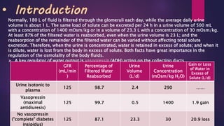
- 1. Normally, 180 L of fluid is filtered through the glomeruli each day, while the average daily urine volume is about 1 L. The same load of solute can be excreted per 24 h in a urine volume of 500 mL with a concentration of 1400 mOsm/kg or in a volume of 23.3 L with a concentration of 30 mOsm/kg. At least 87% of the filtered water is reabsorbed, even when the urine volume is 23 L; and the reabsorption of the remainder of the filtered water can be varied without affecting total solute excretion. Therefore, when the urine is concentrated, water is retained in excess of solute; and when it is dilute, water is lost from the body in excess of solute. Both facts have great importance in the regulation of the osmolality of the body fluids. A key regulator of water output is vasopressin (ADH) acting on the collecting ducts. Gain or Loss of Water in Excess of Solute (L/d) Urine Concentration (mOsm/kg H2O) Urine Volume (L/d) Percentage of Filtered Water Reabsorbed GFR (mL/min ) ……2902.498.7125 Urine isotonic to plasma 1.9 gain14000.599.7125 Vasopressin (maximal antidiuresis) 20.9 loss3023.387.1125 No vasopressin ("complete" diabetes insipidus)
- 2. I. Proximal Convoluted Tubule In the Proximal convoluted tubule, water reabsorption is always linked to Na+ reabsorption, and the mechanism is described as isosmotic. The routes of solute and water reabsorption are shown by the following steps: 1. Na+ enters the cell across the luminal membrane. Because the luminal membrane is very permeable to water, water follows the solute to maintain isosmolarity. 2. Na+ is pumped out of the cell by the Na+-K+ ATPase, which is located in the peritubular or basolateral membranes. ("Basal" refers to the cell membranes facing the peritubular capillary [2a], and "lateral" refers to the cell membranes facing the lateral intercellular spaces between cells [2b].) As Na+ is pumped out of the cell, water again follows passively. 3. The lateral intercellular space is an important route for reabsorption of solute and water. Isosmotic fluid accumulates in these spaces between the proximal tubule cells, as described in step 2. (Electron micrographs show the spaces actually widening when there is increased proximal tubule reabsorption.) This isosmotic fluid in the spaces is then acted upon by Starling forces in the peritubular capillary. The major Starling force driving reabsorption is the high oncotic pressure (πc) of peritubular capillary blood. Recall that glomerular filtration
- 3. • Important Notes: The entire proximal tubule reabsorbs 65% of the filtered water. The tight coupling between Na+ and water reabsorption is called isosmotic reabsorption. This bulk reabsorption of Na+ and water (the major constituents of ECF) is critically important for maintaining ECF volume. The proximal tubule is the site of glomerulotubular balance, a mechanism for coupling reabsorption to the GFR. Aquaporin-1 is localized to both the basolateral and apical membrane of the proximal tubules and its presence allows water to move rapidly out of the tubule along the osmotic gradients set up by active transport of solutes, and isotonicity is maintained.
- 4. Changes in ECF Volume A. ECF volume expansion produces a decrease in fractional reabsorption in the proximal tubule. When ECF volume is increased (e.g., by infusion of isotonic NaCl), the plasma protein concentration is decreased by dilution, and the capillary hydrostatic pressure (Pc) is increased. For the peritubular capillaries, these changes result in a decrease in πc and an increase in Pc. Both of these changes in Starling forces in the peritubular capillary produce a decrease in fractional reabsorption of isosmotic fluid in the proximal tubule. A portion of the fluid that would have been reabsorbed instead leaks back into the lumen of the tubule (across the tight junction) and is excreted. This alteration of glomerulotubular balance is one of several mechanisms that aids in the excretion of excess NaCl and water when there is ECF volume expansion. Glomerulotubular balance ensures that normally 65% of the filtered Na+ and water is reabsorbed in the proximal tubule. This balance is maintained because the glomerulus communicates with the proximal tubule via changes in the πc of peritubular capillary blood. However, glomerulotubular balance can be altered by changes in ECF volume. The mechanisms underlying these changes can be explained by the Starling forces in the peritubular capillaries B. ECF volume contraction produces an increase in fractional reabsorption in the proximal tubule . When ECF volume is decreased (e.g., diarrhea or vomiting), the plasma protein concentration increases (is concentrated) and the capillary hydrostatic pressure decreases. As a result, there is an increase in πc and a decrease in Pc of peritubular capillary blood. These changes in Starling forces in the peritubular capillaries produce an increase in fractional reabsorption of isosmotic fluid. This alteration of glomerulotubular balance is a logical protective mechanism, as the kidneys are trying to restore ECF volume by reabsorbing more solute and water than usual. In addition to the Starling forces, a second mechanism contributes to the increased proximal tubule reabsorption that occurs in ECF volume contraction. A decrease in ECF volume causes a decrease in blood volume and arterial pressure that activates the renin-angiotensin- aldosterone system. Angiotensin II stimulates Na+-H+ exchange in the proximal tubule, and thereby stimulates reabsorption of Na+, HCO3-, and water. Because the angiotensin II mechanism specifically
- 5. II. Loop of Henle A. Thin Descending Limb and Thin Ascending Limb The thin descending limb is permeable to water and small solutes such as NaCl and urea. In countercurrent multiplication, water moves out of the thin descending limb, solutes move into the thin descending limb, and the tubular fluid becomes progressively hyperosmotic as it flows down the descending limb. The thin ascending limb also is permeable to NaCl, but it is impermeable to water. During countercurrent multiplication, solute moves out of the thin ascending limb without water, and the tubular fluid becomes progressively hyposmotic as it flows up the ascending limb. B. Thick Ascending Limb The cells of the thick ascending limb are impermeable to water, clearly an unusual characteristic since virtually all other cell membranes are highly permeable to water. As a consequence of the water impermeability, NaCl is reabsorbed by the thick ascending limb, but water is not reabsorbed along with it. For this reason, the thick ascending limb also is called the diluting segment: Solute is reabsorbed, but water remains behind, diluting the tubular fluid. C. DISTAL TUBULE AND COLLECTING DUCT The early distal tubule is impermeable to water. Thus, it reabsorbs solute but leaves water behind, which then dilutes the tubular fluid. For this reason, the early distal tubule is called the cortical diluting segment . A key regulator of water output in the distal tubule and the collecting duct is vasopressin (ADH) acting on H2O channels ( aquaporine-2) present in Principal cells . This is called facultative water reabsorption.
- 6. Summary Of Tubular Handling Of Water Hormone ActionsCellular MechanismMajor FunctionSegment/Cell Type ...................Passive diffusion Isosmotic reabsorption of solute and water Proximal Tubule ADH stimulates Na+- K+-2Cl- cotransport Na+-K+-2Cl- cotransport Reabsorption of NaCl without water Dilution of tubular fluid Thick Ascending Limb of The Loop of the Henle PTH stimulates Ca2+ reabsorption Na+-Cl- cotransport Reabsorption of NaCl without water Early Distal Tubule ADH stimulates water reabsorption Water channels ( aquaporine-2) Variable water reabsorption Late Distal Tubule and Collecting Ducts (principal cells)