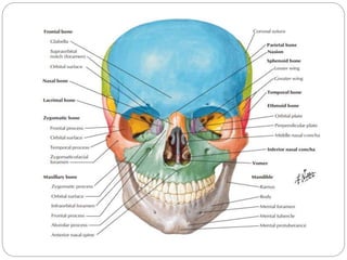
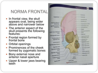
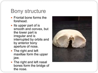
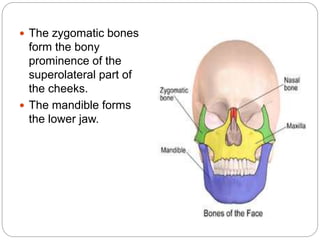
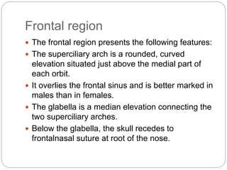
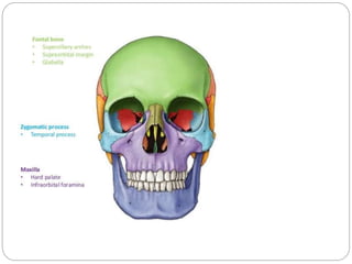
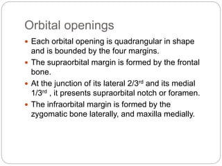
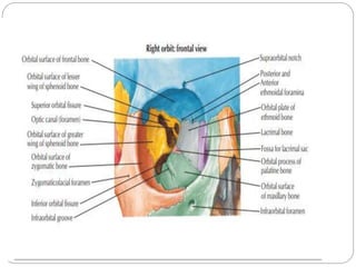
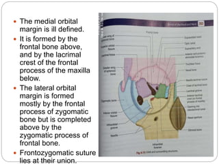
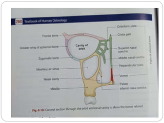
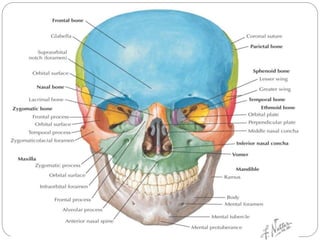
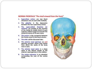
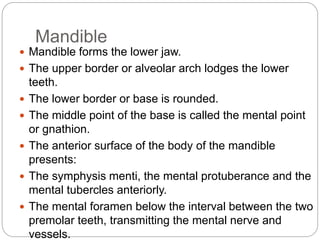
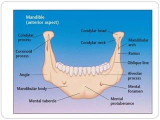
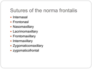
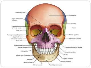
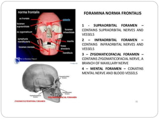
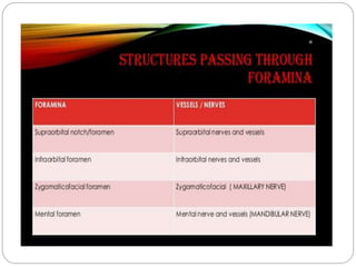
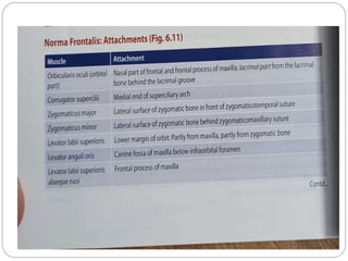
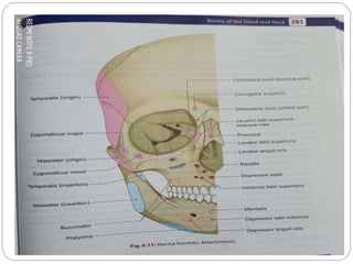
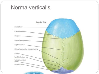
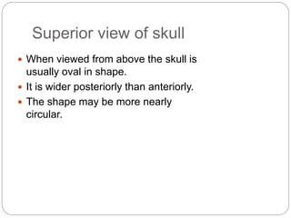
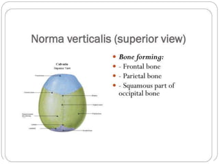
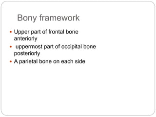
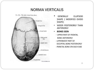
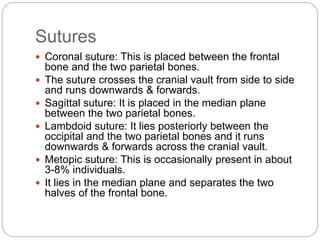
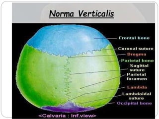
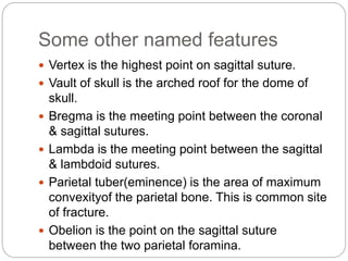
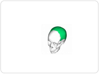
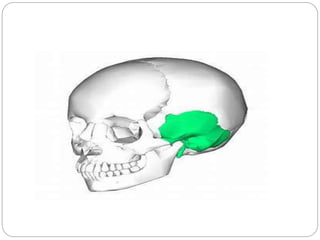
S M BADAR HAYAT
TUTOR
DEPARTMENT OF ANATOMY
NMCH, JAMUHAR
Norma frontalis &
Norma verticalis
NORMA FRONTALIS
 In frontal view, the skull
appears oval, being wider
above and narrower below
 The anterior aspect of the
skull presents the following
features:
 Frontal region formed by
frontal bone
 Orbital openings
 Prominences of the cheek
formed by zygomatic bones
 Bony external nose and
anterior nasal aperture
 Upper & lower jaws bearing
teeth
Bony structure
 Frontal bone forms the
forehead.
 Its upper part of is
smooth and convex, but
the lower part is
irregular and is
interrupted by orbits and
by anterior bony
aperture of nose.
 The right and left
maxillae form the upper
jaw.
 The right and left nasal
bones form the bridge of
the nose.
 The zygomatic bones
form the bony
prominence of the
superolateral part of
the cheeks.
 The mandible forms
the lower jaw.
Frontal region
 The frontal region presents the following features:
 The superciliary arch is a rounded, curved
elevation situated just above the medial part of
each orbit.
 It overlies the frontal sinus and is better marked in
males than in females.
 The glabella is a median elevation connecting the
two superciliary arches.
 Below the glabella, the skull recedes to
frontalnasal suture at root of the nose.
 The nasion is a median point at
the root of the nose where the
internasal suture meets with the
frontonasal suture.
 The frontal tuber or eminence is a
low rounded elevation above the
superciliary arch, one on each
side
Orbital openings
 Each orbital opening is quadrangular in shape
and is bounded by the four margins.
 The supraorbital margin is formed by the frontal
bone.
 At the junction of its lateral 2/3rd and its medial
1/3rd , it presents supraorbital notch or foramen.
 The infraorbital margin is formed by the
zygomatic bone laterally, and maxilla medially.
 The medial orbital
margin is ill defined.
 It is formed by the
frontal bone above,
and by the lacrimal
crest of the frontal
process of the maxilla
below.
 The lateral orbital
margin is formed
mostly by the frontal
process of zygomatic
bone but is completed
above by the
zygomatic process of
frontal bone.
 Frontozygomatic suture
lies at their union.
Anterior bony aperture of the
nose
 The anterior bony aperture is pear-shaped, being wide
below and narrow above.
 Boundaries:
 Above : by the lower border of the nasal bones.
 Below : by the nasal notch of the body of maxilla on
each side.
Features
 Articulations of the nasal bone:
 Anteriorly, with the opposite bone at the internasal
suture.
 Posteriorly, with the frontal process of the maxilla.
 Superiorly, with the frontal bone at the frontonasal
suture.
 Inferiorly, the upper nasal cartilage is attached to it.
 The anterior nasal spine is a sharp projection in the
median plane in the lower boundary of the piriform
aperture.
Lower part of the face
 Maxilla :
 Maxilla contributes a large share in the formation
of the facial skeleton.
 The anterior surface of the body of the maxilla
presents:
 The nasal notch medially
 The anterior nasal spine
 The infra orbital foramen, 1cm below the infra
orbital margin
 The incisive fossa above the incisor teeth, and
 The canine fossa lateral to the canine eminence
 The frontal process of the maxilla is directed
upwards.
 It articulates anteriorly with the nasal bone,
posteriorly with the lacrimal bone, and superiorly
with the frontal bone.
 The zygomatic process of the maxilla is short but
stout and articulates with the zygomatic bone.
 The alveolar process of maxilla bears sockets for
the upper teeth.
Zygomatic bone
 Zygomatic bone forms
the prominence of the
cheek.
 The zygomaticofacial
foramen is seen on its
surface.
Mandible
 Mandible forms the lower jaw.
 The upper border or alveolar arch lodges the lower
teeth.
 The lower border or base is rounded.
 The middle point of the base is called the mental point
or gnathion.
 The anterior surface of the body of the mandible
presents:
 The symphysis menti, the mental protuberance and the
mental tubercles anteriorly.
 The mental foramen below the interval between the two
premolar teeth, transmitting the mental nerve and
vessels.
Sutures of the norma frontalis
 Internasal
 Frontonasl
 Nasomaxillary
 Lacrimomaxillary
 Frontomaxillary
 Intermaxillary
 Zygomaticomaxillary
 zygomaticofrontal
Norma verticalis
Superior view of skull
 When viewed from above the skull is
usually oval in shape.
 It is wider posteriorly than anteriorly.
 The shape may be more nearly
circular.
Bony framework
 Upper part of frontal bone
anteriorly
 uppermost part of occipital bone
posteriorly
 A parietal bone on each side
Sutures
 Coronal suture: This is placed between the frontal
bone and the two parietal bones.
 The suture crosses the cranial vault from side to side
and runs downwards & forwards.
 Sagittal suture: It is placed in the median plane
between the two parietal bones.
 Lambdoid suture: It lies posteriorly between the
occipital and the two parietal bones and it runs
downwards & forwards across the cranial vault.
 Metopic suture: This is occasionally present in about
3-8% individuals.
 It lies in the median plane and separates the two
halves of the frontal bone.
Some other named features
 Vertex is the highest point on sagittal suture.
 Vault of skull is the arched roof for the dome of
skull.
 Bregma is the meeting point between the coronal
& sagittal sutures.
 Lambda is the meeting point between the sagittal
& lambdoid sutures.
 Parietal tuber(eminence) is the area of maximum
convexityof the parietal bone. This is common site
of fracture.
 Obelion is the point on the sagittal suture
between the two parietal foramina.
Skull nf
Skull nf
Skull nf
Skull nf
Skull nf

Skull nf

  • 1.
    S M BADARHAYAT TUTOR DEPARTMENT OF ANATOMY NMCH, JAMUHAR Norma frontalis & Norma verticalis
  • 3.
    NORMA FRONTALIS  Infrontal view, the skull appears oval, being wider above and narrower below  The anterior aspect of the skull presents the following features:  Frontal region formed by frontal bone  Orbital openings  Prominences of the cheek formed by zygomatic bones  Bony external nose and anterior nasal aperture  Upper & lower jaws bearing teeth
  • 4.
    Bony structure  Frontalbone forms the forehead.  Its upper part of is smooth and convex, but the lower part is irregular and is interrupted by orbits and by anterior bony aperture of nose.  The right and left maxillae form the upper jaw.  The right and left nasal bones form the bridge of the nose.
  • 5.
     The zygomaticbones form the bony prominence of the superolateral part of the cheeks.  The mandible forms the lower jaw.
  • 6.
    Frontal region  Thefrontal region presents the following features:  The superciliary arch is a rounded, curved elevation situated just above the medial part of each orbit.  It overlies the frontal sinus and is better marked in males than in females.  The glabella is a median elevation connecting the two superciliary arches.  Below the glabella, the skull recedes to frontalnasal suture at root of the nose.
  • 8.
     The nasionis a median point at the root of the nose where the internasal suture meets with the frontonasal suture.  The frontal tuber or eminence is a low rounded elevation above the superciliary arch, one on each side
  • 9.
    Orbital openings  Eachorbital opening is quadrangular in shape and is bounded by the four margins.  The supraorbital margin is formed by the frontal bone.  At the junction of its lateral 2/3rd and its medial 1/3rd , it presents supraorbital notch or foramen.  The infraorbital margin is formed by the zygomatic bone laterally, and maxilla medially.
  • 11.
     The medialorbital margin is ill defined.  It is formed by the frontal bone above, and by the lacrimal crest of the frontal process of the maxilla below.  The lateral orbital margin is formed mostly by the frontal process of zygomatic bone but is completed above by the zygomatic process of frontal bone.  Frontozygomatic suture lies at their union.
  • 13.
    Anterior bony apertureof the nose  The anterior bony aperture is pear-shaped, being wide below and narrow above.  Boundaries:  Above : by the lower border of the nasal bones.  Below : by the nasal notch of the body of maxilla on each side.
  • 14.
    Features  Articulations ofthe nasal bone:  Anteriorly, with the opposite bone at the internasal suture.  Posteriorly, with the frontal process of the maxilla.  Superiorly, with the frontal bone at the frontonasal suture.  Inferiorly, the upper nasal cartilage is attached to it.  The anterior nasal spine is a sharp projection in the median plane in the lower boundary of the piriform aperture.
  • 17.
    Lower part ofthe face  Maxilla :  Maxilla contributes a large share in the formation of the facial skeleton.  The anterior surface of the body of the maxilla presents:  The nasal notch medially  The anterior nasal spine  The infra orbital foramen, 1cm below the infra orbital margin  The incisive fossa above the incisor teeth, and  The canine fossa lateral to the canine eminence
  • 19.
     The frontalprocess of the maxilla is directed upwards.  It articulates anteriorly with the nasal bone, posteriorly with the lacrimal bone, and superiorly with the frontal bone.  The zygomatic process of the maxilla is short but stout and articulates with the zygomatic bone.  The alveolar process of maxilla bears sockets for the upper teeth.
  • 20.
    Zygomatic bone  Zygomaticbone forms the prominence of the cheek.  The zygomaticofacial foramen is seen on its surface.
  • 21.
    Mandible  Mandible formsthe lower jaw.  The upper border or alveolar arch lodges the lower teeth.  The lower border or base is rounded.  The middle point of the base is called the mental point or gnathion.  The anterior surface of the body of the mandible presents:  The symphysis menti, the mental protuberance and the mental tubercles anteriorly.  The mental foramen below the interval between the two premolar teeth, transmitting the mental nerve and vessels.
  • 23.
    Sutures of thenorma frontalis  Internasal  Frontonasl  Nasomaxillary  Lacrimomaxillary  Frontomaxillary  Intermaxillary  Zygomaticomaxillary  zygomaticofrontal
  • 30.
  • 31.
    Superior view ofskull  When viewed from above the skull is usually oval in shape.  It is wider posteriorly than anteriorly.  The shape may be more nearly circular.
  • 33.
    Bony framework  Upperpart of frontal bone anteriorly  uppermost part of occipital bone posteriorly  A parietal bone on each side
  • 35.
    Sutures  Coronal suture:This is placed between the frontal bone and the two parietal bones.  The suture crosses the cranial vault from side to side and runs downwards & forwards.  Sagittal suture: It is placed in the median plane between the two parietal bones.  Lambdoid suture: It lies posteriorly between the occipital and the two parietal bones and it runs downwards & forwards across the cranial vault.  Metopic suture: This is occasionally present in about 3-8% individuals.  It lies in the median plane and separates the two halves of the frontal bone.
  • 37.
    Some other namedfeatures  Vertex is the highest point on sagittal suture.  Vault of skull is the arched roof for the dome of skull.  Bregma is the meeting point between the coronal & sagittal sutures.  Lambda is the meeting point between the sagittal & lambdoid sutures.  Parietal tuber(eminence) is the area of maximum convexityof the parietal bone. This is common site of fracture.  Obelion is the point on the sagittal suture between the two parietal foramina.