
Recommended
More Related Content
Similar to Introduction to Skeletal system oh human
Similar to Introduction to Skeletal system oh human (20)
Recently uploaded
Recently uploaded (20)
Introduction to Skeletal system oh human
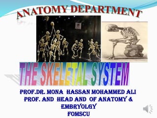
- 1. PROF.Dr. MONA HASSAN MOHAMMED ALI prof. AND HEAD AND of Anatomy & Embryolgy FOMSCU
- 2. At the end of the lecture, the students should be able to: Enumerate the functions of bone Classify bones according to shape, structure & development Enumerate the bones of axial and their functions INTENDED LEARNING OUTCOMES
- 3. Definition of bone: - it is a hard type of connective tissue which forms the skeleton. BONE
- 4. 1. Support: • Due to its rigidity and hardness, it provides the strength that keep the rigid framework that support the body. 2. Protection: • Protect vital delicate internal organs vital for life. • Examples: the skull protects the brain and the thoracic cage protects heart and lungs from external shocks. 3. Levers: • Providing anchoring points for muscles to facilitate their movements at the joints. FUNCTIONS OF BONE
- 5. • Serves as a reservoir for minerals (calcium and phosphate) • Occurs within the marrow cavities of certain bones like the sternum and heads of the tibia FUNCTIONS OF BONE
- 6. • The vertebral column transmits the weight of the head and the trunk to the bony pelvis then to the bones of the lower limb to the feet and lastly to the ground. • To make important part of the locomotor system FUNCTIONS OF BONE
- 7. • It is the system of bones associated with cartilages and joints of the human body. • Together these structures form the human skeleton. • It describes the form and organization of the body parts. SKELETAL SYSTEM
- 8. • It consists of 206 bones. • It can be studied in two parts: • A- Exoskeleton: Nail, enamel of the teeth. • B- Endoskelton: 1- Axial skeleton 2- Appendicular skeleton HUMAN SKELETON
- 9. • It consists of 80 bones • Consists of the bones that lie around the longitudinal axis of the human body. - Skull, mandible - Ossicles of the middle ear - Hyoid bone - Thoracic cage - Vertebral column 1- AXIAL SKELETON
- 11. Consists of the (126) bones of the upper and lower limbs (extremities): Upper Extremity (64) Shoulder Girdle Skeleton of the upper limb Lower Extremity (62) Pelvic Girdle Skeleton of the lower limb 2- APPENDICULAR SKELETON
- 12. 1- According to the microscopic structure: Forms outer layer of the long bone Very hard and dense Consist of delicate trabeculae filled with red or yellow marrow Withstand stress and support shifts in weight CLASSIFICATIONS OF BONES
- 13. Compact and spongy bones CLASSIFICATIONS OF BONES
- 14. Compact bones Cancellous bones CLASSIFICATIONS OF BONES
- 15. 2- According to development (bone formation): Ossification: Process of converting other tissues to bone Two types of ossification processes occur during embryological formation: ▪ Membranous ▪ Cartilagenous CLASSIFICATIONS OF BONES
- 16. Bones develop in membrane from mesenchymal cells Examples: most bones of the skull cap and the clavicle CLASSIFICATIONS OF BONES
- 17. Occurs in long bone, vertebrae, ribs. The mesenchyme is changed first into a cartilage model, then the cartilage is dissolved and disappear and changed into bone. CLASSIFICATIONS OF BONES
- 18. 3- According to the shape of bones : Long bones Short bones Flat bones Irregular bones Sesamoid bones Pneumatic bones CLASSIFICATIONS OF BONES
- 19. Length greater than width Composed of two ends (epiphyses) and a shaft (diaphysis) e.g. femur, radius, ulna, etc. CLASSIFICATIONS OF BONES
- 20. Consist of spongy bone covered by a thin layer of compact bone. Cube shaped e.g. carpal, tarsal bones CLASSIFICATIONS OF BONES
- 21. Cont.. Thin, flattened, a bit curve e.g. ribs, sternum, skull bones, scapula, etc CLASSIFICATIONS OF BONES
- 22. Cont.. Complicated irregular shapes e.g. verterbrae, facial bones, hip bone. CLASSIFICATIONS OF BONES
- 23. Embedded in some muscle tendons , (e.g: patella) Diminish the friction between the tendon and the underlying bones. CLASSIFICATIONS OF BONES
- 24. These are bones containing air cavities Example: some bones of the skull: Maxilla, Sphenoid, Ethmoid CLASSIFICATIONS OF BONES
- 25. CLASSIFICATIONS OF BONES According to bone marrow: Red. Yellow.
- 26. Gross Anatomy of a Long Bone Diaphysis Shaft Composed of compact bone Epiphysis Ends of the bone Composed mostly of spongy bone Figure 5.2a
- 27. Structures of a Long Bone Periosteum Outside covering of the diaphysis Fibrous connective tissue membrane Sharpey’s fibers Secure periosteum to underlying bone Arteries Supply bone cells with nutrients Figure 5.2c
- 28. Structures of a Long Bone Articular cartilage Covers the external surface of the epiphyses Made of hyaline cartilage Decreases friction at joint surfaces Figure 5.2a
- 29. Structures of a Long Bone Medullary cavity Cavity of the shaft Contains yellow marrow (mostly fat) in adults Contains red marrow (for blood cell formation) in infants Figure 5.2a
- 30. MAJOR BONES OF SKELETON
- 31. SKULL • It is made up of 22 bones and 6 ear ossicles: • Paired bones Unpaired bones • Temporal Frontal • Parietal Occipital • Maxilla Sphenoid • Lacrimal Ethmoid • Palatine Vomer • Zygomatic Mandible • Nasal • Inferior concha • Bones of middle ear cavity • Incus • Malleus • Stapes.
- 32. VERTEBRAL COLUMN • It is made up of 33 vertebrae, namely, 7 cervical, 12 thoracic, 5 lumbar, 5 sacral and 4 coccygeal vertebrae.
- 33. THORACIC CAGE • It consists of 12 thoracic vertebrae, 12 pairs of ribs with their costal cartilages, sternum and xiphoid process.
- 35. Clinical Anatomy by Systems, 8th Edition, Richard S. Snell. B.D . Chaurasia’s General anatomy. REFERENCES
- 36. THANK YOU