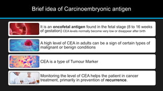
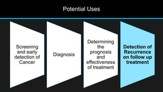
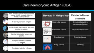
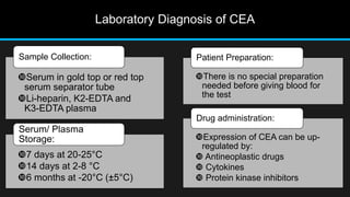
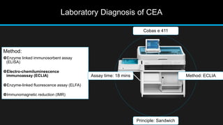
Serum CEA as Tumour Marker
Saswata Chakraborty | DMLT | Tata Medical Centre Date: 10.03.2023
Brief idea of Carcinoembryonic antigen
It is an oncofetal antigen found in the fetal stage (8 to 16 weeks
of gestation) CEA levels normally become very low or disappear after birth
A high level of CEA in adults can be a sign of certain types of
malignant or benign conditions
CEA is a type of Tumour Marker
Monitoring the level of CEA helps the patient in cancer
treatment, primarily in prevention of recurrence.
Tumour is an abnormal mass of tissue that results from
excessive cell division that is uncontrolled and progressive
DIAGNOSIS OF TUMOUR
Patient history and Clinical
examination
Imaging: X-Ray,
Ultrasonography, CT, MRI
Biochemical assays:
Laboratory analysis of
Tumour marker
Histological and Cytological
Molecular Method.
Tumour Marker
Tumour Marker:
Substances present in the tumour
or produced by the tumour or by
the host.
It provides an information about
biological characteristics of the
tumour.
DIAGNOSIS OF TUMOUR
Patient history and Clinical
examination
Imaging: X-Ray,
Ultrasonography, CT, MRI
Biochemical assays: Laboratory
analysis of Tumour marker
Histological and
Cytological
Molecular Method.
Characteristics of an ideal Tumour Marker
High sensitivity and
specificity
• Should be able to differentiate
between malignant and benign
disease
• Should show positive correlation
with tumor volume and extent
Early prediction of
recurrence
Must show indication of
changes in a patient
receiving treatment
Cost effective and easy to
assay in laboratory
Potential Uses
Screening
and early
detection of
Cancer
Diagnosis
Determining
the
prognosis
and
effectiveness
of treatment
Detection of
Recurrence
on follow up
treatment
Carcinoembryonic Antigen (CEA)
First isolated from human colorectal
cancer (CRC) tissue in 1965 by
Gold and Freedman
It is a Glycoprotein antigen
Found on the cell surfaces and shed
into the circulation
Can be detected in serum and
plasma
Biological reference interval:
<3.8 ng/mL
Circulation half-life: 1-5 days
Elevated in Malignancy Elevated in Benign
Conditions
Colorectal cancer Chronic obstructive
pulmonary disease
Pancreatic cancer Peptic bowel disease
Breast cancer Crohn’s disease
Lung cancer Smoking
Laboratory Diagnosis of CEA
Serum in gold top or red top
serum separator tube
Li-heparin, K2-EDTA and
K3-EDTA plasma
Sample Collection:
7 days at 20-25°C
14 days at 2-8 °C
6 months at -20°C (±5°C)
Serum/ Plasma
Storage:
There is no special preparation
needed before giving blood for
the test
Patient Preparation:
Expression of CEA can be up-
regulated by:
 Antineoplastic drugs
 Cytokines
 Protein kinase inhibitors
Drug administration:
Laboratory Diagnosis of CEA
Method:
Enzyme linked immunosorbent assay
(ELISA)
Electro-chemiluminescence
immunoassay (ECLIA)
Enzyme-linked fluorescence assay (ELFA)
Immunomagnetic reduction (IMR)
Cobas e 411
Method: ECLIA
Principle: Sandwich
Assay time: 18 mins
TEST PRINCIPLE
1st incubation:
10 µL of sample, a Biotinylated
monoclonal CEA-specific
antibody and a Monoclonal
CEA-specific antibody labeled
with a ruthenium complex react
to form a sandwich complex.
2nd incubation:
After addition of streptavidin-
coated microparticles, the
complex becomes bound to the
solid phase via interaction of
biotin and streptavidin.
The reaction mixture is aspirated
into the measuring cell where the
microparticles are magnetically
captured onto the surface of the
electrode.
Unbound substances are then
removed with ProCell/ProCell M.
Application of a voltage to the
electrode then induces
chemiluminescent emission which
is measured by a photomultiplier.
Expected Values
All subjects Non-smokers
(past/never
smokers)
Smokers (current)
Age (years) 20-69 40-69 20-69 40-69 20-69 40-69
95th percentile
(ng/mL)
4.7 5.2 3.8 5.0 5.5 6.5
N 352 203 242 154 110 49
Studies with the Elecsys CEA assay were performed on 352 healthy subjects. The
following results were obtained:
Calibration and Quality Control
Calibration Quality Control
Controls for the various concentration ranges
should be run individually at least once every
24 hours when the test is in use
Once per reagent kit
Following each calibration
• Calibration must be performed once per
reagent lot using fresh reagent
• After 28 days when using the same
reagent lot
• After 7 days (when using the same
reagent kit on the analyzer)
• Situations where quality control findings
are outside of the defined limits
Renewed
calibration is
recommended
as follows:
Quality Control: Level 1
N: 24
Mean: 25.14
SD: 0.57 CV(%): 2.2
Quality Control: Level 2
N: 24
Mean: 53.39
SD: 1.32 CV(%): 2.47
CEA in management of recurrence
Source: A Review of the Role of Carcinoembryonic Antigen in Clinical Practice
CEA in management of Cancer
Source: A Review of the Role of Carcinoembryonic Antigen in Clinical Practice
Things to remember
Healthy individuals has a low level of CEA (<5 ng/mL)
CEA level elevates in both Malignant and Benign conditions
Smokers may have an elevated level of CEA
CEA not used for screening or diagnostic purpose
CEA measurements helps to prevent Recurrence of colorectal cancer
References
• Colorectal Cancer in India: An Audit from a Tertiary Center in a Low Prevalence Area
• Tumor markers in clinical practice: General principles and guidelines
• Laboratory Testing for Tumour Markers
• Elecsys CEA
• Carcinoembryonic Antigen as a Marker for Colorectal Cancer: Is It Clinically Useful?
• A Review of the Role of Carcinoembryonic Antigen in Clinical Practice
Thank You

Serum CEA as Tumor Marker

  • 1.
    Serum CEA asTumour Marker Saswata Chakraborty | DMLT | Tata Medical Centre Date: 10.03.2023
  • 2.
    Brief idea ofCarcinoembryonic antigen It is an oncofetal antigen found in the fetal stage (8 to 16 weeks of gestation) CEA levels normally become very low or disappear after birth A high level of CEA in adults can be a sign of certain types of malignant or benign conditions CEA is a type of Tumour Marker Monitoring the level of CEA helps the patient in cancer treatment, primarily in prevention of recurrence.
  • 3.
    Tumour is anabnormal mass of tissue that results from excessive cell division that is uncontrolled and progressive DIAGNOSIS OF TUMOUR Patient history and Clinical examination Imaging: X-Ray, Ultrasonography, CT, MRI Biochemical assays: Laboratory analysis of Tumour marker Histological and Cytological Molecular Method.
  • 4.
    Tumour Marker Tumour Marker: Substancespresent in the tumour or produced by the tumour or by the host. It provides an information about biological characteristics of the tumour. DIAGNOSIS OF TUMOUR Patient history and Clinical examination Imaging: X-Ray, Ultrasonography, CT, MRI Biochemical assays: Laboratory analysis of Tumour marker Histological and Cytological Molecular Method.
  • 5.
    Characteristics of anideal Tumour Marker High sensitivity and specificity • Should be able to differentiate between malignant and benign disease • Should show positive correlation with tumor volume and extent Early prediction of recurrence Must show indication of changes in a patient receiving treatment Cost effective and easy to assay in laboratory
  • 6.
    Potential Uses Screening and early detectionof Cancer Diagnosis Determining the prognosis and effectiveness of treatment Detection of Recurrence on follow up treatment
  • 7.
    Carcinoembryonic Antigen (CEA) Firstisolated from human colorectal cancer (CRC) tissue in 1965 by Gold and Freedman It is a Glycoprotein antigen Found on the cell surfaces and shed into the circulation Can be detected in serum and plasma Biological reference interval: <3.8 ng/mL Circulation half-life: 1-5 days Elevated in Malignancy Elevated in Benign Conditions Colorectal cancer Chronic obstructive pulmonary disease Pancreatic cancer Peptic bowel disease Breast cancer Crohn’s disease Lung cancer Smoking
  • 8.
    Laboratory Diagnosis ofCEA Serum in gold top or red top serum separator tube Li-heparin, K2-EDTA and K3-EDTA plasma Sample Collection: 7 days at 20-25°C 14 days at 2-8 °C 6 months at -20°C (±5°C) Serum/ Plasma Storage: There is no special preparation needed before giving blood for the test Patient Preparation: Expression of CEA can be up- regulated by:  Antineoplastic drugs  Cytokines  Protein kinase inhibitors Drug administration:
  • 9.
    Laboratory Diagnosis ofCEA Method: Enzyme linked immunosorbent assay (ELISA) Electro-chemiluminescence immunoassay (ECLIA) Enzyme-linked fluorescence assay (ELFA) Immunomagnetic reduction (IMR) Cobas e 411 Method: ECLIA Principle: Sandwich Assay time: 18 mins
  • 10.
    TEST PRINCIPLE 1st incubation: 10µL of sample, a Biotinylated monoclonal CEA-specific antibody and a Monoclonal CEA-specific antibody labeled with a ruthenium complex react to form a sandwich complex. 2nd incubation: After addition of streptavidin- coated microparticles, the complex becomes bound to the solid phase via interaction of biotin and streptavidin. The reaction mixture is aspirated into the measuring cell where the microparticles are magnetically captured onto the surface of the electrode. Unbound substances are then removed with ProCell/ProCell M. Application of a voltage to the electrode then induces chemiluminescent emission which is measured by a photomultiplier.
  • 11.
    Expected Values All subjectsNon-smokers (past/never smokers) Smokers (current) Age (years) 20-69 40-69 20-69 40-69 20-69 40-69 95th percentile (ng/mL) 4.7 5.2 3.8 5.0 5.5 6.5 N 352 203 242 154 110 49 Studies with the Elecsys CEA assay were performed on 352 healthy subjects. The following results were obtained:
  • 12.
    Calibration and QualityControl Calibration Quality Control Controls for the various concentration ranges should be run individually at least once every 24 hours when the test is in use Once per reagent kit Following each calibration • Calibration must be performed once per reagent lot using fresh reagent • After 28 days when using the same reagent lot • After 7 days (when using the same reagent kit on the analyzer) • Situations where quality control findings are outside of the defined limits Renewed calibration is recommended as follows:
  • 13.
    Quality Control: Level1 N: 24 Mean: 25.14 SD: 0.57 CV(%): 2.2
  • 14.
    Quality Control: Level2 N: 24 Mean: 53.39 SD: 1.32 CV(%): 2.47
  • 15.
    CEA in managementof recurrence Source: A Review of the Role of Carcinoembryonic Antigen in Clinical Practice
  • 16.
    CEA in managementof Cancer Source: A Review of the Role of Carcinoembryonic Antigen in Clinical Practice
  • 17.
    Things to remember Healthyindividuals has a low level of CEA (<5 ng/mL) CEA level elevates in both Malignant and Benign conditions Smokers may have an elevated level of CEA CEA not used for screening or diagnostic purpose CEA measurements helps to prevent Recurrence of colorectal cancer
  • 18.
    References • Colorectal Cancerin India: An Audit from a Tertiary Center in a Low Prevalence Area • Tumor markers in clinical practice: General principles and guidelines • Laboratory Testing for Tumour Markers • Elecsys CEA • Carcinoembryonic Antigen as a Marker for Colorectal Cancer: Is It Clinically Useful? • A Review of the Role of Carcinoembryonic Antigen in Clinical Practice
  • 19.

Editor's Notes

  • #4 Summarize your research in three to five points.
  • #5 Summarize your research in three to five points.
  • #6 Summarize your research in three to five points.
  • #7 Summarize your research in three to five points.
  • #11 Summarize your research in three to five points.
  • #16 List all of the steps used in completing your experiment. Remember to number your steps.
  • #17 List all of the steps used in completing your experiment. Remember to number your steps.