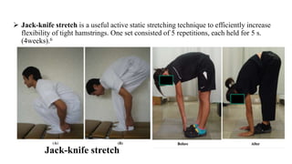
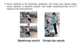
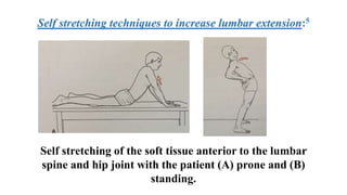
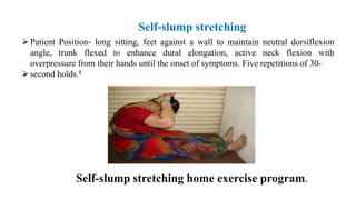
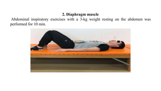
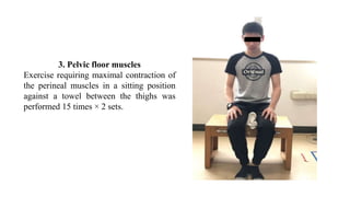
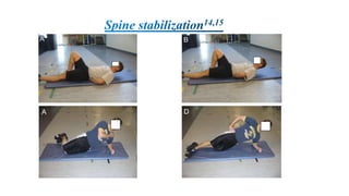
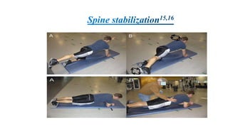
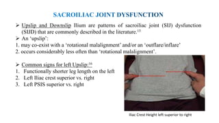
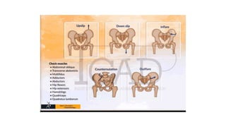
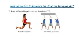
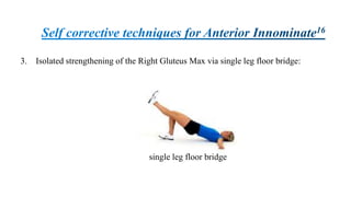
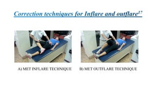
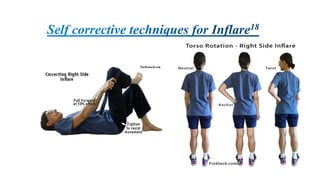
This document discusses various exercises and techniques for rehabilitation of chronic lower back pain (CLBP). It finds that core stability exercises are more effective than stretching alone in improving tonic core muscle strength and endurance for CLBP. Several specific exercises are described to target the transverse abdominis, multifidus, diaphragm and pelvic floor muscles. Stretching techniques for the hamstrings, quadriceps and other muscle groups are also outlined. The document further discusses techniques for addressing sacroiliac joint dysfunction issues like upslips and downslips.
The document provides an overview of gait analysis, including both subjective and objective analysis techniques. It discusses measuring kinematic parameters like joint angles and kinetic parameters like ground reaction forces. Key aspects of gait like temporal-spatial measures, determinants of gait, and gait physiology are explained. Common gait analysis equipment is also outlined, including force plates, motion capture systems, and EMG. The document concludes with an example case study of calculating joint moments and loads at the knee for a person using a trans-tibial prosthesis.
This document discusses the pathomechanics of ankle joint injuries. It begins with the anatomy and ligaments of the ankle joint. It then discusses the muscle groups around the ankle joint and their actions. Next, it explores the mechanics of ankle motion and different types of ankle injuries including lateral and medial ligament injuries, fractures, and muscular imbalances. It provides details on specific muscles like the tibialis anterior and their weaknesses or tightnesses. It concludes with discussing chronic ankle instability and recent literature on lateral ankle sprains and reinjury rates. In summary, the document provides an in-depth overview of ankle joint anatomy, mechanics, common injuries and their pathomechanics, as well as muscular factors.
The document discusses the basics of soft tissue examination for practitioners who treat the human structure using their hands. It emphasizes the importance of functional testing through observation, palpation, and passive and contractile muscle testing to determine the source of soft tissue pain. Dr. James Cyriax developed orthopedic medicine based on examining, diagnosing and treating non-surgical lesions of the musculoskeletal system through assessing soft tissues and their patterns of limitation.
This document discusses strategies to reduce force on the hip joint for individuals with hip osteoarthritis or weak hip abductor muscles. It analyzes using a lateral lean, cane on the same side, or cane on the opposite side. A lateral lean reduces gravitational torque but increases energy expenditure. A cane on the same side provides some relief but a cane on the opposite side may offset gravity's torque, reducing the need for abductor muscle force and joint compression to just body weight. However, the full distance between hand and hip may overestimate the cane's effectiveness.
The document discusses the biomechanics of respiration including the structure and function of the ribs, ribcage muscles, and accessory muscles involved in breathing. The diaphragm is the primary muscle of inspiration and contracts to increase the vertical diameter of the thorax. The external intercostal muscles elevate the ribs during inspiration while the internal intercostals depress the ribs during expiration. Accessory muscles such as the scalenes, sternocleidomastoid, and pectorals assist with forced breathing by stabilizing the ribcage and sternum.
This document discusses various types of pathological and abnormal gaits. It begins by outlining common causes of abnormal gait such as pain, joint limitations, muscle weakness, neurological involvement, and leg length discrepancies. It then describes specific gaits in more detail, including antalgic gait, psoatic gait, gluteus maximus gait, gluteus medius gait, quadriceps gait, genu recurvatum gait, hemiplegic gait, scissoring gait, dragging gait, cerebellar ataxic gait, sensory ataxic gait, short shuffling gait, foot drop gait, equinus gait, calcaneal g
This document provides guidance on checking various aspects of a lower limb prosthesis. It discusses checking the prosthesis and patient's stump in general, as well as when sitting, standing, walking, and with the prosthesis removed. Checks include ensuring proper fit and alignment of socket components, comfort and stability of the patient, and identification of any potential issues. The document focuses on checkouts for above knee prosthetics but also briefly discusses below knee prosthetics. The goal of prosthetic checkouts is to assess proper functioning and make any necessary adjustments before training the patient.
This document provides an overview of biomechanics of the sacroiliac joints. It discusses the osteology, articulating surfaces, ligaments, blood and nerve supply, factors promoting stability, kinematics, and functional considerations of the sacroiliac joints. It also covers clinical anatomy and sacroiliac dysfunction. The sacroiliac joints connect the sacrum to the iliac bones and allow for slight motion including rotation and translation while maintaining stability through interlocking surfaces and strong ligaments. Proper functioning of the sacroiliac joints is important for load transfer and movements including those related to childbirth.
The document discusses various aspects of competitive swimming strokes and biomechanics. It describes the phases and techniques of the front crawl, backstroke, breaststroke, and butterfly strokes. Key factors that influence swimming speed and efficiency are also examined, such as stroke length, stroke frequency, propulsive forces, and streamlining techniques.
The document discusses the biomechanics of the knee joint, including the tibiofemoral joint and patellofemoral joint. It covers the articulating surfaces, degrees of freedom, ligaments, muscles, alignment and weight bearing forces of the knee. It also discusses normal patellar tracking in the trochlear groove during range of motion and the changing contact areas between the patella and femur through different degrees of flexion.
Muscle Testing of the Trunk
Prof. Satyen Bhattacharyya
Associate Professor: BIMLS, Bardhaman
Chief Physio: Fit O Fine
Director: Well O Fit Healthcare PVT. LTD.
Muscle Testing of the Trunk
Trunk Flexion
Rectus abdominis
Muscles contribute to Trunk Flexion Rectus abdominis
Origin:
Pubic crest and pubic symphysis
Insertion:
5, 6, 7 costal cartilages, medial inferiorcostal margin and posterior aspect of xiphoid
Action:
Trunk Flexion
Nerve supply:
Normal
Position:
Supine with hands behind neck.
Stabilization:
Stabilize legs firmly.
Desired Motion:
Patient flexes thorax on pelvis through ROM
Normal
Note:
If hip flexor muscles are weak, stabilize pelvis.
A curl up is emphasized, and flexion is possible until scapulae are raised from table.
Tests for neck flexion should precede those for trunk flexion
Good
Position:
Back lying with arms at sides.
Stabilization:
Stabilize legs firmly.
Desired Motion:
Patient flexes thorax on pelvis through range of motion.
If hip flexor muscles are weak, stabilize pelvis.
Flexion is possible until scapula are raised from table.
Fair
Position:
Supine with arms at sides.
Stabilization:
Stabilize legs firmly.
Desired Motion:
Patient flexes thorax on pelvis through partial range of motion.
Head, tips of shoulders and cranial borders of scapulae should clear table with inferior angle remaining in contact with table.
If hip flexor muscles are weak, stabilize pelvis
Poor
Position:
Supine with arms at sides
Desired Motion:
Patient flexes cervical spine.
Caudal portion of thorax is depressed, and pelvis is tilted until the lumbar area of spine is flat on table.
Palpation will help to determine smoothness of contraction
Trace & Zero
Position:
Supine
Observation:
A slight contraction may be determined by palpation over anterior abdominal wall as patient attempts to cough (also during rapid exhalation or as patient attempts to lift head).
Observe deviation of umbilicus.
Cranial movement indicates stronger contraction of upper section of muscle, and caudal movement, stronger contraction of lower section (not illustrated.)
Note
Factors Limiting Motion:
1- Tension of posterior longitudinal ligament, ligamenta flava, and interspinal and supraspinal ligaments
2- Tension of spinal extensor muscles
3-Apposition of caudal lips of vertebra bodies anteriorly with surfaces of subjacent vertebrae
4-Compression of ventral part of intervertebral fibrocartilages
5-Contact of last ribs with abdomen
Fixation:
1-Reverse action of hip flexor muscles
2-Weight of legs and pelvis
Trunk Extension
Muscles contribute to Trunk Extension Erector spinae – Spinalis
Origin:
Spinous processes
Insertion:
Spinous processes six levels above
Action:
Trunk Extension
Nerve supply:
Dorsal rami of spinal nerves
Muscles contribute to Trunk Extension Erector spinae – lliocostalis
Origin:
Iliac crest, sacrum, lumbar vertebrae
Insertion:
Ribs, cervical transverse processes
Action:
Trunk Extension
Nerve supply:
Dorsal ram
Ligaments connect bone to bone, while tendons connect muscle to bone. Both have a hierarchical structure consisting of fascicles containing fibrils and fibroblasts. Ligaments are more elastic and flexible than tendons, which have great tensile strength to transfer forces from muscles to bones. Both ligaments and tendons are viscoelastic and exhibit nonlinear mechanical properties like stress relaxation and creep. Their biomechanical properties can be affected by factors like aging, pregnancy, mobilization, and various medical conditions or treatments.
This document discusses different types of postures including inactive, active, static, and dynamic postures. It defines posture as the alignment of the body with support during muscular activity or movement. Active postures require integrated muscle function to maintain a specific posture, either statically or dynamically. Good posture allows maximum efficiency with minimum effort while poor posture fails to benefit the intended function. Factors like illness, fatigue, pain and improper mental conditioning can contribute to poor posture.
1. Gait is the series of rhythmic, alternating movements of the trunk and limbs that result in forward body progression. It involves a repeated cycle of controlled falls as the body's center of gravity moves forward over a stable foot.
2. The gait cycle is defined as the period from initial contact of one foot to the subsequent initial contact of the same foot. It consists of the stance phase, where the foot is on the ground, and the swing phase, where the foot is off the ground.
3. Normal gait involves specific motion patterns at the hips, knees, ankles, and trunk to minimize the vertical and horizontal excursion of the body's center of gravity, thereby reducing energy
This topic contains the core topic in Biomechanics which serves as the foundation for curriculum of Undergraduate and Post graduate Physiotherapy students.This presentation aims at the clarification of foundational concepts.
The document discusses the biomechanics of sit-to-stand (STS) movement. STS is an important daily activity that requires moving the center of mass from a stable seated position to an unstable standing position. It involves four phases - flexion momentum, momentum transfer, extension, and stabilization. Kinematics include pelvic tilt, trunk extension, hip and knee flexion/extension. Kinetics involve using leg, back and arm muscles to generate momentum to rise from sitting to standing and stabilize in the upright position. Proper timing and coordination of body segments is important for effective STS.
The document discusses core stability and core muscles. The core, or lumbo-pelvic-hip complex, consists of 29 muscles including the abdominal muscles, back muscles, and hip muscles. It is the center of gravity and where all movement originates. Core training exercises like planks, bridges, and exercises using a Swiss ball can improve posture, muscle balance, stabilization, and prevent low back pain by developing efficient neuromuscular control since all movement originates from the core. Sample exercises described are planks, bridges, planks on a Swiss ball, Swiss ball curls, supermans, and more.
Human locomotion, or walking, involves alternating between a stance phase where one foot is on the ground and a swing phase where the foot is off the ground. The gait cycle can be divided into these two phases and further subdivided. There are many types of gait deviations that can occur due to injuries, diseases or other impairments affecting the nervous system, muscles or bones. Common causes of gait deviations include stroke, cerebral palsy, spinal cord injuries or tumors, neurological conditions like Parkinson's disease or multiple sclerosis, and musculoskeletal problems. Kinesiotherapists evaluate gait to identify deviations and their causes in order to develop treatment plans.
This document discusses anatomical planes and axes of movement. There are three planes: the sagittal plane which divides the body into right and left parts and involves movements like walking; the frontal plane which divides the body into front and back, like in star jumps; and the transverse plane which divides the body into superior and inferior parts and involves rotations. There are also three axes: the vertical axis which runs top to bottom; the sagittal axis from left to right; and the frontal axis from back to front. Certain movements occur within each plane, like flexion and extension in the sagittal plane.
The document provides information on rolling and crawling/creeping movements. It defines rolling as moving forward along a surface by revolving on an axis or repeatedly turning over, and defines crawling as moving slowly on hands and knees while creeping is slow movement by dragging the prone body. The document outlines the typical development sequence of these movements in infants from 2-10 months. It describes mat activities to strengthen muscles and mobilize the body that incorporate these movements in different positions.
The normal ROM for each hip motion is provided along with positioning details for accurate goniometric measurement. Precautions and common limiting factors are also outlined to ensure safe assessment.
This document discusses the biomechanics of lifting. It defines lifting as moving an object from one location to another, generally involving vertical and horizontal movement. There are two main types of lifts discussed: stoop lifting, which involves trunk flexion without knee bending, and squat lifting, which keeps the spine erect and bends at the hips and knees. Squat lifting is preferable as it reduces pressure on the discs of the spine compared to stoop lifting. Proper lifting technique involves keeping loads close to the body, using both hands to hold the load securely, and choosing the appropriate lift type based on the load and situation.
Hello everyone here I upload mckenzie exrercise basic details and some of its position.Its technique for use to cervical, Lumabar pain relief via particular position.Thank you.
Postural Restoration (PRITM) is a neurological based movement approach developed by Ron Hruska to identify and address postural dysfunctions. It uses objective tests to categorize clients into patterns based on overactive muscle chains and imbalances between right and left sides of the body. Treatment focuses on repositioning the pelvis, ribcage, and head to achieve neutral alignment and restoring symmetrical, reciprocal movement. Key concepts include the diaphragm's role in directing spine position, imbalances between the right and left halves of the diaphragm, and achieving the zone of apposition for optimal diaphragm and respiratory function. The goal is to improve neuromotor balance and integrate all systems of the
This document discusses different types of crawling exercises used in physiotherapy. It describes five types of crawls - dog's crawl, low dog's crawl, arm stretch crawl, leg shift crawl, and low 'S' crawl. Each type is explained, including starting position and technique. Crawling exercises are used to improve coordination, endurance, spinal mobility, and control excessive mobility. They also help build reciprocal arm and leg movement like walking. Resistance can be added and different starting positions impact the effects. Crawling provides a safe way to move for those with poor balance.
This document discusses the physiotherapy management of lower limb deformities resulting from polio. It covers strengthening weakened muscles, stretching shortened muscles, use of orthotics and splints, gait training, surgical correction of deformities if conservative treatment is not effective, and physiotherapy after surgeries like tendon transfers. Specific deformities around the hip, knee and ankle are described along with their causes and management approaches. Surgical options discussed include soft tissue releases, osteotomies, arthrodesis and tendon transfers. The overall goal of treatment is to improve strength, prevent deformities, achieve functional mobility and independence.
Abdominal Exercises: A Review Study For Training Prescriptioninventionjournals
The abdominal muscles are composed of four muscles that must be trained in the same way as the other muscles of the body.This mucular group are very important for postural control in exercises and prevention of low back pain. The objective of this study was to review some questions about abdominal exercises and their practical application, helping the work of the professional training prescription.We used articles in the database: Scielo, Pubmed and SciencDirect resulting in a total of 24 articles used to produce this paper. Knowledge of the issues related to the Electromyographic Activity; stabilisation exercises and abdominal therapeutic exercises is of fundamental importance for professionals working with human movement.
This document discusses hip disorders and treatment techniques including muscle energy technique (MET), soft tissue technique, and Mulligan technique. It provides details on hip anatomy, ligaments, muscles and movements. It then describes MET techniques for various muscles like the quadriceps, illiopsoas, hamstrings, adductors, and tensor fascia lata/iliotibial band. Soft tissue techniques like effleurage, stripping, pin and stretch, and friction are explained. Specific conditions like piriformis syndrome, sacroiliac joint dysfunction, trochanteric bursitis, and anterior/posterior/lateral pelvic tilts are addressed with relevant soft tissue techniques.
This document discusses exercises to build arm muscles. It begins by describing the muscles of the arms, including the biceps and triceps. It then discusses important arm exercises like triceps extensions, biceps curls, and chin-ups. Details are provided on how to perform each exercise properly with good form. Variations that make exercises easier or more difficult are also described. The goal is to provide a well-rounded workout for both the biceps and triceps muscles.
This document discusses the anatomy and biomechanics of the shoulder as well as osteopathic manipulative techniques for treating shoulder issues. It outlines the static and dynamic stabilizers of the shoulder including bones, ligaments, muscles, and soft tissues. Two specific techniques are described in detail - the Spencer technique which uses articulatory motions to increase shoulder range of motion, and myofascial release of the scapula to improve scapular motion and stability. The goal of osteopathic manipulation is to maintain efficient muscle balance and flexibility in the shoulder complex.
The HAPI 'Hip Arthroscopy Pre-habilitation Intervention' Study : Does pre-habilitation affect outcomes in patients undergoing hip arthroscopy for femoro-acetabular impingement?
Patient exercise guide document
The core muscles can be categorized as stabilizers or mobilizers. Stabilizers like the transversus abdominis and multifidus are deeply placed, have slow twitch fibers, and help provide stability. Mobilizers like the rectus abdominis are more superficial and have fast twitch fibers for movement. Chronic low back pain is associated with weakness in the transversus abdominis and multifidus as well as decreased flexibility. A core strengthening program focuses on training these local stabilizer muscles in three stages: 1) local segmental control, 2) closed chain exercises, and 3) open chain exercises and functional progression. Exercises target the transversus abdominis, multifidus, and glute
This case study describes the physical therapy treatment of a 61-year-old male with a partially healed proximal humeral fracture and rotator cuff tear in his right shoulder. He had limited range of motion and pain with movement after 8 weeks of immobilization. The treatment plan involved joint mobilization techniques to increase shoulder range of motion, as well as strengthening exercises to improve muscular strength. The goals were to restore normal motion and strength without exacerbating pain.
JOINT MOBILTY IN PHYSIOTHERAPY PPT FILESbharti pawar
This document provides information about joint mobility techniques for various joints of the body. It begins with definitions of joint mobility and common causes of limited mobility. It then describes techniques for assessing and improving range of motion in major joints like the shoulder, elbow, wrist, hip, knee, ankle, cervical spine, and lumbar spine. Diagrams show hand placement and procedures for passive range of motion exercises targeting specific motions, like flexion, extension, lateral flexion, and rotation. The goal is to safely increase range of motion and maintain or restore mobility.
This document defines thoracic kyphosis as an excessive backward curvature of the spine in the thoracic region. It is caused by factors like poor posture, arthritis, lung issues, and diseases affecting the vertebrae. There are different types including round and angular kyphosis. Management involves exercises to improve mobility, posture training, manual mobilization, and bracing for more severe cases. Rehabilitation approaches aim to stretch tightened areas and strengthen weakened muscles to reduce the deformity.
This document discusses the biomechanics of the hip joint. It begins by defining biomechanics as the science examining forces acting on biological structures. It then describes the hip as both mobile and stable due to its strong bones, powerful muscles, and ligaments. The document goes on to discuss topics such as the femoral neck angle, acetabular version, muscles, joint reaction forces, gait biomechanics, and the effects of conditions like osteoarthritis. It concludes by covering the history and principles of hip biomechanics in total hip arthroplasty, including how procedures aim to decrease joint reaction forces.
This document provides descriptions of various standing, kneeling, sitting and lying positions used in kinesiology. It discusses the key muscle groups engaged in each position and their effects and uses. Some of the positions described include standing, stride standing, half kneeling, kneel sitting, prone kneeling, stride sitting and cross sitting. For each one, the document outlines the muscles worked, the biomechanical advantages of the position, and how it can be used for specific exercises.
USMLE MSK L002 Back Ligamnets and muscles of back.pdfAHMED ASHOUR
The anatomy of the back is complex and involves a combination of bones, muscles, nerves, and other structures that provide support, protection, and mobility.
The back is generally divided into several regions, including the cervical, thoracic, lumbar, sacral, and coccygeal regions.
Understanding the anatomy of the back is essential for healthcare professionals, including orthopedic specialists, physical therapists, and chiropractors, as well as for individuals interested in maintaining back health and preventing injuries.
ACTIVITY AND EXERCISE NURSING FOUNDATION OF NURSINGKrishna Gandhi
This document discusses various types of exercises that can be performed during physical therapy or rehabilitation. It describes active, passive, resistive, isometric, and active-assistive exercises. Active exercises are performed independently by the patient without assistance in order to increase strength. Passive exercises are carried out by the nurse without patient effort. Resistive exercises involve the patient pushing or pulling against resistance. Isometric exercises involve contracting muscles without joint movement. Active-assistive exercises are performed with some nurse assistance to encourage normal muscle function. Range of motion exercises are also discussed.
Effect of yogic asana on Adhesive capsulitis (frozen shoulder) to increasing ...Sports Journal
This document summarizes a study that examined the effects of sleeper stretch exercises and yogic asanas on pain level and range of motion (ROM) for internal rotation in patients with frozen shoulder. Forty patients were randomly assigned to two groups - Group A received sleeper stretches daily for a month, while Group B performed various yogic asanas daily for a month. Pain levels and ROM were measured before and after the interventions. The study found that Group B saw a statistically significant reduction in pain and improvement in ROM, while Group B saw no significant changes. The study concluded that yogic asanas were more effective than sleeper stretches for treating frozen shoulder by increasing flexibility and strengthening muscles.
This document discusses spinal muscle anatomy and function. It notes that traditional anatomy descriptions only viewed muscles from the back view rather than how they create movement. The core muscles work synergistically to stiffen the trunk and prevent motion rather than initiate it. True stability comes from balanced stiffening from the entire torso musculature, not singular muscles. It provides examples of potential "energy leaks" that can lead to injury and discusses exercises to train the core muscles while avoiding energy leaks.
Similar to Self correction techniques for biomechanical problems related to spine (20)
Let's Talk About It: Breast Cancer (What is Mindset and Does it Really Matter?)bkling
Your mindset is the way you make sense of the world around you. This lens influences the way you think, the way you feel, and how you might behave in certain situations. Let's talk about mindset myths that can get us into trouble and ways to cultivate a mindset to support your cancer survivorship in authentic ways. Let’s Talk About It!
Empowering ACOs: Leveraging Quality Management Tools for MIPS and BeyondHealth Catalyst
Join us as we delve into the crucial realm of quality reporting for MSSP (Medicare Shared Savings Program) Accountable Care Organizations (ACOs).
In this session, we will explore how a robust quality management solution can empower your organization to meet regulatory requirements and improve processes for MIPS reporting and internal quality programs. Learn how our MeasureAble application enables compliance and fosters continuous improvement.
Under Pressure : Kenneth Kruk's StrategyKenneth Kruk
Kenneth Kruk's story of transforming challenges into opportunities by leading successful medical record transitions and bridging scientific knowledge gaps during COVID-19.
The best massage spa Ajman is Chandrima Spa Ajman, which was founded in 2023 and is exclusively for men 24 hours a day. As of right now, our parent firm has been providing massage services to over 50,000+ clients in Ajman for the past 10 years. It has about 8+ branches. This demonstrates that Chandrima Spa Ajman is among the most reasonably priced spas in Ajman and the ideal place to unwind and rejuvenate. We provide a wide range of Spa massage treatments, including Indian, Pakistani, Kerala, Malayali, and body-to-body massages. Numerous massage techniques are available, including deep tissue, Swedish, Thai, Russian, and hot stone massages. Our massage therapists produce genuinely unique treatments that generate a revitalized sense of inner serenely by fusing modern techniques, the cleanest natural substances, and traditional holistic therapists.
KEY Points of Leicester travel clinic In London doc.docxNX Healthcare
In order to protect visitors' safety and wellbeing, Travel Clinic Leicester offers a wide range of travel-related health treatments, including individualized counseling and vaccines. Our team of medical experts specializes in getting people ready for international travel, with a particular emphasis on vaccines and health consultations to prevent travel-related illnesses. We provide a range of travel-related services, such as health concerns unique to a trip, prevention of malaria, and travel-related medical supplies. Our clinic is dedicated to providing top-notch care, keeping abreast of the most recent recommendations for vaccinations and travel health precautions. The goal of Travel Clinic Leicester is to keep you safe and well-rested no matter what kind of travel you choose—business, pleasure, or adventure.
Hypertension and it's role of physiotherapy in it.Vishal kr Thakur
This particular slides consist of- what is hypertension,what are it's causes and it's effect on body, risk factors, symptoms,complications, diagnosis and role of physiotherapy in it.
This slide is very helpful for physiotherapy students and also for other medical and healthcare students.
Here is summary of hypertension -
Hypertension, also known as high blood pressure, is a serious medical condition that occurs when blood pressure in the body's arteries is consistently too high. Blood pressure is the force of blood pushing against the walls of blood vessels as the heart pumps it. Hypertension can increase the risk of heart disease, brain disease, kidney disease, and premature death.
Trauma Outpatient Center is a comprehensive facility dedicated to addressing mental health challenges and providing medication-assisted treatment. We offer a diverse range of services aimed at assisting individuals in overcoming addiction, mental health disorders, and related obstacles. Our team consists of seasoned professionals who are both experienced and compassionate, committed to delivering the highest standard of care to our clients. By utilizing evidence-based treatment methods, we strive to help our clients achieve their goals and lead healthier, more fulfilling lives.
Our mission is to provide a safe and supportive environment where our clients can receive the highest quality of care. We are dedicated to assisting our clients in reaching their objectives and improving their overall well-being. We prioritize our clients' needs and individualize treatment plans to ensure they receive tailored care. Our approach is rooted in evidence-based practices proven effective in treating addiction and mental health disorders.
International Cancer Survivors Day is celebrated during June, placing the spotlight not only on cancer survivors, but also their caregivers.
CANSA has compiled a list of tips and guidelines of support:
https://cansa.org.za/who-cares-for-cancer-patients-caregivers/
Healthy Eating Habits:
Understanding Nutrition Labels: Teaches how to read and interpret food labels, focusing on serving sizes, calorie intake, and nutrients to limit or include.
Tips for Healthy Eating: Offers practical advice such as incorporating a variety of foods, practicing moderation, staying hydrated, and eating mindfully.
Benefits of Regular Exercise:
Physical Benefits: Discusses how exercise aids in weight management, muscle and bone health, cardiovascular health, and flexibility.
Mental Benefits: Explains the psychological advantages, including stress reduction, improved mood, and better sleep.
Tips for Staying Active:
Encourages consistency, variety in exercises, setting realistic goals, and finding enjoyable activities to maintain motivation.
Maintaining a Balanced Lifestyle:
Integrating Nutrition and Exercise: Suggests meal planning and incorporating physical activity into daily routines.
Monitoring Progress: Recommends tracking food intake and exercise, regular health check-ups, and provides tips for achieving balance, such as getting sufficient sleep, managing stress, and staying socially active.
Chandrima Spa Ajman is one of the leading Massage Center in Ajman, which is open 24 hours exclusively for men. Being one of the most affordable Spa in Ajman, we offer Body to Body massage, Kerala Massage, Malayali Massage, Indian Massage, Pakistani Massage Russian massage, Thai massage, Swedish massage, Hot Stone Massage, Deep Tissue Massage, and many more. Indulge in the ultimate massage experience and book your appointment today. We are confident that you will leave our Massage spa feeling refreshed, rejuvenated, and ready to take on the world.
Visit : https://massagespaajman.com/
Call : 052 987 1315
TEST BANK FOR Health Assessment in Nursing 7th Edition by Weber Chapters 1 - ...rightmanforbloodline
TEST BANK FOR Health Assessment in Nursing 7th Edition by Weber Chapters 1 - 34.
At Apollo Hospital, Lucknow, U.P., we provide specialized care for children experiencing dehydration and other symptoms. We also offer NICU & PICU Ambulance Facility Services. Consult our expert today for the best pediatric emergency care.
For More Details:
Map: https://cutt.ly/BwCeflYo
Name: Apollo Hospital
Address: Singar Nagar, LDA Colony, Lucknow, Uttar Pradesh 226012
Phone: 08429021957
Opening Hours: 24X7
Rate Controlled Drug Delivery Systems, Activation Modulated Drug Delivery Systems, Mechanically activated, pH activated, Enzyme activated, Osmotic activated Drug Delivery Systems, Feedback regulated Drug Delivery Systems systems are discussed here.
Self correction techniques for biomechanical problems related to spine
1.
2. It is discovered that core stability exercise promotes improvement in tonic core
muscle strength and endurance whereas muscular stretching does not in CLBP
rehabilitation.1
The core also known as lumbo-pelvic-hip complex is attached to local (postural,
tonic) muscles which are responsible for providing segmental stability and directly
controlling of the lumbar segments during movement.
Weakness and/or tightness of these groups of muscles will cause many disorganized
movements that leads to lower back injury.1
3. This approach will offer a more biomechanical efficient for the entire kinetic chain of
the lumbo-pelvic-hip complex thus allowing the body to decelerate gravity, ground
reaction forces and momentum at the right joint, in the right plane and at the right
time.2
Increased stability of the trunk will enable the individual to maintain the spine and
pelvis in the most comfortable & acceptable mechanical position that control the
forces of repetitive micro trauma and protect the structures of the back from further
damage.
4. ADIM(Abdominal drwaing in manuever)
side-bridge
quadruped exercises can be used.2
For the ADIM, 3 sets of 10 contractions, with a 10 second hold and 15 second rest,
can be used. (Figure 1).
5.
6. For the side-bridge exercise, the patient will start by lying on their right side, with the
weight-bearing elbow flexed and both knees flexed.2
The position will be held for 10 seconds, with a 15-second rest. 3 sets of ten
contractions can be performed. (Figure 2).
7.
8. Lastly, for the quadruped exercise, the patient will adopt quadruped position.
Keeping a flat back, the patient will perform the ADIM, holding the contraction for 10
seconds with 15 seconds rest between contractions.2 (Figure 3).
9.
10. 1. In a supine position, the subject placed a ball below the neck, bent the knees, and
crossed and bent 90° the arms so that the crossed arms came to the eye level.
While breathing out, the subject slowly raised each lower limb in turn. The subject
performed the motion of bending the hip joint and the knee joint 90° five times, for
10 seconds each time.3
11. 2. In a supine position, the subject placed a ball below the pelvis, bent the knees, and
crossed and bent 90° the arms so that the crossed arms came to the eye level. The
subject performed the motion of pressing the ball below the pelvis slowly five times,
for 10 seconds each time.3
12. 3. In a crawling position, the subject placed a ball below one knee and kept the toes away
from contact with the floor. The subject balanced first to stabilize the posture and
slowly raised the other lower limb. This exercise was performed in turn for the two
lower limbs 10 times, for 10 seconds each time.3
13. 4. After assuming a prone position, the subject placed a ball in front of the pelvis and
raised both lower limbs. As if kicking, the subjects repeatedly raised and lowered the
two lower limbs alternately. The subject performed this exercise 10 times, for five sets,
taking a rest of at least 15 seconds between each set.3
14. Stretching is widely used in physical therapy which helps in alleviating CLBP by
progressively stretching the muscle groups which are assumed to be too short,
especially the lumbar spinal muscle and the hip flexors and extensor.6,7
Stretching
15. Self stretching techniques to increase lumbar flexion5,6
Hamstrings
stretch
Lumbar erector spinae muscle
and tissue posterior to the spine
16. Jack-knife stretch is a useful active static stretching technique to efficiently increase
flexibility of tight hamstrings. One set consisted of 5 repetitions, each held for 5 s.
(4weeks).6
Jack-knife stretch
17. Active stretching to the hamstrings, quadriceps, and triceps surae muscle reduce
muscle tightness in paediatric patients with lumbar spondylolysis.One set of 5
repetition with 10seconds hold.7
Quadriceps stretch Triceps slae muscle
18. Self stretching techniques to increase lumbar extension:5
Self stretching of the soft tissue anterior to the lumbar
spine and hip joint with the patient (A) prone and (B)
standing.
19. Self-slump stretching home exercise program.
Patient Position- long sitting, feet against a wall to maintain neutral dorsiflexion
angle, trunk flexed to enhance dural elongation, active neck flexion with
overpressure from their hands until the onset of symptoms. Five repetitions of 30-
second holds.8
Self-slump stretching
20. Self stretch techniques to increase lateral flexibility of
the spine
Patient with a right thoracic left lumbar curve. Patient actively stretches
thoracic curve by reaching upward on side of concavity and downward on side of
convexity.
21. Self stretch techniques to increase lateral flexibility of
the spine
Heel-sitting position with patient
reaching the arms overhead and
then walk the hands toward the
convex side.
Side-lying with a rolled towel at
the apex of the convexity
22. Self stretching techniques to increase thoracic
extension:
A) Touchdown position
B) With the shouldes abducted and laterally rotated.
C) Adducting the scapula and
extending the thoracic spine
against the back of the chair.
23. Positional Traction: Lumbar spine
Patient position:
Side lying, with the side to be stretched uppermost.
A rolled blanket or thick towel is placed under the spine at a level where the
traction force is desired.
Side bending over a 6 to 8 inch roll causes longitudinal
traction to the segments on the upward side.
24. Rotation is added to isolate a distraction force to the desired level.
Flex the patient's upper most thigh, again palpating the spinous processes until
flexion of the lower portion of the spine occurs at the desired level. The segment at
which two opposing forces meet now has maximum positional distraction forces.
Side bending with rotation adds a distraction force to the facets on the
upward side.
25. Self Traction: Cervical spine
Patient Position:
Siting or lying down.
Have the patient place his or he hands behind the neck with the fingers
interlocking.
The patient then gives a lifting motion to the head. The head and spine
maybe placed in flexion, extension, side bending or rotation for more
isolated effects. He or she may apply the traction intermittently or in a
sustaied manner.
Recommendations varies from few min to 40 minutes. 15-30seconds hold
with 5-10 seconds rest.
Saunders et al advised 8-10min of spinal traction in disc protrusion
whereas Hickling et al advised 20-40min of spinal traction in disc
protrusion.19,20
27. Shih-Lin Hsu et al in their study found that exercises for the transverse abdominis,
multifidus, diaphragm, and pelvic floor muscles helps to improve core strength.
Core strength training
1. Transverse abdominis and multifidus muscle,
Hand-knee bird dog exercise with draw-in can be performed for 30 s ×10 sets.
28. 2. Diaphragm muscle
Abdominal inspiratory exercises with a 3-kg weight resting on the abdomen was
performed for 10 min.
29. 3. Pelvic floor muscles
Exercise requiring maximal contraction of
the perineal muscles in a sitting position
against a towel between the thighs was
performed 15 times × 2 sets.
30. A series of studies showed that 3 forms of exercise produced stabilizing patterns,
specifically for flexion dominant challenges using a form of the curl-up, frontal
plane challenges using the side-bridge, and extensor dominant challenges using
the birddog.10,11
Specific transverse abdominis and multifidus training are not as effective as
multimuscle therapeutic exercises. More recently, Suni et al showed that the
position of the spine (neutral in this case) when performing exercise resulted in
better outcome.12
31. Fritz et al showed that those patients with stiff backs did better with mobilizing
approaches, whereas those with unstable backs did better with stabilization
exercise.13
Hicks et al16 have shown that testing for shear instability (using the test described
by Magee17) was a good predictor of those who would do well with stabilization
exercise approaches.14
32.
33.
34. Upslip and Downslip Ilium are patterns of sacroiliac joint (SIJ) dysfunction
(SIJD) that are commonly described in the literature.15
An ‘upslip’:
1. may co-exist with a ‘rotational malalignment’ and/or an ‘outflare/inflare’
2. occurs considerably less often than ‘rotational malalignment’.
Common signs for left Upslip:16
1. Functionally shorter leg length on the left
2. Left Iliac crest superior vs. right
3. Left PSIS superior vs. right
Iliac Crest Height left superior to right
35.
36. A unilateral ‘downslip’ occurs rarely and the diagnosis is frequently delayed or
missed altogether.16
Typically, there is a history of excessive traction on an extremity.
In posterior innominate rotation, the anterior superior iliac spine is carried
superior and posterior, the posterior superior iliac spine is carried anterior and
inferior, and the ischial tuberosity is carried anterior and inferior
37. Common signs for right anterior Innominate:
Functionally longer leg length on the right
Anteriorly rotated ilium (Right ASIS appears
inferior)
Right PSIS superior vs. left..
ASIS Height Right inferior to left
38. Treatment of Upslip:
1. trigger point release to the quadratus
lumborum
2. Muscle energy technique to the
Quadrautus Lumborum
3. Isolated strengthening of right
Quadratus Lumborum via right hip hike
(approximation).
Treatment of anterior innominate:
1. trigger point release to the hip flexors
2. Muscle energy technique to the
Quadrautus Lumborum
3. Isolated strengthening of the Right
Gluteus Max via single leg floor bridge.
Common trigger point (QL)
Trigger points for TFL and Rectus
Femoris
39. 1. Self myofascial release to quadratus lumborum: Once tender spot is located
lean back in to the ball with moderate pressure and hold for 30-60 seconds.
Myofascial release tools such as a Thera-Cane or Backnobber can be used to
release the QL.
41. 3. Isolated strengthening of right Quadratus Lumborum via right hip hike
(approximation): Stand on a 6-12” box or step, with the right leg hanging off
and weight shifted over the left leg. Slowly pull up or hike the right leg. The
right iliac crest should move toward the right 12th rib. Hold the up position
for 2 seconds and slowly return to the starting position. Repeat for 10-15
repetitions. For increased intensity, add a cuff weight to the ankle.
42. 1. Self Myofascial Release to the right Rectus Femoris and right TFL via foam
roll:
Rectus femoris : lie flat on the foam roll, the roll should be perpendicular
to the front of the thigh. Begin at the hip and slowly roll toward the knee. Hold
the tender spot for 30-60 seconds.
Foam Roll Rectus Femoris
43. TFL :lie down on the foam roll and rotate to approximately 45 degrees to
the side, so that the TFL is on the foam roll (near the area of the front
pocket). Once the tender spot is found hold pressure on the spot for 30-60
seconds.
Foam Roll TFL
44. 2. Static self stretching of the rectus femoris and TFL.
Rectus femoris stretch Tensor fascia lata stretch
45. 3. Isolated strengthening of the Right Gluteus Max via single leg floor bridge:
single leg floor bridge
48. 1. Ebby Waqqash, Rahmat Adnan, Sarina Md Yusof, Shariman Ismadi Ismail. fficacy of
core stability exercise and muscular stretching on chronic lower back pain. Proceedings
of the International Colloquium on Sports Science, Exercise, Engineering and
Technology 2014 (ICoSSEET 2014).
2. Noelle M. Selkow, , Molly R. Eck, Stephen Rivas. TRANSVERSUS ABDOMINIS
ACTIVATION AND TIMING IMPROVES FOLLOWING CORE STABILITY
TRAINING: A RANDOMIZED TRIAL. IJSPT. The International Journal of Sports
Physical Therapy | Volume 12, Number 7 | December 2017 | Page 1048-56. DOI:
10.16603/ijspt20171048.
3. SinHo Chung, JuSang Lee, JangSoon Yoon. Effects of Stabilization Exercise Using a
Ball on Mutifidus Cross-Sectional Area in Patients with Chronic Low Back Pain.
Journal of Sports Science and Medicine (2013) 12, 533-541.
REFERENCES
49. 4. Geraldine I. Pellecchia, Lumbar Traction: A Review of the Literature. JOSPT.
20:5:1994.
5. Carolyn Kisner, Lynn Allen Colby. Therapeutic exercises. Sixth edition.
6. Sairyo K, Kawamura T, Mase Y, Hada Y, Sakai T, Hasebe K, Dezawa A. Jack-
knife stretching promotes flexibility of tight hamstrings after 4 weeks: a pilot
study. Eur J Orthop Surg Traumatol. 2013 Aug;23(6):657-63. doi:
10.1007/s00590-012-1044-6.
7. Masahiro Sato, Yasuyoshi Mase, and Koichi Sairyo. Active stretching for
lower extremity muscle tightness in pediatric patients with lumbar
spondylolysis. The Journal of Medical Investigation Vol. 64 2017. 136-139.
8. Amit Vinayak Nagrale, Shubhangi Pandurang Patil, Rita Amarchand Gandhi,
Ken Learman. Effect of slump stretching versus lumbar mobilization with
exercise in subjects with non-radicular low back pain: a randomized clinical
trial. Journal of Manual and Manipulative Therapy 2012 VOL. 20 NO. 1 35-
42.
50. 9. Shih-Lin Hsu, Harumi Oda, Saya Shirahata, Mana Watanabe, Makoto Sasaki. Effects
of core strength training on core stability. J. Phys. Ther. Sci. 30: 1014–1018, 2018.
10. Stuart M. McGill, Amy Karpowicz. Exercises for Spine Stabilization: Motion/Motor
Patterns, Stability Progressions, and Clinical Technique. Arch Phys Med Rehabil Vol
90, January 2009; 118-126.
11. Kavcic N, Grenier S, McGill SM. Quantifying tissue loads and spine stability while
performing commonly prescribed low back stabilization exercises. Spine
2004;29:2319-2.
12. Koumantakis GA, Watson PJ, Oldham JA. Trunk muscle stabilization training plus
general exercise versus general exercise only:randomized controlled trial of patients
with recurrent low back pain. Phys Ther 2005;85:209-25.
13. Suni J, Rinne M, Natri A, Statistisian MP, Parkkari J, Alaranta H. Control of the
lumbar neutral zone decreases low back pain and improves self-evaluated work
ability: a 12-month randomized controlled study. Spine 2006;31:E611-20.
51. 14. Fritz JM, Whitman JM, Childs JD. Lumbar spine segmental mobility assessment: an
examination of validity for determining intervention strategies in patients with low
back pain. Arch Phys Med Rehabil 2005;86:1745-52.
15. https://booksite.elsevier.com/samplechapters/9780443069291/9780443069291.pdf.
Chapter 2.
16. http://stoneathleticmedicine.com/2014/05/pelvic-upslip-and-rotation-evaluation-and-
treatment/
17. https://www.dynamicchiropractic.com/mpacms/dc/article.php?id=9073.
18. Malarvizhi D*, Harshavardhan S, Sivakumar VPR. IJKS. Effectiveness of Muscle
Energy Technique to Quadratus Lumborum for Treating Innominate Up-Slip
Sacroiliac Joint Dysfunction: A Single Case Study.
19. Saunders HD, Saunders R: Evaluation, Treatment and prevention of musculoskeletal
disorders, bloomington,MN: Educational opportunities,1993.
20. Hickling J: Spinal traction techniques.Physiother.58:58-63:1972.