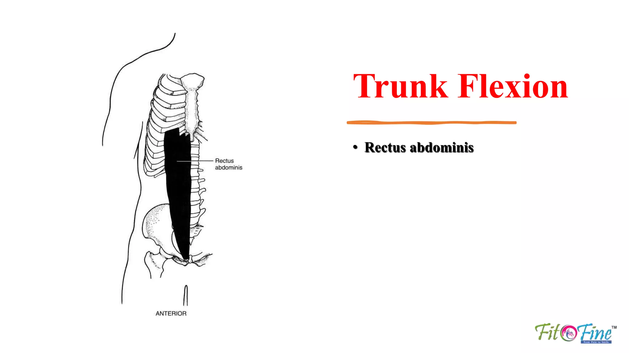
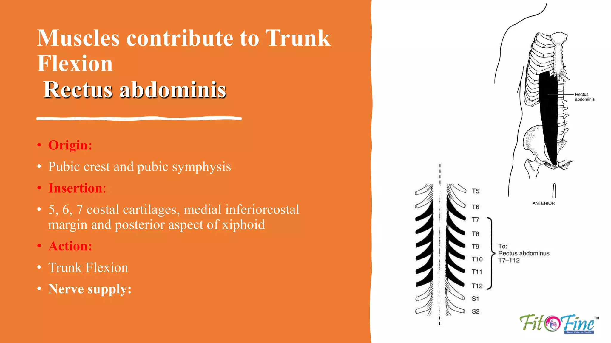
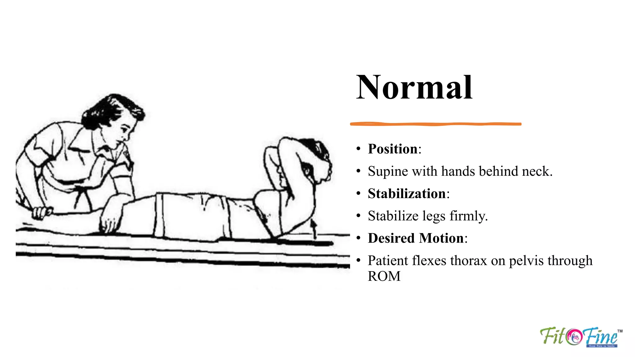
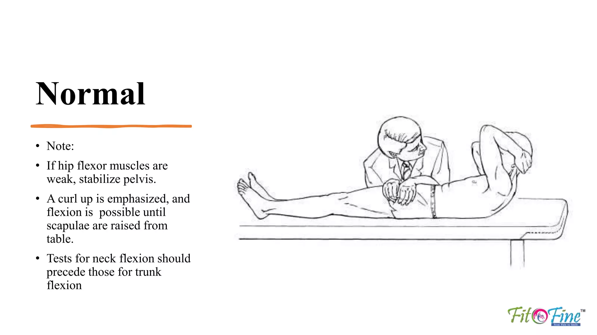
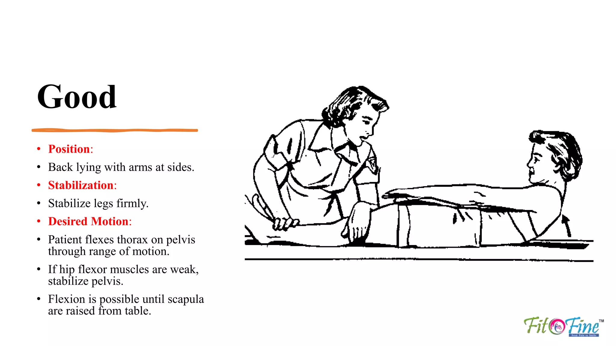
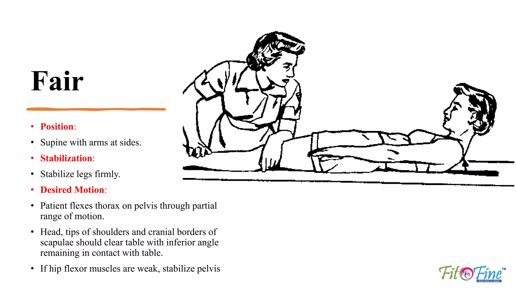
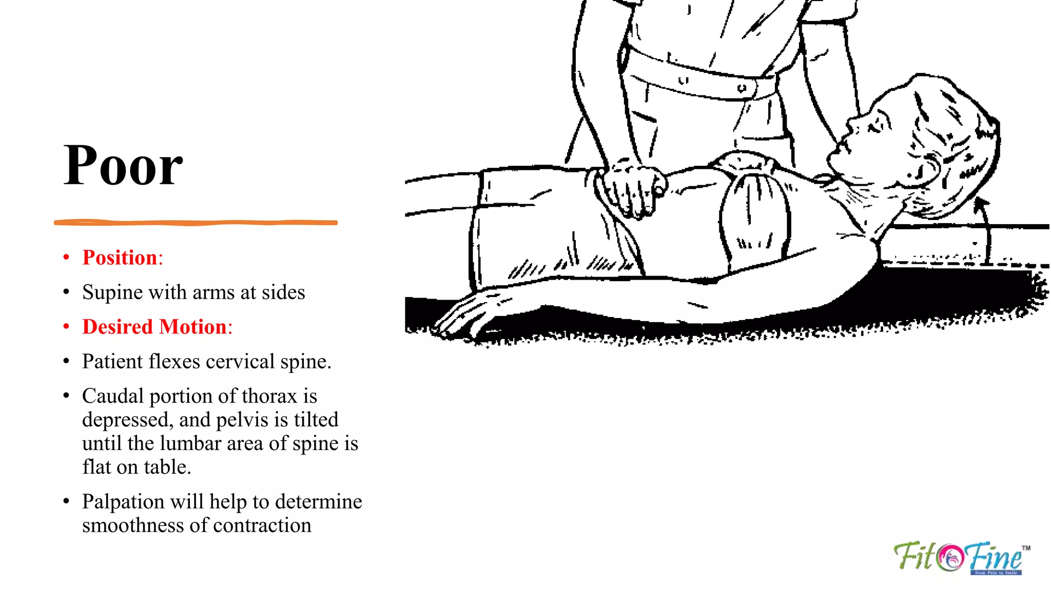
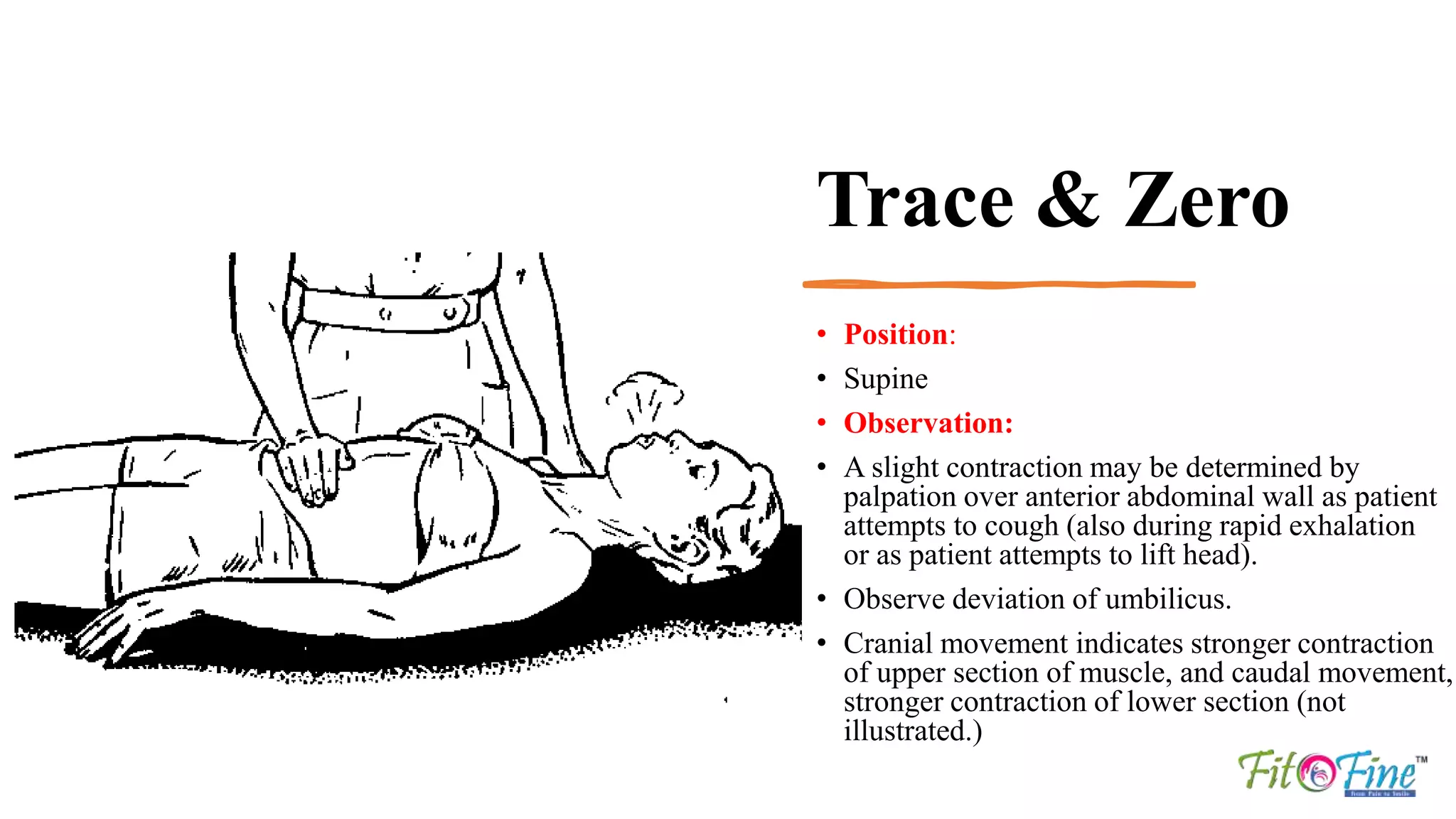
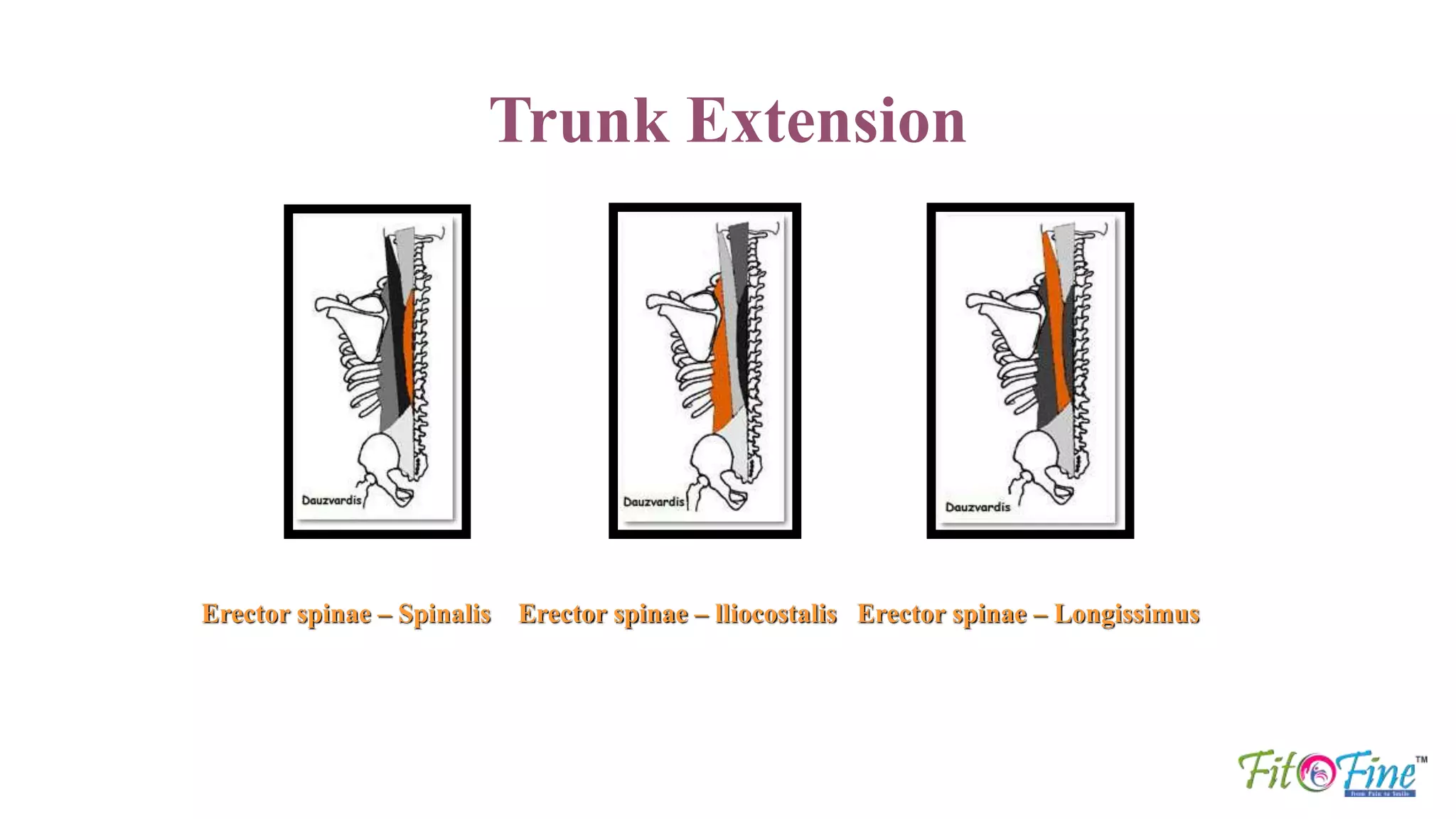
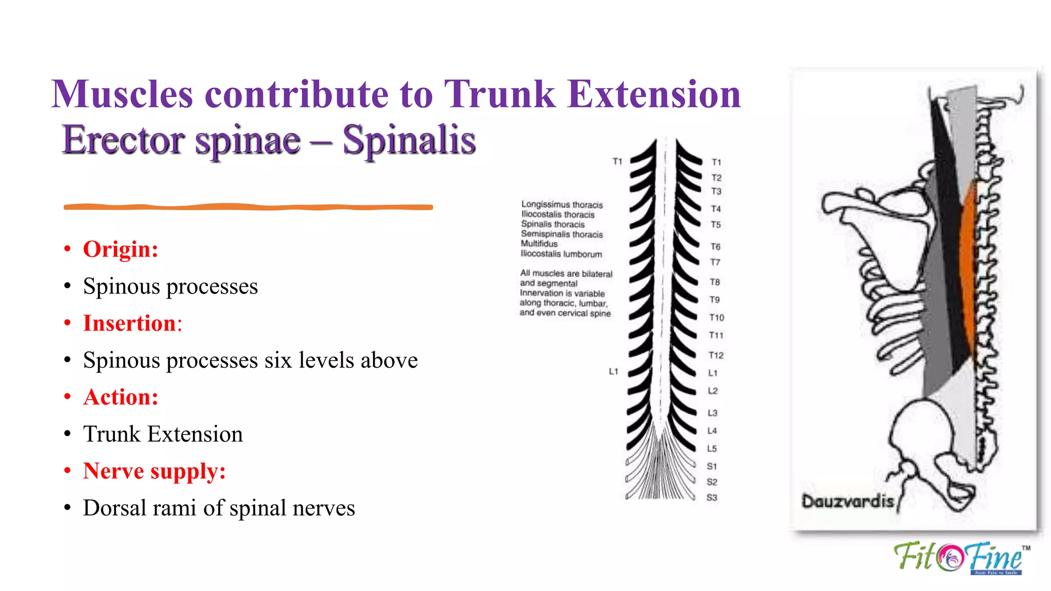
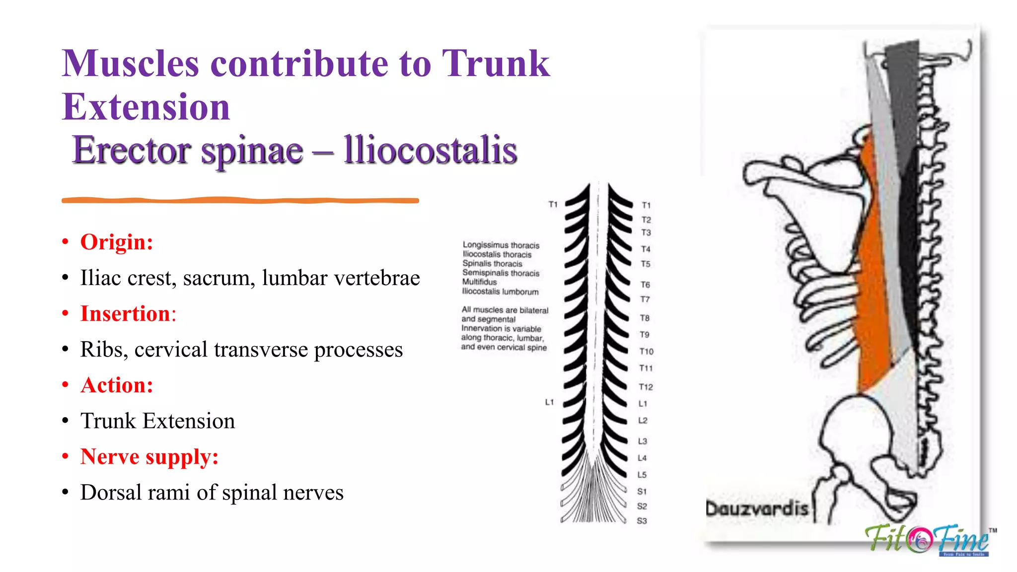
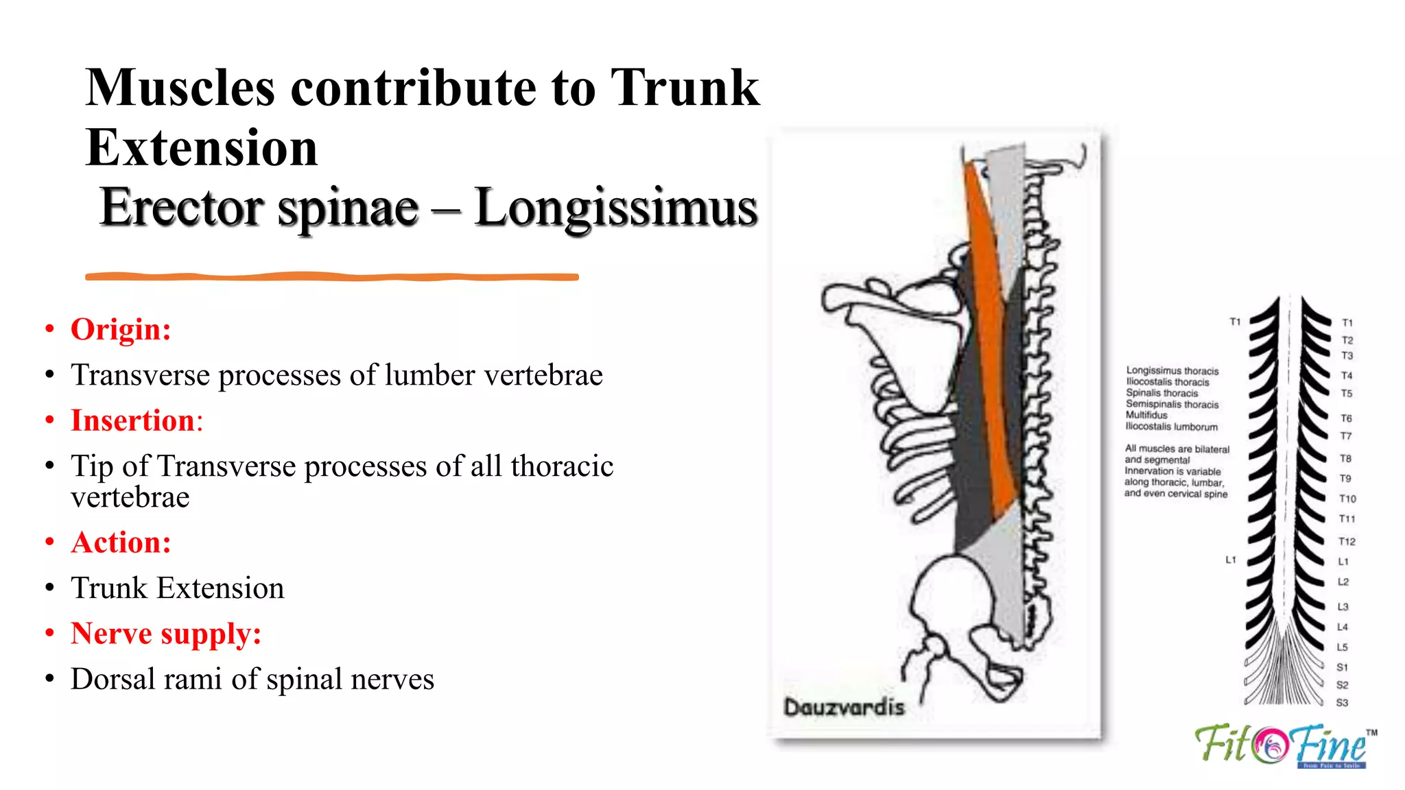
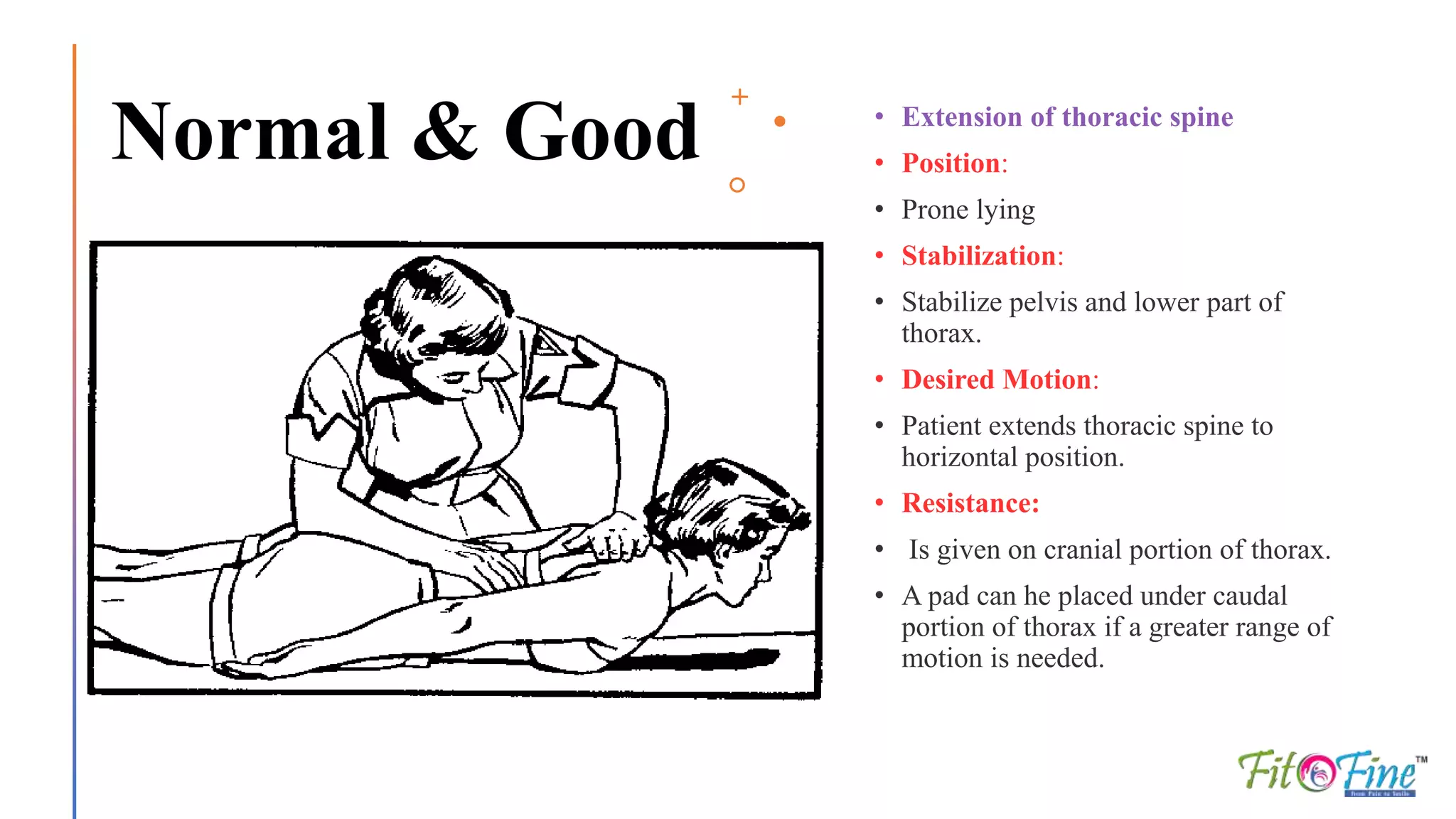
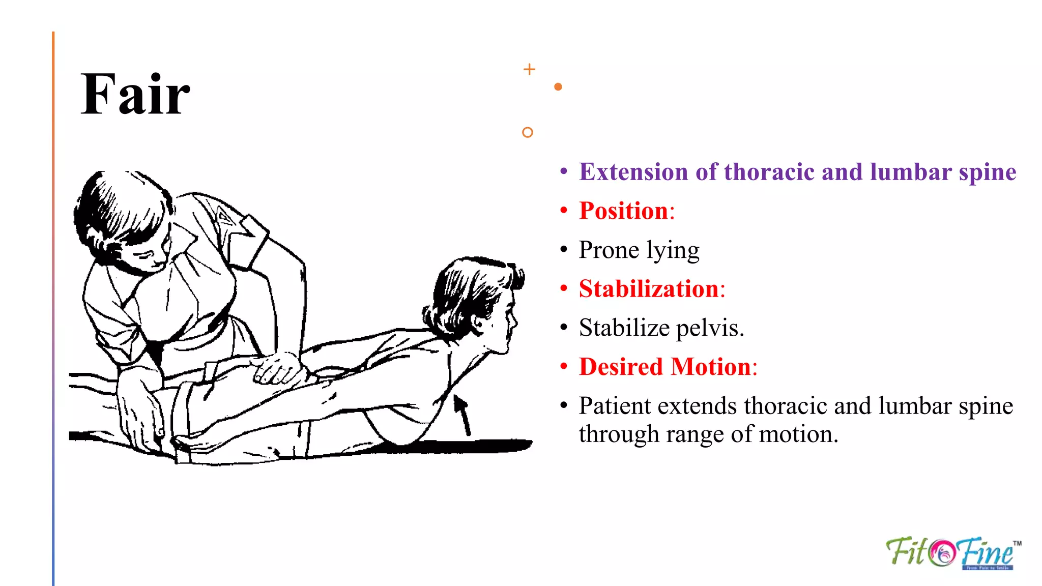
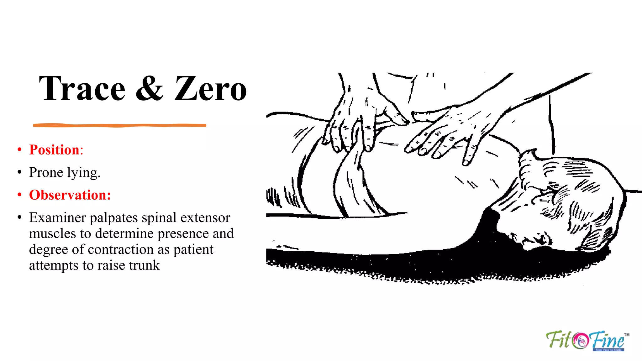
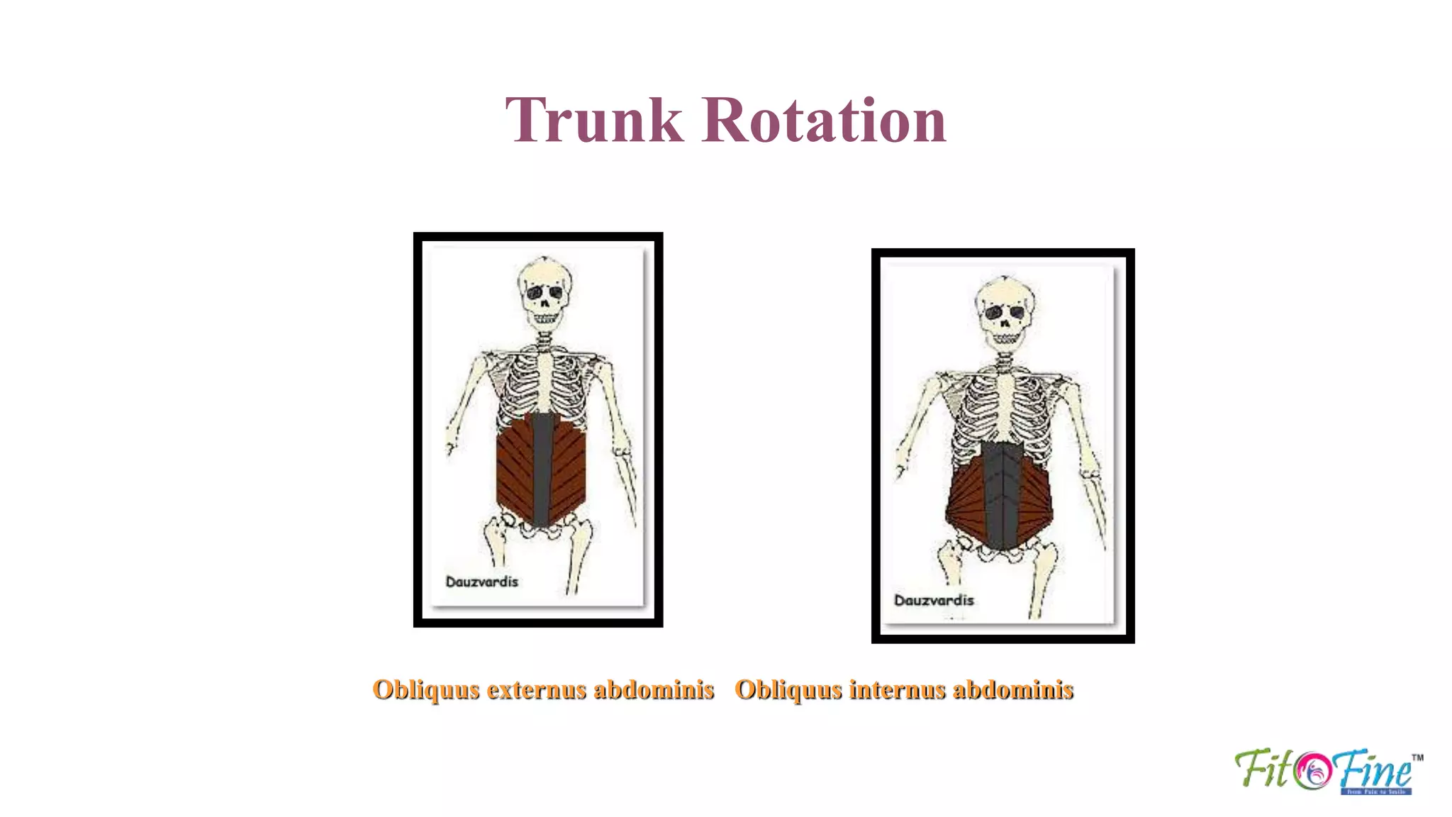
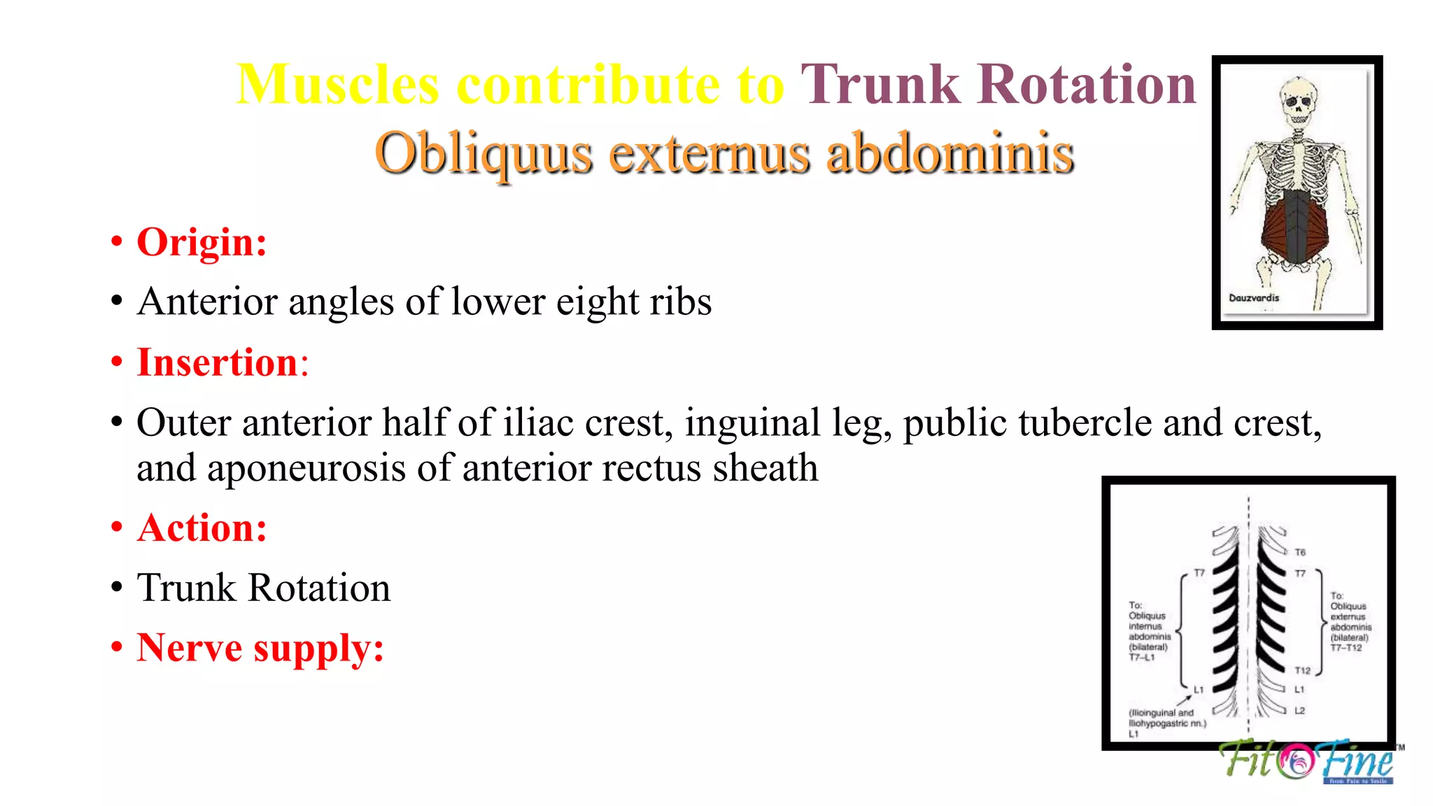
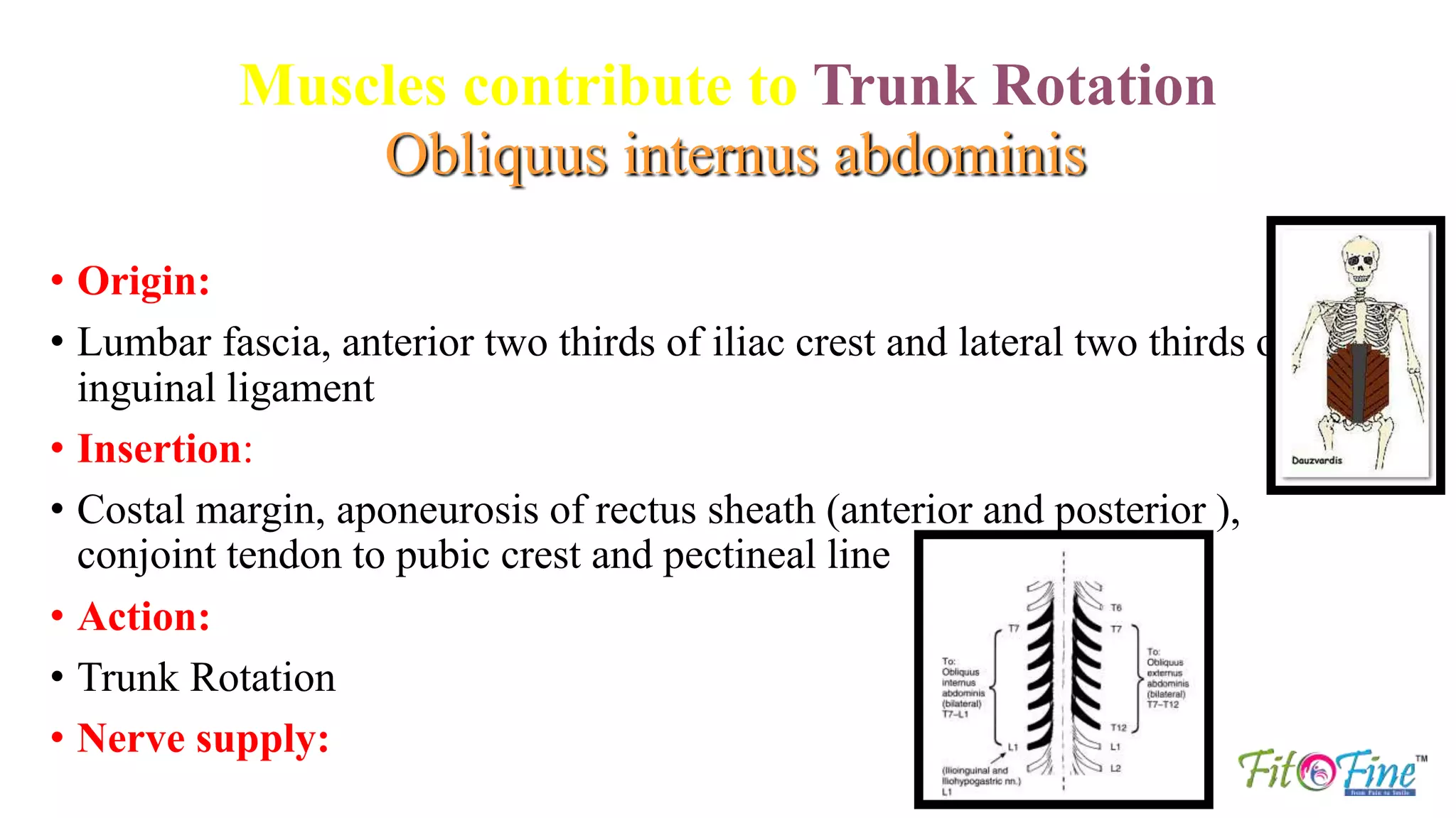
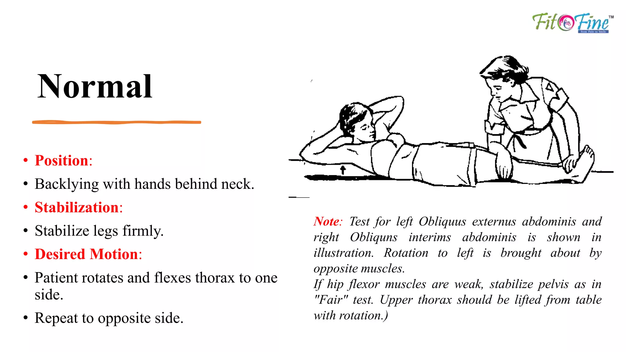
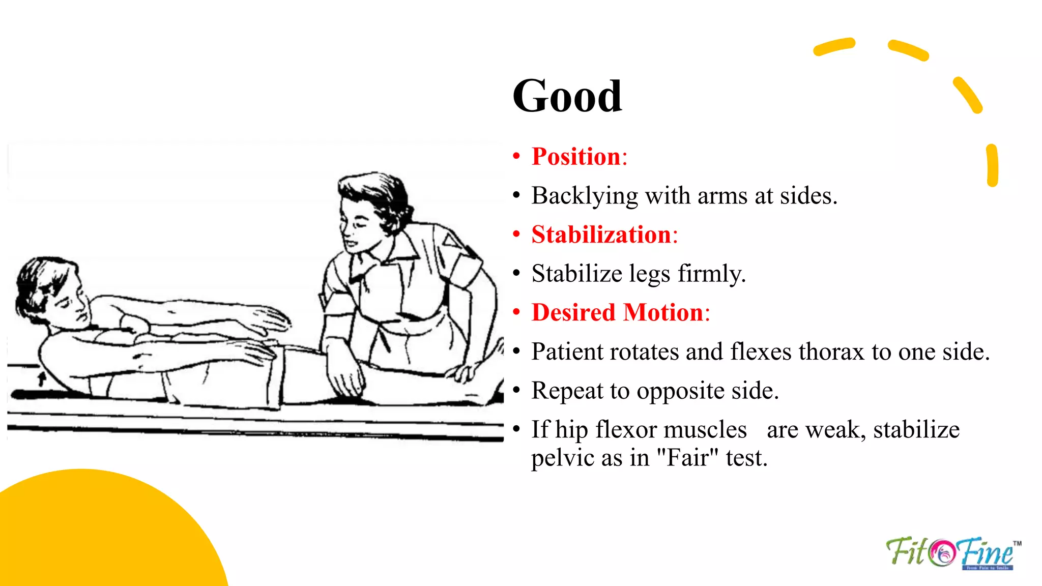
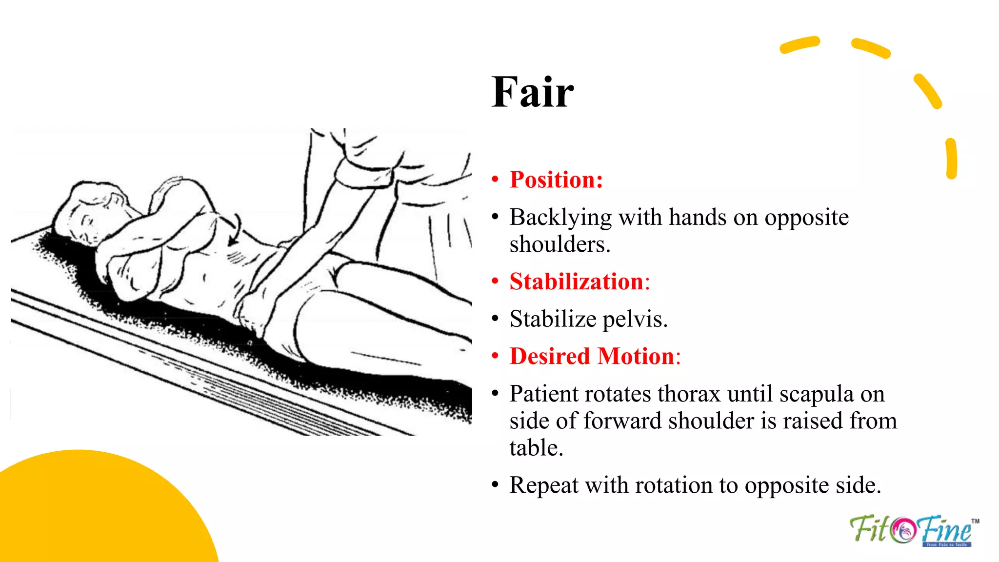
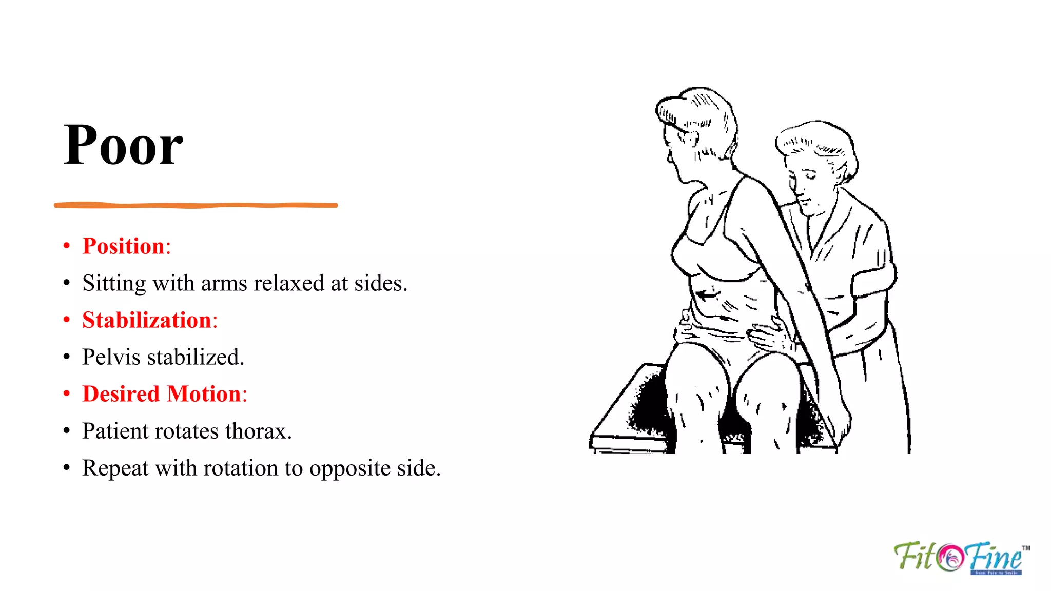
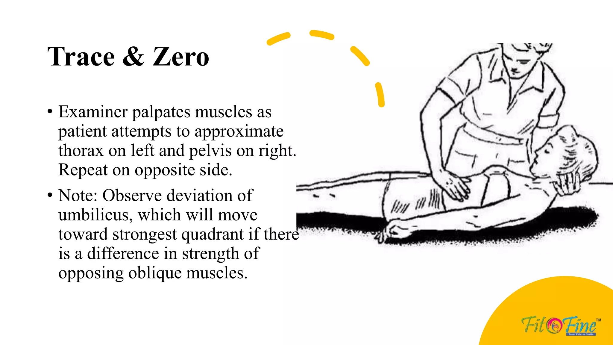
Prof. Satyen Bhattacharyya
Associate Professor: BIMLS, Bardhaman
Chief Physio: Fit O Fine
Director: Well O Fit Healthcare PVT. LTD.
Muscle Testing of the
Trunk
Muscle Testing of the Trunk
Trunk Flexion
• Rectus abdominis
Muscles contribute to Trunk
Flexion
Rectus abdominis
• Origin:
• Pubic crest and pubic symphysis
• Insertion:
• 5, 6, 7 costal cartilages, medial inferiorcostal
margin and posterior aspect of xiphoid
• Action:
• Trunk Flexion
• Nerve supply:
Normal
• Position:
• Supine with hands behind neck.
• Stabilization:
• Stabilize legs firmly.
• Desired Motion:
• Patient flexes thorax on pelvis through
ROM
Normal
• Note:
• If hip flexor muscles are
weak, stabilize pelvis.
• A curl up is emphasized, and
flexion is possible until
scapulae are raised from
table.
• Tests for neck flexion should
precede those for trunk
flexion
Good
• Position:
• Back lying with arms at sides.
• Stabilization:
• Stabilize legs firmly.
• Desired Motion:
• Patient flexes thorax on pelvis
through range of motion.
• If hip flexor muscles are weak,
stabilize pelvis.
• Flexion is possible until scapula
are raised from table.
Fair
• Position:
• Supine with arms at sides.
• Stabilization:
• Stabilize legs firmly.
• Desired Motion:
• Patient flexes thorax on pelvis through partial
range of motion.
• Head, tips of shoulders and cranial borders of
scapulae should clear table with inferior angle
remaining in contact with table.
• If hip flexor muscles are weak, stabilize pelvis
Poor
• Position:
• Supine with arms at sides
• Desired Motion:
• Patient flexes cervical spine.
• Caudal portion of thorax is
depressed, and pelvis is tilted
until the lumbar area of spine is
flat on table.
• Palpation will help to determine
smoothness of contraction
Trace & Zero
• Position:
• Supine
• Observation:
• A slight contraction may be determined by
palpation over anterior abdominal wall as patient
attempts to cough (also during rapid exhalation
or as patient attempts to lift head).
• Observe deviation of umbilicus.
• Cranial movement indicates stronger contraction
of upper section of muscle, and caudal movement,
stronger contraction of lower section (not
illustrated.)
Note
• Factors Limiting Motion:
• 1- Tension of posterior longitudinal ligament, ligamenta flava, and interspinal and supraspinal
ligaments
• 2- Tension of spinal extensor muscles
• 3-Apposition of caudal lips of vertebra bodies anteriorly with surfaces of subjacent vertebrae
• 4-Compression of ventral part of intervertebral fibrocartilages
• 5-Contact of last ribs with abdomen
• Fixation:
• 1-Reverse action of hip flexor muscles
• 2-Weight of legs and pelvis
Trunk Extension
Erector spinae – Spinalis Erector spinae – lliocostalis Erector spinae – Longissimus
Muscles contribute to Trunk Extension
Erector spinae – Spinalis
• Origin:
• Spinous processes
• Insertion:
• Spinous processes six levels above
• Action:
• Trunk Extension
• Nerve supply:
• Dorsal rami of spinal nerves
Muscles contribute to Trunk
Extension
Erector spinae – lliocostalis
• Origin:
• Iliac crest, sacrum, lumbar vertebrae
• Insertion:
• Ribs, cervical transverse processes
• Action:
• Trunk Extension
• Nerve supply:
• Dorsal rami of spinal nerves
Muscles contribute to Trunk
Extension
Erector spinae – Longissimus
• Origin:
• Transverse processes of lumber vertebrae
• Insertion:
• Tip of Transverse processes of all thoracic
vertebrae
• Action:
• Trunk Extension
• Nerve supply:
• Dorsal rami of spinal nerves
Normal & Good
Normal & Good
Fair
• Extension of thoracic and lumbar spine
• Position:
• Prone lying
• Stabilization:
• Stabilize pelvis.
• Desired Motion:
• Patient extends thoracic and lumbar spine
through range of motion.
Poor
Trace & Zero
• Position:
• Prone lying.
• Observation:
• Examiner palpates spinal extensor
muscles to determine presence and
degree of contraction as patient
attempts to raise trunk
Note
• Factors Limiting Motion:
1-Tension of anterior longitudinal ligament of spine
2-Tension of anterior abdominal muscles
3-Contact of spinous processes
4-Contact of caudal articular margins with laminae
• Fixation:
1-Contraction of Glutens maximums and
2-Hamstring muscles
3-Weight of pelvis and legs
Trunk Rotation
Obliquus externus abdominis Obliquus internus abdominis
Muscles contribute to Trunk Rotation
Obliquus externus abdominis
• Origin:
• Anterior angles of lower eight ribs
• Insertion:
• Outer anterior half of iliac crest, inguinal leg, public tubercle and crest,
and aponeurosis of anterior rectus sheath
• Action:
• Trunk Rotation
• Nerve supply:
Muscles contribute to Trunk Rotation
Obliquus internus abdominis
• Origin:
• Lumbar fascia, anterior two thirds of iliac crest and lateral two thirds of
inguinal ligament
• Insertion:
• Costal margin, aponeurosis of rectus sheath (anterior and posterior ),
conjoint tendon to pubic crest and pectineal line
• Action:
• Trunk Rotation
• Nerve supply:
Normal
• Position:
• Backlying with hands behind neck.
• Stabilization:
• Stabilize legs firmly.
• Desired Motion:
• Patient rotates and flexes thorax to one
side.
• Repeat to opposite side.
Note: Test for left Obliquus externus abdominis and
right Obliquns interims abdominis is shown in
illustration. Rotation to left is brought about by
opposite muscles.
If hip flexor muscles are weak, stabilize pelvis as in
"Fair" test. Upper thorax should be lifted from table
with rotation.)
Good
• Position:
• Backlying with arms at sides.
• Stabilization:
• Stabilize legs firmly.
• Desired Motion:
• Patient rotates and flexes thorax to one side.
• Repeat to opposite side.
• If hip flexor muscles are weak, stabilize
pelvic as in "Fair" test.
Fair
• Position:
• Backlying with hands on opposite
shoulders.
• Stabilization:
• Stabilize pelvis.
• Desired Motion:
• Patient rotates thorax until scapula on
side of forward shoulder is raised from
table.
• Repeat with rotation to opposite side.
Poor
• Position:
• Sitting with arms relaxed at sides.
• Stabilization:
• Pelvis stabilized.
• Desired Motion:
• Patient rotates thorax.
• Repeat with rotation to opposite side.
Trace & Zero
• Examiner palpates muscles as
patient attempts to approximate
thorax on left and pelvis on right.
Repeat on opposite side.
• Note: Observe deviation of
umbilicus, which will move
toward strongest quadrant if there
is a difference in strength of
opposing oblique muscles.
Reference
• Daniels and Wortingham’s Muscle Testing
Techniques of Manual Examination
• Physiopedia

MMT Trunk.pptx

  • 1.
    Prof. Satyen Bhattacharyya AssociateProfessor: BIMLS, Bardhaman Chief Physio: Fit O Fine Director: Well O Fit Healthcare PVT. LTD. Muscle Testing of the Trunk
  • 2.
  • 3.
  • 4.
    Muscles contribute toTrunk Flexion Rectus abdominis • Origin: • Pubic crest and pubic symphysis • Insertion: • 5, 6, 7 costal cartilages, medial inferiorcostal margin and posterior aspect of xiphoid • Action: • Trunk Flexion • Nerve supply:
  • 5.
    Normal • Position: • Supinewith hands behind neck. • Stabilization: • Stabilize legs firmly. • Desired Motion: • Patient flexes thorax on pelvis through ROM
  • 6.
    Normal • Note: • Ifhip flexor muscles are weak, stabilize pelvis. • A curl up is emphasized, and flexion is possible until scapulae are raised from table. • Tests for neck flexion should precede those for trunk flexion
  • 7.
    Good • Position: • Backlying with arms at sides. • Stabilization: • Stabilize legs firmly. • Desired Motion: • Patient flexes thorax on pelvis through range of motion. • If hip flexor muscles are weak, stabilize pelvis. • Flexion is possible until scapula are raised from table.
  • 8.
    Fair • Position: • Supinewith arms at sides. • Stabilization: • Stabilize legs firmly. • Desired Motion: • Patient flexes thorax on pelvis through partial range of motion. • Head, tips of shoulders and cranial borders of scapulae should clear table with inferior angle remaining in contact with table. • If hip flexor muscles are weak, stabilize pelvis
  • 9.
    Poor • Position: • Supinewith arms at sides • Desired Motion: • Patient flexes cervical spine. • Caudal portion of thorax is depressed, and pelvis is tilted until the lumbar area of spine is flat on table. • Palpation will help to determine smoothness of contraction
  • 10.
    Trace & Zero •Position: • Supine • Observation: • A slight contraction may be determined by palpation over anterior abdominal wall as patient attempts to cough (also during rapid exhalation or as patient attempts to lift head). • Observe deviation of umbilicus. • Cranial movement indicates stronger contraction of upper section of muscle, and caudal movement, stronger contraction of lower section (not illustrated.)
  • 11.
    Note • Factors LimitingMotion: • 1- Tension of posterior longitudinal ligament, ligamenta flava, and interspinal and supraspinal ligaments • 2- Tension of spinal extensor muscles • 3-Apposition of caudal lips of vertebra bodies anteriorly with surfaces of subjacent vertebrae • 4-Compression of ventral part of intervertebral fibrocartilages • 5-Contact of last ribs with abdomen • Fixation: • 1-Reverse action of hip flexor muscles • 2-Weight of legs and pelvis
  • 12.
    Trunk Extension Erector spinae– Spinalis Erector spinae – lliocostalis Erector spinae – Longissimus
  • 13.
    Muscles contribute toTrunk Extension Erector spinae – Spinalis • Origin: • Spinous processes • Insertion: • Spinous processes six levels above • Action: • Trunk Extension • Nerve supply: • Dorsal rami of spinal nerves
  • 14.
    Muscles contribute toTrunk Extension Erector spinae – lliocostalis • Origin: • Iliac crest, sacrum, lumbar vertebrae • Insertion: • Ribs, cervical transverse processes • Action: • Trunk Extension • Nerve supply: • Dorsal rami of spinal nerves
  • 15.
    Muscles contribute toTrunk Extension Erector spinae – Longissimus • Origin: • Transverse processes of lumber vertebrae • Insertion: • Tip of Transverse processes of all thoracic vertebrae • Action: • Trunk Extension • Nerve supply: • Dorsal rami of spinal nerves
  • 16.
  • 17.
  • 18.
    Fair • Extension ofthoracic and lumbar spine • Position: • Prone lying • Stabilization: • Stabilize pelvis. • Desired Motion: • Patient extends thoracic and lumbar spine through range of motion.
  • 19.
  • 20.
    Trace & Zero •Position: • Prone lying. • Observation: • Examiner palpates spinal extensor muscles to determine presence and degree of contraction as patient attempts to raise trunk
  • 21.
    Note • Factors LimitingMotion: 1-Tension of anterior longitudinal ligament of spine 2-Tension of anterior abdominal muscles 3-Contact of spinous processes 4-Contact of caudal articular margins with laminae • Fixation: 1-Contraction of Glutens maximums and 2-Hamstring muscles 3-Weight of pelvis and legs
  • 22.
    Trunk Rotation Obliquus externusabdominis Obliquus internus abdominis
  • 23.
    Muscles contribute toTrunk Rotation Obliquus externus abdominis • Origin: • Anterior angles of lower eight ribs • Insertion: • Outer anterior half of iliac crest, inguinal leg, public tubercle and crest, and aponeurosis of anterior rectus sheath • Action: • Trunk Rotation • Nerve supply:
  • 24.
    Muscles contribute toTrunk Rotation Obliquus internus abdominis • Origin: • Lumbar fascia, anterior two thirds of iliac crest and lateral two thirds of inguinal ligament • Insertion: • Costal margin, aponeurosis of rectus sheath (anterior and posterior ), conjoint tendon to pubic crest and pectineal line • Action: • Trunk Rotation • Nerve supply:
  • 25.
    Normal • Position: • Backlyingwith hands behind neck. • Stabilization: • Stabilize legs firmly. • Desired Motion: • Patient rotates and flexes thorax to one side. • Repeat to opposite side. Note: Test for left Obliquus externus abdominis and right Obliquns interims abdominis is shown in illustration. Rotation to left is brought about by opposite muscles. If hip flexor muscles are weak, stabilize pelvis as in "Fair" test. Upper thorax should be lifted from table with rotation.)
  • 26.
    Good • Position: • Backlyingwith arms at sides. • Stabilization: • Stabilize legs firmly. • Desired Motion: • Patient rotates and flexes thorax to one side. • Repeat to opposite side. • If hip flexor muscles are weak, stabilize pelvic as in "Fair" test.
  • 27.
    Fair • Position: • Backlyingwith hands on opposite shoulders. • Stabilization: • Stabilize pelvis. • Desired Motion: • Patient rotates thorax until scapula on side of forward shoulder is raised from table. • Repeat with rotation to opposite side.
  • 28.
    Poor • Position: • Sittingwith arms relaxed at sides. • Stabilization: • Pelvis stabilized. • Desired Motion: • Patient rotates thorax. • Repeat with rotation to opposite side.
  • 29.
    Trace & Zero •Examiner palpates muscles as patient attempts to approximate thorax on left and pelvis on right. Repeat on opposite side. • Note: Observe deviation of umbilicus, which will move toward strongest quadrant if there is a difference in strength of opposing oblique muscles.
  • 30.
    Reference • Daniels andWortingham’s Muscle Testing Techniques of Manual Examination • Physiopedia