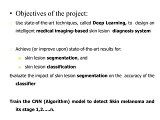
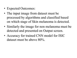
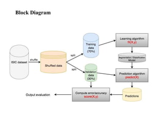
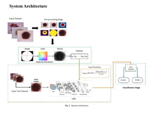
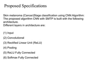
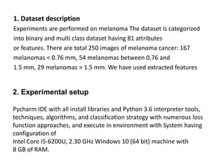
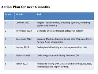
“Machine learning approach in melanoma
cancer stage detection”.
Name of Project Group members
1.
2.
3.
4.
Name of Guide:
• Objectives of the project:
□ Use state-of-the-art techniques, called Deep Learning, to design an
intelligent medical imaging-based skin lesion diagnosis system
□ Achieve (or improve upon) state-of-the-art results for:
■ skin lesion segmentation, and
■ skin lesion classification
Evaluate the impact of skin lesion segmentation on the accuracy of the
classifier
Train the CNN (Algorithm) model to detect Skin melanoma and
its stage 1,2…..n.
• Expected Outcomes:
• The input image from dataset must be
processed by algorithms and classified based
on which stage of Skin melanoma is detected.
• Similarly the image for non melanoma must be
detected and presented on Output screen.
• Accuracy for trained CNN model for ISIC
dataset must be above 80%.
Block Diagram
Problem Definition:
Discriminating between benign and malignant OR
stage classification skin lesions is challenging
Without computer-based assistance: 60~80%
detection accuracy
Social Relevance of the project:
The cancer of any kind must be identified on
right time with precision to avoid any delay in
treatment. And this system gives better option
to test the skin lesions of any person without
any expensive lab setup.
Literature Survey/Market Survey:
Sr.
No.
Title Authors Journal
-Year
Outcomes
1 Skin cancer diagnosis based on
optimized convolutional neural
network
Zhang, Ni, Cai, Yi-Xin,
Wang, Yong-Yong, Tian, Yi-
Tao, Wang, Xiao-Li,
Badami,Benjamin
2020 A new image processing based
method has been proposed for the
early detection of skin cancer.
2 Automatic Skin Cancer Detection
in Dermoscopy Images Based on
Ensemble Lightweight Deep
Learning Network
Lisheng wei , Kun ding, and
Huosheng hu
2020 Designed a discriminant
dermoscopy image lesion
recognition model.
3 Dermoscopy Image Classification
Based on StyleGANs and Decision
Fusion
Gong, A., Yao, X., Lin, W. 2020 propose a decision fusion method.
Through transfer learning, based on
multiple pre-trained convolutional
neural networks (CNNs)
4 Noninvasive Real-Time
Automated Skin Lesion Analysis
System for Melanoma Early
Detection and Prevention
Omar Abuzaghleh; Buket D.
Barkana; Miad Faezipour
2015 presented the components of a
system to aid in
the malignant melanoma
prevention and early detection
5 Two methodologies for
identification of stages and
different types of melanoma
detection
M. Reshma; B. Priestly Shan 2017 the identification of Skin lesion
Melanoma at different Stages based
on Total Dermoscopic score (TDS)
using ABCD features.
System Architecture
Proposed Specifications
Skin melanoma (Cancer)Stage classification using CNN Algorithm:
The proposed algorithm CNN with SMTP is built with the following
architecture.
Different layers in architecture are:
(1) Input
(2) Convolutional
(3) Rectified Linear Unit (ReLU)
(4) Pooling
(5) ReLU Fully Connected
(6) Softmax Fully Connected
1. Dataset description
Experiments are performed on melanoma The dataset is categorized
into binary and multi class dataset having 81 attributes
or features. There are total 250 images of melanoma cancer: 167
melanomas < 0.76 mm, 54 melanomas between 0.76 and
1.5 mm, 29 melanomas > 1.5 mm. We have used extracted features
2. Experimental setup
Pycharm IDE with all install libraries and Python 3.6 interpreter tools,
techniques, algorithms, and classification strategy with numerous loss
function approaches, and execute in environment with System having
configuration of
Intel Core i5-6200U, 2.30 GHz Windows 10 (64 bit) machine with
8 GB of RAM.
Hardware:
System having configuration of Intel Core i5-6200U, 2.30 GHz
Windows 10 (64 bit) machine with 8 GB of RAM.
Software:
• Pycharm IDE latest version
• Python 3.6 compiler/ interpreter
• Open CV, Scikit learn libraray packages
• Dataset: ISIC for skin Melonoma images
• OS: Windows 10 (64 bit)
List of hardware and software simulation tools
Work Done
Dataset creation for CNN model training is Done
• Dataset consist of training and testing data
for stage 1 and stage 2 of skin melanoma
detection
• Training CNN Model for Stage classification
and detection is Done.
Action Plan for next 6 months
Sr. no. Month Task
1 October 2022 Project Topic Selection, preparing Synopsis, collecting
papers and review 1
2 November 2022 Generate or create Dataset, categories dataset
3 December 2022 Learning Machine learning basics with CNN algorithms,
Review 2 and presentation
4 January 2023 Coding Model training and testing on random data
5 February 2023 Code integration and adding front end GUI
6 March 2023 Final code testing with Dataset and recording Accuracy,
Final review and Report writing.
References
Abuzaghleh, O., Barkana, B.D., Faezipour, M., 2015. Noninvasive real-time
automated skin lesion analysis system for melanoma early detection and
prevention 4300212 IEEE J. Transl. Eng. Health Med. 3, 1–12. https://doi.org/
10.1109/JTEHM.2015.2419612.
Barata, C., Ruela, M., Francisco, M., Mendonça, T., Marques, J.S., 2014. Two systems
for the detection of melanomas in dermoscopy images using texture and color
features. IEEE Syst. J., 965–979
Breslow, A., 1970. Thickness, cross-sectional areas and depth of invasion in the
prognosis of cutaneous melanoma. Ann. Surg. 172 (5), 902–908.
Chim, H., Deng, X., 2010. Efficient phrase-based document similarity for clustering.
IEEE Trans. Knowl. Data Eng. 20 (9), 1217–1229.
• Gong, A., Yao, X., Lin, W., 2020. Dermoscopy image classification based on
StyleGANs and decision fusion. IEEE Access 8, 70640–70650. https://doi.org/
10.1109/ACCESS.2020.2986916.
• Jaworek-Korjakowska, J., Kleczek, P., Gorgon, M., 2019. Melanoma thickness
prediction based on convolutional neural network with VGG-19 model
transfer learning, in: 2019 IEEE/CVF Conference on Computer Vision and
Pattern Recognition Workshops (CVPRW), Long Beach, CA, USA, pp. 2748–2756,
http://dx.doi.org/10.1109/CVPRW.2019.00333.
Ma, Z., Tavares, J.M.R.S., 2016. A novel approach to segment skin lesions in
dermoscopic images based on a deformable model. IEEE J. Biomed. Health
Inform. 20 (2), 615–623.
• Patil, R.R., Bellary, S., 2017. Review: melanoma detection & classification based on
thickness using dermascopic images. IJCTA 10 (8), 821–825.
Pehamberger, H., Steiner, A., Wolff, K., 1987. In vivo epiluminescence microscopy of
pigmented skin lesions. Pattern analysis of pigmented skin lesions. J. Am. Acad.
Dermatol. 17 (4), 571–583.
• Reshma, M., Shan, B.P., 2017. Two methodologies for identification of stages and
different types of melanoma detection, in: 2017 Conference on Emerging
Devices and Smart Systems (ICEDSS), Tiruchengode, 2017, pp. 257–259, http://
dx.doi.org/10.1109/ICEDSS.2017.8073689.
Rubegni, Pietro et al., 2010. Evaluation of cutaneous melanoma thickness by
digital dermoscopy analysis: a retrospective study. Melanoma Res. 20, 212–
217.Sangve, S.M., Patil, R.R., 2014. Competitive analysis for the detection of melanomas
in dermoscopy images. IJERT 3 (6), 351–354.
• Wang, X., Jiang, X., Ding, H., Liu, J., 2020. Bi-directional dermoscopic feature learning
and multi-scale consistent decision fusion for skin lesion segmentation. IEEE
Trans. Image Process. 29, 3039–3051. https://doi.org/10.1109/
TIP.2019.2955297.
• Wei, L., Ding, K., Hu, H., 2020. Automatic skin cancer detection in
dermoscopy images based on ensemble lightweight deep learning
network. In: IEEE Access vol. 8, 99633–99647, http://dx.doi.org/10.1109/
ACCESS.2020.2997710.
• Zhang, Ni, Cai, Yi-Xin, Wang, Yong-Yong, Tian, Yi-Tao, Wang, Xiao-Li, Badami,
Benjamin, 2020. Skin cancer diagnosis based on optimized convolutional neural
network. Artificial Intelligence Med. 102, 101756. https://doi.org/10.1016/j.
artmed.2019.101756.
THANK YOU

Second PPT.ppt

  • 1.
    “Machine learning approachin melanoma cancer stage detection”. Name of Project Group members 1. 2. 3. 4. Name of Guide:
  • 2.
    • Objectives ofthe project: □ Use state-of-the-art techniques, called Deep Learning, to design an intelligent medical imaging-based skin lesion diagnosis system □ Achieve (or improve upon) state-of-the-art results for: ■ skin lesion segmentation, and ■ skin lesion classification Evaluate the impact of skin lesion segmentation on the accuracy of the classifier Train the CNN (Algorithm) model to detect Skin melanoma and its stage 1,2…..n.
  • 3.
    • Expected Outcomes: •The input image from dataset must be processed by algorithms and classified based on which stage of Skin melanoma is detected. • Similarly the image for non melanoma must be detected and presented on Output screen. • Accuracy for trained CNN model for ISIC dataset must be above 80%.
  • 4.
  • 5.
    Problem Definition: Discriminating betweenbenign and malignant OR stage classification skin lesions is challenging Without computer-based assistance: 60~80% detection accuracy Social Relevance of the project: The cancer of any kind must be identified on right time with precision to avoid any delay in treatment. And this system gives better option to test the skin lesions of any person without any expensive lab setup.
  • 6.
    Literature Survey/Market Survey: Sr. No. TitleAuthors Journal -Year Outcomes 1 Skin cancer diagnosis based on optimized convolutional neural network Zhang, Ni, Cai, Yi-Xin, Wang, Yong-Yong, Tian, Yi- Tao, Wang, Xiao-Li, Badami,Benjamin 2020 A new image processing based method has been proposed for the early detection of skin cancer. 2 Automatic Skin Cancer Detection in Dermoscopy Images Based on Ensemble Lightweight Deep Learning Network Lisheng wei , Kun ding, and Huosheng hu 2020 Designed a discriminant dermoscopy image lesion recognition model. 3 Dermoscopy Image Classification Based on StyleGANs and Decision Fusion Gong, A., Yao, X., Lin, W. 2020 propose a decision fusion method. Through transfer learning, based on multiple pre-trained convolutional neural networks (CNNs) 4 Noninvasive Real-Time Automated Skin Lesion Analysis System for Melanoma Early Detection and Prevention Omar Abuzaghleh; Buket D. Barkana; Miad Faezipour 2015 presented the components of a system to aid in the malignant melanoma prevention and early detection 5 Two methodologies for identification of stages and different types of melanoma detection M. Reshma; B. Priestly Shan 2017 the identification of Skin lesion Melanoma at different Stages based on Total Dermoscopic score (TDS) using ABCD features.
  • 7.
  • 8.
    Proposed Specifications Skin melanoma(Cancer)Stage classification using CNN Algorithm: The proposed algorithm CNN with SMTP is built with the following architecture. Different layers in architecture are: (1) Input (2) Convolutional (3) Rectified Linear Unit (ReLU) (4) Pooling (5) ReLU Fully Connected (6) Softmax Fully Connected
  • 9.
    1. Dataset description Experimentsare performed on melanoma The dataset is categorized into binary and multi class dataset having 81 attributes or features. There are total 250 images of melanoma cancer: 167 melanomas < 0.76 mm, 54 melanomas between 0.76 and 1.5 mm, 29 melanomas > 1.5 mm. We have used extracted features 2. Experimental setup Pycharm IDE with all install libraries and Python 3.6 interpreter tools, techniques, algorithms, and classification strategy with numerous loss function approaches, and execute in environment with System having configuration of Intel Core i5-6200U, 2.30 GHz Windows 10 (64 bit) machine with 8 GB of RAM.
  • 10.
    Hardware: System having configurationof Intel Core i5-6200U, 2.30 GHz Windows 10 (64 bit) machine with 8 GB of RAM. Software: • Pycharm IDE latest version • Python 3.6 compiler/ interpreter • Open CV, Scikit learn libraray packages • Dataset: ISIC for skin Melonoma images • OS: Windows 10 (64 bit) List of hardware and software simulation tools
  • 11.
    Work Done Dataset creationfor CNN model training is Done • Dataset consist of training and testing data for stage 1 and stage 2 of skin melanoma detection • Training CNN Model for Stage classification and detection is Done.
  • 12.
    Action Plan fornext 6 months Sr. no. Month Task 1 October 2022 Project Topic Selection, preparing Synopsis, collecting papers and review 1 2 November 2022 Generate or create Dataset, categories dataset 3 December 2022 Learning Machine learning basics with CNN algorithms, Review 2 and presentation 4 January 2023 Coding Model training and testing on random data 5 February 2023 Code integration and adding front end GUI 6 March 2023 Final code testing with Dataset and recording Accuracy, Final review and Report writing.
  • 13.
    References Abuzaghleh, O., Barkana,B.D., Faezipour, M., 2015. Noninvasive real-time automated skin lesion analysis system for melanoma early detection and prevention 4300212 IEEE J. Transl. Eng. Health Med. 3, 1–12. https://doi.org/ 10.1109/JTEHM.2015.2419612. Barata, C., Ruela, M., Francisco, M., Mendonça, T., Marques, J.S., 2014. Two systems for the detection of melanomas in dermoscopy images using texture and color features. IEEE Syst. J., 965–979 Breslow, A., 1970. Thickness, cross-sectional areas and depth of invasion in the prognosis of cutaneous melanoma. Ann. Surg. 172 (5), 902–908. Chim, H., Deng, X., 2010. Efficient phrase-based document similarity for clustering. IEEE Trans. Knowl. Data Eng. 20 (9), 1217–1229. • Gong, A., Yao, X., Lin, W., 2020. Dermoscopy image classification based on StyleGANs and decision fusion. IEEE Access 8, 70640–70650. https://doi.org/ 10.1109/ACCESS.2020.2986916. • Jaworek-Korjakowska, J., Kleczek, P., Gorgon, M., 2019. Melanoma thickness prediction based on convolutional neural network with VGG-19 model transfer learning, in: 2019 IEEE/CVF Conference on Computer Vision and Pattern Recognition Workshops (CVPRW), Long Beach, CA, USA, pp. 2748–2756, http://dx.doi.org/10.1109/CVPRW.2019.00333. Ma, Z., Tavares, J.M.R.S., 2016. A novel approach to segment skin lesions in dermoscopic images based on a deformable model. IEEE J. Biomed. Health Inform. 20 (2), 615–623. • Patil, R.R., Bellary, S., 2017. Review: melanoma detection & classification based on thickness using dermascopic images. IJCTA 10 (8), 821–825. Pehamberger, H., Steiner, A., Wolff, K., 1987. In vivo epiluminescence microscopy of pigmented skin lesions. Pattern analysis of pigmented skin lesions. J. Am. Acad. Dermatol. 17 (4), 571–583.
  • 14.
    • Reshma, M.,Shan, B.P., 2017. Two methodologies for identification of stages and different types of melanoma detection, in: 2017 Conference on Emerging Devices and Smart Systems (ICEDSS), Tiruchengode, 2017, pp. 257–259, http:// dx.doi.org/10.1109/ICEDSS.2017.8073689. Rubegni, Pietro et al., 2010. Evaluation of cutaneous melanoma thickness by digital dermoscopy analysis: a retrospective study. Melanoma Res. 20, 212– 217.Sangve, S.M., Patil, R.R., 2014. Competitive analysis for the detection of melanomas in dermoscopy images. IJERT 3 (6), 351–354. • Wang, X., Jiang, X., Ding, H., Liu, J., 2020. Bi-directional dermoscopic feature learning and multi-scale consistent decision fusion for skin lesion segmentation. IEEE Trans. Image Process. 29, 3039–3051. https://doi.org/10.1109/ TIP.2019.2955297. • Wei, L., Ding, K., Hu, H., 2020. Automatic skin cancer detection in dermoscopy images based on ensemble lightweight deep learning network. In: IEEE Access vol. 8, 99633–99647, http://dx.doi.org/10.1109/ ACCESS.2020.2997710. • Zhang, Ni, Cai, Yi-Xin, Wang, Yong-Yong, Tian, Yi-Tao, Wang, Xiao-Li, Badami, Benjamin, 2020. Skin cancer diagnosis based on optimized convolutional neural network. Artificial Intelligence Med. 102, 101756. https://doi.org/10.1016/j. artmed.2019.101756.
  • 15.