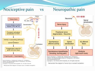
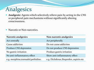
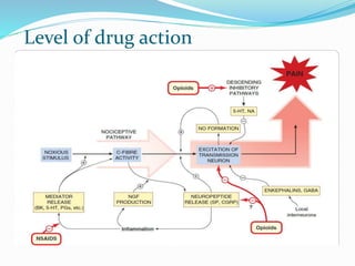
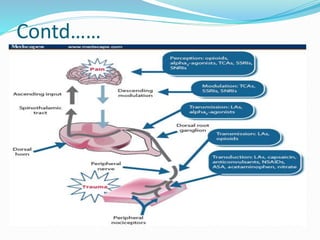
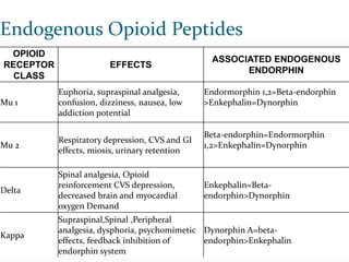
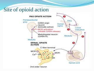
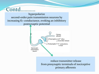
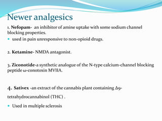
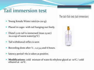
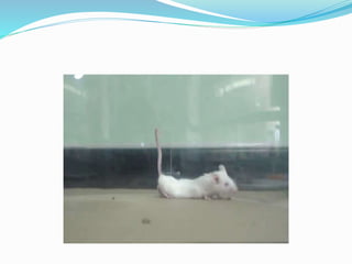
Dr. Sourav Chakrabarty
Post-graduate trainee
Department of Pharmacology
B.S. Medical College
“But pain is a perfect misery
The worst of evils
Excessive overturns
All patience”
John Milton - Paradise Lost
Overview
 Introduction
 Pain physiology
 Analgesics- Narcotic & Non-narcotic.
 Evaluation of analgesics.
 In vitro methods
 In vivo methods
 Discussion
 Conclusion.
Introduction
What is Pain?
Sherrington- “the physical adjunct of an imperative protective reflex.”
 Medical Definition
“Pain is an unpleasant sensory and emotional experience associated
with actual or potential tissue damage or described in terms of such
damage”- IASP
 Operative Definition
“Pain is whatever the experiencing person says it is, existing whenever
he/she says it does.”
 5th vital sign.
Contd……
 Types-
1. Acute pain/physiologic pain/good pain
2. Chronic pain/pathologic pain/bad pain
 But from pharmacological point of view
1. Nociceptive pain
2. Neuropathic pain
 Two components- sensory & affective.
Some basic terminologies
 Nociception: neural processes of encoding and processing noxious
stimuli.
 Hyperalgesia: an exaggerated response to a noxious stimulus
 Allodynia : a sensation of pain in response to a normally innocuous
stimulus.
 Nociceptive pain: pain arising from activation of nocieptors.
 Neuropathic pain: pain arising as a direct consequence of a lesion in the
neuron or disease affecting the somatosensory system.
 Pain threshold: the minimal intensity of stimulus that is perceived as
painful.
Pain pathway
Chemical signalling in the
nociceptive pathway
Modulation of pain
Nociceptive pain vs Neuropathic pain
Analgesics
 Analgesic: Agents which selectively relieve pain by acting in the CNS
or peripheral pain mechanisms without significantly altering
consciousness.
 Narcotic or Non-narcotics.
Narcotic analgesics Non-narcotic analgesics
Act centrally Act peripherally
Cause addiction Do not cause addiction
Produce CNS depression Do not produce CNS depression
No gastric irritation Produce gastric irritation
No anti-inflammatory effect Show anti-inflammatory effect
e.g. morphine,tramadol,pethidine. e.g. Diclofenac,Ibuprofen, aspirin etc
Level of drug action
Contd……
Endogenous Opioid Peptides
OPIOID
RECEPTOR
CLASS
EFFECTS
ASSOCIATED ENDOGENOUS
ENDORPHIN
Mu 1
Euphoria, supraspinal analgesia,
confusion, dizziness, nausea, low
addiction potential
Endormorphin 1,2=Beta-endorphin
>Enkephalin=Dynorphin
Mu 2
Respiratory depression, CVS and GI
effects, miosis, urinary retention
Beta-endorphin=Endormorphin
1,2>Enkephalin=Dynorphin
Delta
Spinal analgesia, Opioid
reinforcement CVS depression,
decreased brain and myocardial
oxygen Demand
Enkephalin=Beta-
endorphin>Dynorphin
Kappa
Supraspinal,Spinal ,Peripheral
analgesia, dysphoria, psychomimetic
effects, feedback inhibition of
endorphin system
Dynorphin A=beta-
endorphin>Enkephalin
Site of opioid action
Contd………..
reduce transmitter release
from presynaptic terminals of nociceptive
primary afferents
hyperpolarize
second-order pain transmission neurons by
increasing K+ conductance, evoking an inhibitory
postsynaptic potential
Newer analgesics
1. Nefopam- an inhibitor of amine uptake with some sodium channel
blocking properties.
 used in pain unresponsive to non-opioid drugs.
2. Ketamine- NMDA antagonist.
3. Ziconotide-a synthetic analogue of the N-type calcium-channel blocking
peptide ω-conotoxin MVIIA.
4. Sativex -an extract of the cannabis plant containing Δ9-
tetrahydrocannabinol (THC) .
 Used in multiple sclerosis
5. Botulinum toxin
6. Ropinirole, pramipexole and rotigotine-used to treat restless leg
syndrome
7. cGRP antagonist- used in migraine.
8. Flupirtine
Future aspects
 monoclonal antibodies to NGF or its receptor TrkA .
 Trp channel ligands.
 Retigabine-Kv7 (M-current) opener (see Ch. 45) inhibits C-fibre-and
Aδ-fibre-mediated nociceptive responses in dorsal horn neurons in
both naive and neuropathic rats.
 Agonists at nicotinic acetylcholine receptors, based on epibatidine
 Antagonists of metabotropic glutamate receptors, mGluR1 and
mGluR5,
Evaluation of analgesics
 In vitro and In vivo screening
 In vitro tests can only partially substitute for animal experiments
involving pain
 In vivo tests should detect central & peripheral analgesic specifically
 What to measure?
 Change in pain threshold
 Change in the latency period of reaction
In vitro tests
1. 3H-Naloxone binding assay
2. 3H-Dihydromorphine binding to μ opiate receptors
3. 3H-Bremazocine binding to κ opiate receptors
4. Inhibition of enkephalinase
5. Receptor binding of nociceptin
6. Vasoactive intestinal polypeptide (VIP) and pituitary adenylate cyclase-
activating peptide(PACAP) binding assay.
7. Cannabinoid receptor binding assay.
In-vivo tests
 Models for acute pain
1. Thermal stimulus- Hot plate method/Tail-flick method/Cold tail flick
test
2. Electric stimulus- Tooth pulp test/ Tail stimulation test/Money shock
titration test
3. Chemical stimulus- Formalin test/ Writhing test/Rat sigmoid colon
model/ Inflammatory uterine pain model
4. Mechanical stimulus- Tail clip method/Randal Selitto test
Contd…...
 Models for chronic pain
1. Neuropathic pain model- mechanical/vincristine induced/diabetic
neuropathy
2. Chronic post thoracotomy pain model
3. Incisional pain model
4. Cancer pain model
Hot plate method
 Purpose- to evaluate central analgesics.
 Methods-
1. Mice weighing 18-22g are used.
2. Standard or test drug given orally/ sc
3. Animals placed on hot plate(55-56° C.)
4. Response- jumping/paw withdrawal/ licking of paws.
5. Latency period is measured after 20, 60 & 90 min.
6. Those increasing the latency period at least 50% are taken positive.
7. ED-50 values are calculated.
 Drawback- not for peripheral analgesics.
 False positive results with sedative/ muscle relaxants/ psychotomimetic.
Tail flick test
 Use of light beam exerting radiant heat, focused to proximal 1/3rd of
the tail.
 Nociceptive spinal reflex response- flicking tail away from heat source.
 Escape reaction- turning the head away.(more reliable)
 Latency period is compared.
 Minimal inter animal variation.
Tail immersion test
 Young female Wister rats(170-210 g).
 Placed in cages with tail hanging out freely.
 Distal 5 cm tail is immersed (max 15 sec)
in a cup of warm water(55°C)
 Tail withdrawal reflex is seen
 Recording done after ½ , 1,2,3,4 and 6 hours.
 latency period ˃6s is taken as positive.
 Modifications- cold mixture of water & ethylene glycol at -10°C./ cold
ethanol at -20°C.
Electric stimulus models
 Tooth pulp test-
1. Rabbits(2-3kg) are anaesthetized with thiopental at 15 mg/kg i.v.
2. Clamping electrodes are placed into tooth pulp chamber through drilled
holes.
3. After 30 min, stimulus given by rectangular current(50 Hz) upto 1 sec
4. Animals starts licking- current threshold is measured.
 Modification: stimulus given via subcutaneous electrodes at the tail
 Monkey shock titration test: shock given via Coulbourn Instrument
Programmable shocker at tail
Grid - shock test
 Drugs like Morphine, Acetylsalicylic acid can be measured by the Flinch –
jump response.
 Male mice (18-20g)
 The floor of the box is wired with stainless steel wire
 The stimulus in the form of
square wave pulses ( 30 cps).
 The output of stimulator is connected
to alternate wires of grid.
 The fixed resistance is placed with
the grid & parallel to an oscilloscope
to allow calibration in mill amperes.
Contd…..
 With increase in shock intensities the mice flinch, exhibit startling
reaction & increase locomotion or attempt to jump.
 The behavior is accurately reflected on the oscilloscope by marked
fluctuations of the displayed pulse.
 Pain thresholds are determined in each individual mouse twice before &
after the administration of the test drug.
 The average pain threshold values for each group at each time interval are
calculated and statistically compared with the control values.
Chemical stimulus models
 An irritant/ algogenic chemical agent given as nociceptive stimulus
 Slower mode of stimulation.- progressive & persisting for longer duration
 Both for central & peripheral analgesics
 Formalin test-
o 10% formalin injected at paw.
o Biphasic response
 Early: immediately due to chemical stimulation of nociceptors causing
C-fibre activation
 Late: after 10-15 min due to combination of an inflammatory reaction and
functional changes at dorsal horn of spinal cord.
 Opioid effective in both phase, but NSAIDs are effective in only second
phase.
Writhing test
 Model for visceral/peritoneal pain
 Used to detect peripheral analgesic activity of a compound.
 Mice(20-25g) are given i.p injection an aliquot of 0.25 ml of
phenylquinone(0.02%) suspended in 1% suspension of carboxy
methylcellulose
 A characteristic stereotyped behavior known as writhing is seen.
 Series of contraction along abdominal wall, turning movements of the
body and extension of hind limbs.
 Number of writhes ( stretching of the abdomen with at least one hind
limb) are recorded for 10 min
 Other chemicals- acetic acid, acetylcholine, bradykinin, PGE1, 4%
NaCl, ethacrynic acid.
Mechanical stimulus models
 Tail-clip method:
1. Noxious stimulus by using artery clip placed at the root of tail.
2. Response- biting the clip/tail.
3. Reaction time is noted.
4. Cut off time- average reaction time plus 3 times the sd of the
combined latencies of the control mice at all time period.
 Randal Selitto test: induction of hyperalgesia by producing
inflammation via sc injection of Brewar’s yeast at hind paw, followed by
application of pressure.
 Mechanical visceral pain: chronic intestinal distension by a
indwelling intraduodenal balloon catheter.
Chronic pain models
 Neuropathic pain- mechanical
1. Allodynia induced by partial somatosensory sciatic nerve injury.
2. Male Sprague-Dawley rat anesthetized with 4% halothane.
3. Sciatic nerves of both sides are exposed at the level of mid-thigh.
4. Right sciatic nerve is tied loosely with square knot using 4-0 chromic
gut sutures.
5. Layer by layer closure done.
6. Mild aversion of right paw & foot drop.
1. Thermal nociceptive threshold is measured separately in each paws.
2. Drugs & vehicle given intrathecally.
3. PWL(paw withdrawal latency) is recorded.
4. Difference score(DS)=maximum PWL(LT)-maximum PWL(RT).
5. Dose is plotted against the change in DS.
 Vincristine induced neuropathy- By vincristine(100µg/kg) daily for
two weeks.
Diabetic neuropathy
 Streptozotocin(STZ) 75mg/kg ip for developing Diabetes.(BG≥14mM)
 4 weeks follow up with record of pain threshold, hyperglycemia &
clinical symptoms.
 Threshold to noxious heat stimuli/ non-painful thermal/
mechanical stimuli is observed.
 After 4 weeks, formalin injection is given to ascertain hyperalgesia.
 Ox-carbazepine can be evaluated in this model.
Models for cancer pain
 Induction of bone cancer by the syngeneic MRMT-1 mammary tumour
cell line.
 Sprague-Dawley rats are given intra-tibial injections of tumor cells.
 Control rats receive heat-killed cells or vehicle.
 Development of bone tumor and structural damage to the bone are
monitored.
 Mechanical allodynia & reduced weight bearing on affected limb,
beginning on day 12-14.
 Drugs are evaluated on the basis of their efficacy to reduce allodynia.
Discussion
 In humans, pain has a polymorphic nature.
 In animals, we focus on the latency period of response to noxious stimuli
or the change in the threshold of response.
 Limitations:
1. In most tests, responses are monitored around a nociceptive threshold,
whereas clinical pain is more severe.
2. Different scenario, when responses are evoked from healthy and
inflamed tissue.
3. Threshold to stimuli continue to increase in intensity
Conclusion
 No model is ideal, except chemical stimuli model, most closely mimicking
acute clinical pain.
 Neuropathic pain, very difficult to design the ideal model both for the
technical and ethical reasons.
 But still, animal models are very important to assess the analgesic activity
of drugs.
 Continued search should be maintained to find out novel drug targets & to
develop ideal animal model for their assessment.
THANK YOU

Evaluation of analgesic agents

  • 1.
    Dr. Sourav Chakrabarty Post-graduatetrainee Department of Pharmacology B.S. Medical College
  • 2.
    “But pain isa perfect misery The worst of evils Excessive overturns All patience” John Milton - Paradise Lost
  • 3.
    Overview  Introduction  Painphysiology  Analgesics- Narcotic & Non-narcotic.  Evaluation of analgesics.  In vitro methods  In vivo methods  Discussion  Conclusion.
  • 4.
    Introduction What is Pain? Sherrington-“the physical adjunct of an imperative protective reflex.”  Medical Definition “Pain is an unpleasant sensory and emotional experience associated with actual or potential tissue damage or described in terms of such damage”- IASP  Operative Definition “Pain is whatever the experiencing person says it is, existing whenever he/she says it does.”  5th vital sign.
  • 5.
    Contd……  Types- 1. Acutepain/physiologic pain/good pain 2. Chronic pain/pathologic pain/bad pain  But from pharmacological point of view 1. Nociceptive pain 2. Neuropathic pain  Two components- sensory & affective.
  • 6.
    Some basic terminologies Nociception: neural processes of encoding and processing noxious stimuli.  Hyperalgesia: an exaggerated response to a noxious stimulus  Allodynia : a sensation of pain in response to a normally innocuous stimulus.  Nociceptive pain: pain arising from activation of nocieptors.  Neuropathic pain: pain arising as a direct consequence of a lesion in the neuron or disease affecting the somatosensory system.  Pain threshold: the minimal intensity of stimulus that is perceived as painful.
  • 7.
  • 8.
    Chemical signalling inthe nociceptive pathway
  • 9.
  • 10.
    Nociceptive pain vsNeuropathic pain
  • 11.
    Analgesics  Analgesic: Agentswhich selectively relieve pain by acting in the CNS or peripheral pain mechanisms without significantly altering consciousness.  Narcotic or Non-narcotics. Narcotic analgesics Non-narcotic analgesics Act centrally Act peripherally Cause addiction Do not cause addiction Produce CNS depression Do not produce CNS depression No gastric irritation Produce gastric irritation No anti-inflammatory effect Show anti-inflammatory effect e.g. morphine,tramadol,pethidine. e.g. Diclofenac,Ibuprofen, aspirin etc
  • 12.
  • 13.
  • 14.
    Endogenous Opioid Peptides OPIOID RECEPTOR CLASS EFFECTS ASSOCIATEDENDOGENOUS ENDORPHIN Mu 1 Euphoria, supraspinal analgesia, confusion, dizziness, nausea, low addiction potential Endormorphin 1,2=Beta-endorphin >Enkephalin=Dynorphin Mu 2 Respiratory depression, CVS and GI effects, miosis, urinary retention Beta-endorphin=Endormorphin 1,2>Enkephalin=Dynorphin Delta Spinal analgesia, Opioid reinforcement CVS depression, decreased brain and myocardial oxygen Demand Enkephalin=Beta- endorphin>Dynorphin Kappa Supraspinal,Spinal ,Peripheral analgesia, dysphoria, psychomimetic effects, feedback inhibition of endorphin system Dynorphin A=beta- endorphin>Enkephalin
  • 15.
  • 16.
    Contd……….. reduce transmitter release frompresynaptic terminals of nociceptive primary afferents hyperpolarize second-order pain transmission neurons by increasing K+ conductance, evoking an inhibitory postsynaptic potential
  • 17.
    Newer analgesics 1. Nefopam-an inhibitor of amine uptake with some sodium channel blocking properties.  used in pain unresponsive to non-opioid drugs. 2. Ketamine- NMDA antagonist. 3. Ziconotide-a synthetic analogue of the N-type calcium-channel blocking peptide ω-conotoxin MVIIA. 4. Sativex -an extract of the cannabis plant containing Δ9- tetrahydrocannabinol (THC) .  Used in multiple sclerosis
  • 18.
    5. Botulinum toxin 6.Ropinirole, pramipexole and rotigotine-used to treat restless leg syndrome 7. cGRP antagonist- used in migraine. 8. Flupirtine
  • 19.
    Future aspects  monoclonalantibodies to NGF or its receptor TrkA .  Trp channel ligands.  Retigabine-Kv7 (M-current) opener (see Ch. 45) inhibits C-fibre-and Aδ-fibre-mediated nociceptive responses in dorsal horn neurons in both naive and neuropathic rats.  Agonists at nicotinic acetylcholine receptors, based on epibatidine  Antagonists of metabotropic glutamate receptors, mGluR1 and mGluR5,
  • 20.
    Evaluation of analgesics In vitro and In vivo screening  In vitro tests can only partially substitute for animal experiments involving pain  In vivo tests should detect central & peripheral analgesic specifically  What to measure?  Change in pain threshold  Change in the latency period of reaction
  • 21.
    In vitro tests 1.3H-Naloxone binding assay 2. 3H-Dihydromorphine binding to μ opiate receptors 3. 3H-Bremazocine binding to κ opiate receptors 4. Inhibition of enkephalinase 5. Receptor binding of nociceptin 6. Vasoactive intestinal polypeptide (VIP) and pituitary adenylate cyclase- activating peptide(PACAP) binding assay. 7. Cannabinoid receptor binding assay.
  • 22.
    In-vivo tests  Modelsfor acute pain 1. Thermal stimulus- Hot plate method/Tail-flick method/Cold tail flick test 2. Electric stimulus- Tooth pulp test/ Tail stimulation test/Money shock titration test 3. Chemical stimulus- Formalin test/ Writhing test/Rat sigmoid colon model/ Inflammatory uterine pain model 4. Mechanical stimulus- Tail clip method/Randal Selitto test
  • 23.
    Contd…...  Models forchronic pain 1. Neuropathic pain model- mechanical/vincristine induced/diabetic neuropathy 2. Chronic post thoracotomy pain model 3. Incisional pain model 4. Cancer pain model
  • 24.
    Hot plate method Purpose- to evaluate central analgesics.  Methods- 1. Mice weighing 18-22g are used. 2. Standard or test drug given orally/ sc 3. Animals placed on hot plate(55-56° C.) 4. Response- jumping/paw withdrawal/ licking of paws. 5. Latency period is measured after 20, 60 & 90 min. 6. Those increasing the latency period at least 50% are taken positive. 7. ED-50 values are calculated.  Drawback- not for peripheral analgesics.  False positive results with sedative/ muscle relaxants/ psychotomimetic.
  • 25.
    Tail flick test Use of light beam exerting radiant heat, focused to proximal 1/3rd of the tail.  Nociceptive spinal reflex response- flicking tail away from heat source.  Escape reaction- turning the head away.(more reliable)  Latency period is compared.  Minimal inter animal variation.
  • 26.
    Tail immersion test Young female Wister rats(170-210 g).  Placed in cages with tail hanging out freely.  Distal 5 cm tail is immersed (max 15 sec) in a cup of warm water(55°C)  Tail withdrawal reflex is seen  Recording done after ½ , 1,2,3,4 and 6 hours.  latency period ˃6s is taken as positive.  Modifications- cold mixture of water & ethylene glycol at -10°C./ cold ethanol at -20°C.
  • 27.
    Electric stimulus models Tooth pulp test- 1. Rabbits(2-3kg) are anaesthetized with thiopental at 15 mg/kg i.v. 2. Clamping electrodes are placed into tooth pulp chamber through drilled holes. 3. After 30 min, stimulus given by rectangular current(50 Hz) upto 1 sec 4. Animals starts licking- current threshold is measured.  Modification: stimulus given via subcutaneous electrodes at the tail  Monkey shock titration test: shock given via Coulbourn Instrument Programmable shocker at tail
  • 28.
    Grid - shocktest  Drugs like Morphine, Acetylsalicylic acid can be measured by the Flinch – jump response.  Male mice (18-20g)  The floor of the box is wired with stainless steel wire  The stimulus in the form of square wave pulses ( 30 cps).  The output of stimulator is connected to alternate wires of grid.  The fixed resistance is placed with the grid & parallel to an oscilloscope to allow calibration in mill amperes.
  • 29.
    Contd…..  With increasein shock intensities the mice flinch, exhibit startling reaction & increase locomotion or attempt to jump.  The behavior is accurately reflected on the oscilloscope by marked fluctuations of the displayed pulse.  Pain thresholds are determined in each individual mouse twice before & after the administration of the test drug.  The average pain threshold values for each group at each time interval are calculated and statistically compared with the control values.
  • 30.
    Chemical stimulus models An irritant/ algogenic chemical agent given as nociceptive stimulus  Slower mode of stimulation.- progressive & persisting for longer duration  Both for central & peripheral analgesics  Formalin test- o 10% formalin injected at paw. o Biphasic response  Early: immediately due to chemical stimulation of nociceptors causing C-fibre activation  Late: after 10-15 min due to combination of an inflammatory reaction and functional changes at dorsal horn of spinal cord.  Opioid effective in both phase, but NSAIDs are effective in only second phase.
  • 31.
    Writhing test  Modelfor visceral/peritoneal pain  Used to detect peripheral analgesic activity of a compound.  Mice(20-25g) are given i.p injection an aliquot of 0.25 ml of phenylquinone(0.02%) suspended in 1% suspension of carboxy methylcellulose  A characteristic stereotyped behavior known as writhing is seen.
  • 32.
     Series ofcontraction along abdominal wall, turning movements of the body and extension of hind limbs.  Number of writhes ( stretching of the abdomen with at least one hind limb) are recorded for 10 min  Other chemicals- acetic acid, acetylcholine, bradykinin, PGE1, 4% NaCl, ethacrynic acid.
  • 34.
    Mechanical stimulus models Tail-clip method: 1. Noxious stimulus by using artery clip placed at the root of tail. 2. Response- biting the clip/tail. 3. Reaction time is noted. 4. Cut off time- average reaction time plus 3 times the sd of the combined latencies of the control mice at all time period.
  • 35.
     Randal Selittotest: induction of hyperalgesia by producing inflammation via sc injection of Brewar’s yeast at hind paw, followed by application of pressure.  Mechanical visceral pain: chronic intestinal distension by a indwelling intraduodenal balloon catheter.
  • 36.
    Chronic pain models Neuropathic pain- mechanical 1. Allodynia induced by partial somatosensory sciatic nerve injury. 2. Male Sprague-Dawley rat anesthetized with 4% halothane. 3. Sciatic nerves of both sides are exposed at the level of mid-thigh. 4. Right sciatic nerve is tied loosely with square knot using 4-0 chromic gut sutures. 5. Layer by layer closure done. 6. Mild aversion of right paw & foot drop.
  • 37.
    1. Thermal nociceptivethreshold is measured separately in each paws. 2. Drugs & vehicle given intrathecally. 3. PWL(paw withdrawal latency) is recorded. 4. Difference score(DS)=maximum PWL(LT)-maximum PWL(RT). 5. Dose is plotted against the change in DS.  Vincristine induced neuropathy- By vincristine(100µg/kg) daily for two weeks.
  • 38.
    Diabetic neuropathy  Streptozotocin(STZ)75mg/kg ip for developing Diabetes.(BG≥14mM)  4 weeks follow up with record of pain threshold, hyperglycemia & clinical symptoms.  Threshold to noxious heat stimuli/ non-painful thermal/ mechanical stimuli is observed.  After 4 weeks, formalin injection is given to ascertain hyperalgesia.  Ox-carbazepine can be evaluated in this model.
  • 39.
    Models for cancerpain  Induction of bone cancer by the syngeneic MRMT-1 mammary tumour cell line.  Sprague-Dawley rats are given intra-tibial injections of tumor cells.  Control rats receive heat-killed cells or vehicle.  Development of bone tumor and structural damage to the bone are monitored.  Mechanical allodynia & reduced weight bearing on affected limb, beginning on day 12-14.  Drugs are evaluated on the basis of their efficacy to reduce allodynia.
  • 40.
    Discussion  In humans,pain has a polymorphic nature.  In animals, we focus on the latency period of response to noxious stimuli or the change in the threshold of response.  Limitations: 1. In most tests, responses are monitored around a nociceptive threshold, whereas clinical pain is more severe. 2. Different scenario, when responses are evoked from healthy and inflamed tissue. 3. Threshold to stimuli continue to increase in intensity
  • 41.
    Conclusion  No modelis ideal, except chemical stimuli model, most closely mimicking acute clinical pain.  Neuropathic pain, very difficult to design the ideal model both for the technical and ethical reasons.  But still, animal models are very important to assess the analgesic activity of drugs.  Continued search should be maintained to find out novel drug targets & to develop ideal animal model for their assessment.
  • 42.

Editor's Notes

  • #6 Nociceptive pain-normal’ response to tissue injury. Neuropathic pain-disordered neural function .
  • #8 Aδ and C fibres. Most of the nociceptive afferents terminate in the superficial region of the dorsal horn, the C fibres and some Aδ fibres innervating cell bodies in laminae I and II (also known as the substantia gelatinosa), while other A fibres penetrate deeper into the dorsal horn (lamina V). The nociceptive afferent neurons release glutamate and possibly ATP as the fast neurotransmitters at their central synapses in the dorsal horn. Glutamate acting on AMPA receptors is responsible for fast synaptic transmission at the first synapse in the dorsal horn. There is also a slower NMDA receptor-mediated response, which is important in relation to the phenomenon of ‘wind-up’ (see Fig. 42.2). The nociceptive afferent neurons also contain several neuropeptides (see Ch. 18), particularly substance P, calcitonin gene-related peptide (CGRP) and galanin.
  • #9 trkA-kinase linked receptor. transient receptor potential TRPV1 . BDNF released from primary afferent nerve terminals activates the kinase-linked TrkB receptor on postsynaptic dorsal horn neurons leading to phosphorylation of the NMDA subunit GluN1 and thus sensitisation of these glutamate receptors, resulting in synaptic facilitation, in the dorsal horn. Agonists such as capsaicin open the channel, which is permeable to Na+, Ca2+ and other cations, causing depolarisation and initiation of action potentials. The large influx of Ca2+ into peripheral nerve terminals also results in peptide release (mainly substance P and CGRP), causing intense vascular and other physiological responses. The Ca2+ influx may be enough to cause nerve terminal degeneration, which takes days or weeks to recover . Bradykinin acts on B2 receptors (see Ch. 18) on nociceptive neurons. B2 receptors are coupled to activation of a specific isoform of protein kinase C (PKCε), which phosphorylates TRPV1 and facilitates opening of the TRPV1 channel. ASIC-acid sensitive ion channels.
  • #10 PAG receives inputs from many other brain regions, including the hypothalamus, amygdala and cortex, and is the main pathway through which cortical and other inputs act to control the nociceptive ‘gate’ in the dorsal horn. Two important transmitters in this pathway are 5-hydroxytryptamine (5-HT; serotonin) and the enkephalins, which act directly or via interneurons to inhibit the discharge of spinothalamic neurons . There is also a noradrenergic pathway from the locus coeruleus (LC; see Ch. 39), which has a similar inhibitory effect on transmission in the dorsal horn. Surprisingly, opioids inhibit rather than activate this pathway. The use of tricyclic antidepressants to control pain probably depends on potentiating this pathway.
  • #11 Hyperalgesia involves both sensitisation of peripheral nociceptive nerve terminals and central facilitation of transmission at the level of the dorsal horn and thalamus. The peripheral component is due to the action of mediators such as bradykinin and prostaglandins acting on the nerve terminals. The central component reflects facilitation of synaptic transmission in the dorsal horn of the spinal cord (see Yaksh, 1999). The synaptic responses of dorsal horn neurons to nociceptive inputs display the phenomenon of ‘wind-up’ – i.e. the synaptic potentials steadily increase in amplitude with each stimulus – when repeated stimuli are delivered at physiological frequencies. Substance P and CGRP released from primary afferent neurons produce neurogenic inflammation. Neuropathic pain-This nerve injury pain state may not depend upon the activation of small afferents, but may be initiated by low-threshold sensory afferents (e.g., A fibers). Such nerve injuries result in the development of ectopic activity arising from neuromas formed by nerve injury and the dorsal root ganglia of the injured axons as well as a dororsal horn reorganization, such that low-threshold afferent input carried by A fibers evokes a pain state. This dorsal horn reorganization reflects changes in ongoing inhibition and in the excitability of dorsal horn projection neurons .
  • #16 Top left: Schematic of organization of opiate action in the periaqueductal gray. Top right: Opiate-sensitive pathways in PAG Mu opiate actions block the release of GABA from tonically active systems that otherwise regulate the projections to the medulla (1) leading to an activation of PAG outflow resulting and activation of forebrain (2) and spinal (3) monoamine receptors that regulate spinal cord projections (4) which provide sensory input to higher centers and mood. Bottom left: Schematic of primary afferent synapse with second order dorsal horn spinal neuron, showing pre- and post-synaptic opiate receptors coupled to Ca2+ and K+ channels, respectively. Opiate receptor binding is highly expressed in the superficial spinal dorsal horn (substantia gelatinosa). These receptors are located presynaptically on the terminals of small primary afferents (C fibers) and postsynaptially on second order neurons. Presynaptically, activation of MOR blocks the opening of the voltage sensitve Ca2+ channel, which otherwise initiates transmitter release. Postsynaptically, MOR activation enhances opening of K+ channels, leading to hyperpolarization. Thus, an opiate agonist acting at these sites jointly serves to attenuate the afferent-evoked excitation of the second order neuron. MOR agonists block release of the inhibitory transmitter GABA from tonically active PAG systems that regulate activity in projections to the medulla. PAG projections to the medulla activate medullospinal release of NE and 5-HT at the level of the spinal dorsal horn. This release can attenuate dorsal horn excitability (Yaksh, 1997). Interestingly, this PAG organization can also serve to increase excitability of dorsal raphe and locus coeruleus from which ascending serotonergic and noradrenergic projections to the limbic forebrain originate.
  • #17  The opioids have two well-established direct G protein-coupled actions on neurons: (1) they close voltage-gated Ca2+ channels on presynaptic nerve terminals and thereby reduce transmitter release, and (2) they hyperpolarize and thus inhibit postsynaptic neurons by opening K+ channels.
  • #19 Flupirtine- selective k channel opener, NMDA antagonist, GABA receptor modulator.
  • #21 But in reality, they differ from the clinical scenario. Animal test- drug given before the noxious stimuli.
  • #27 .(normally after 1-5.5 sec)