PRESENTED BY :
SREYA. S
M. Pharmacy
Academic year:2019-2021
DEPT OF PHARMACOLOGY
INTRODUCTION
 Arrhythmia is deviation of heart from normal RHYTHM.
Abnormality in the origin, rate, rhythm, conduction
velocity and sequence of heart activation. It may cause
sudden death or syncope, heart failure, dizzines,
palpitations, or no symptoms at all.
RHYTHM
1) HR- 60-100
2) Should origin from SAN
3) Cardiac impulse should propagate through normal
conduction pathway with normal velocity.
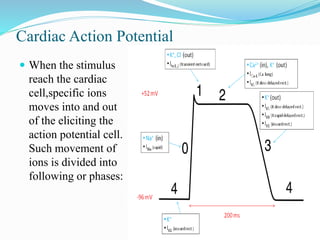
Cardiac Action Potential
 When the stimulus
reach the cardiac
cell,specific ions
moves into and out
of the eliciting the
action potential cell.
Such movement of
ions is divided into
following or phases:
 Phase 0: rapid depolarization of cell membrane during
which theirs is fast entry of Na ions into the cells through
Na channels, this is followed by repolarization.
 Phase 1: is short initial rapid repolarization due to Ka
efflux
 Phase 2: prolonged plateue phase due to slow Ca influx
 Phases 3: rapid repolarization with Ka efflux
 Phase 4: resting phase during which Ka ions return into
the cell while Na and Ka move out of it and resting
membrane potential is stored
Mechanisms of cardiac arrhythmias
•Enhanced/ectopic pacemaker activity
•After-depolarizations
• Reentry
Enhanced/ectopic pacemaker activity – Ectopic impulse
may also result from current of injury. The slope of phase4
depolarization may be increased pathologically in the
automatic fibres
 After-depolarizations –
Early after-depolarization
(EAD) – Delayed
afterdepolarization (DAD
 Reentry – Primarily due to
abnormality of conduction,
an impulse may recirculate
in the heart and cause
repetitive activation without
the need for any new
impulse to be generated.
These are called reentrant
arrhythmias.
CLASSIFICATION OFARRHYTHMIAS
1. Characteristics:
a. Flutter – very rapid but regular contractions
b. Tachyarrhthmia – increased rate
c. Bradyarrhythmia – decreased rate
d. Fibrillation – disorganized contractile activity
 500 Atrial fibrillation
 350 Atrial flutter
 200 Paroxysmal TA
 100-60-Normal range
 40 Mild bradyarrhythmias
 20 moderate BA
 Atrial flutter (AFL) [can be regular or irregular] is an
abnormality of conduction within the atria. The atria beat
more frequently than the ventricles (up to 300 bpm).
 Atrial Fibrillation (AF) [always irregular] there is a
complete absence of a Sino-Atrial stimulus which would
result in a P wave. The atria beat out of coordination
ventricle). This leads to a rapid and irregular heart rhythm.
In AF, the heart rate ranges from 100 to 175 bpm.
SYMPTOMS
 Noticeable arrhythmia symptoms may include:
A fluttering in your chest. A racing heartbeat
(tachycardia) A slow heartbeat (bradycardia)
 Can have no symptoms, but people may experience:
 Pain areas: in the chest
 Whole body: dizziness, fainting, or light-headedness
 Heart: palpitations or slow heart rate
 Also common: shortness of breath
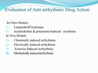
Evaluation of Anti arrhythmic Drug Action
In-Vitro Models:
1) Langendorff technique
2) Acetylcholine & potassium Induced arrythmia
In Vivo Models
1) Chemically induced arrhythmia
2) Electrically induced arrhythmia
3) Exercise Induced Arrhythmia
4) Mechanicallyinducedarrhythmia
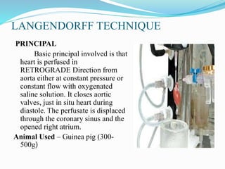
LANGENDORFF TECHNIQUE
PRINCIPAL
Basic principal involved is that
heart is perfused in
RETROGRADE Direction from
aorta either at constant pressure or
constant flow with oxygenated
saline solution. It closes aortic
valves, just in situ heart during
diastole. The perfusate is displaced
through the coronary sinus and the
opened right atrium.
Animal Used – Guinea pig (300-
500g)
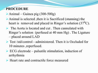
PROCEDURE
Animal – Guinea pig (300-500g)
Animal is selected ,then it is Sacrificed (stunning) the
heart is removed and placed in Ringer’s solution (37⁰C).
 The Aorta is located and cut . Then cannulated with
Ringer’s solution (perfused at 40 mm Hg) . The Ligature
– placed around LAD
 Test /std/control - administered. Then it is Occluded for
10 minutes ,reperfused.
 ECG electrode – pulsatile stimulation, induction of
arrhythmia
 Heart rate and contractile force measured
EVALUATION
Heart rate measured through chronometer coupled to
polygraph, Contractile force measured by force transducer.
Incidence and duration of ventricular fibrillation or
ventricular tachycardia is recorded in the control as well
as test group.
Acetylcholine or Potassium Induced
Arrhythmias
PRINCIPLE
The K+ channel function determines membrane
potential and refractoriness of the myocardium. Both gain
and loss of the K+ channel function can lead to arrhythmia
Animal used- New Zealand white rabbits (0.5-3kg)
PROCEDURE
 New Zealand white rabbits (0.5-3kg) sacrificed and heart
removed
 Atria dissected from other tissue in RL and attached with
electrode
 Fibrillation produced when atria exposed with Acetylcholine or
KCL
 After 5min exposure to Acetylcholine or KCL, atria stimulated
with pulses 0.75 ms , usually of 10V and recorded on
Kymograph
 Control arrhythmia produced for 6-10 min followed by 30 min
rest period
 Test compound were added to the bath
EVALUATION
 Test drug found to be effective If fibrillation is
isdisappears immediately or within 5min
 If fibrillation doesn't disappear within 8-10 min,
preparation is washed to allowed to normal concentration
IN VIVO MODELS
Chemically Induced Arrhythmia
ACONITINE ANTAGONISM IN RATS
Priciple
Aconitine, its plant alkaloid from roots, it Acts
perstintant on Na+ channels resulting ventricular
arrhythmias
Animal used: Male Ivanovas rats (300-400 gm)
Procedure:
Male Ivanovas rats (300-400 gm) anesthetized
intraperitonially with urethane (1.25 g/kg)
Aconitine dissolved (5 mcg/kg) in 0.1 N HNO3 &
infused into rat’s saphenous vein @ 0.1 ml/min
Lead II ECG is recorded every 30 sec.
 Test compound injected orally 5 min before Aconitine
infusion
 Higher dose of aconitine in the test group gives an index
of antiarrhythmic activity.
 The antiarrhythmic effect of test compound is measured
by the amount of aconitine /100g animal
 Higher dose of aconitine is needed as compared with
control group
Evaluation
 The anti-arrhythmic effect of the test compound is
measured by the amount of aconitine/100 gm animal
(infusion duration ), required to precipitate... Ventricular
extra systoles. Ventricular tachycardia. Ventricular
fibrillation and death
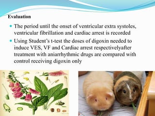
DIGOXIN INDUCED ARRHYTHMIA IN
GUINEA PIGS
 PRINICPLE:
Over dose of cardiac glycosides induces ventricular
extrasystoles, ventricular fibrillation, and finally death.
Animal used:male guinea pigs.(Marioth 350-500 gms)
Standard drugs : lidocaine 3 mg/kg iv
Procedure:
 Anesthetised male guinea pigs.(Marioth 350-500 gms)
 Trachea, jugular vein and carotid artery are catheterized.
 Test grp receives Test drug either orally 1 hr or iv 1 min prior
to the infusion while controlol group receieves digoxin infusion
only at rate of 85 mg / kg in 0.266 ml/min. until cardiac arrest
occurs.
 Lead III ECG recorded
Evaluation
 The period until the onset of ventricular extra systoles,
ventricular fibrillation and cardiac arrest is recorded
 Using Student’s t-test the doses of digoxin needed to
induce VES, VF and Cardiac arrest respectivelyafter
treatment with aniarrhythmic drugs are compared with
control receiving digoxin only
ELECTRICALLY INDUCED ARHYTHMIAS
VENTRICULAR FIBRILLATION
ELECTRICAL THRESHOLD
Principle
Ventricular fibrillations can be induced by various
tec. Of electrical stimulation like single pulse stimulation ,
train of pulse stimulation , continous 50 HZ stimulation
 Ventricular fibrillation threshold :The minimal current
intensity of the pulse train required to induce sustained
ventricular fibrillation.
Animals – Adult dogs (8-12kg)
Anesthetic – Sodium pentobarbital (35mg/kg)
PROCEDURE
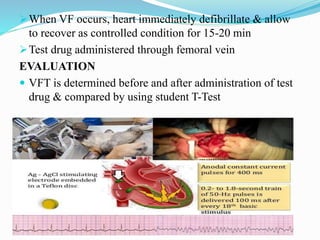
 Dogs (8-12 kg) anesthetized with pentobarbitone
intraperitonially and maintained on artificial respiration
 Chest opened & heart is suspended on pericardial cradle
 SA node crushed & Ag-AgCl stimulating electrode embedded
with Teflon disc sutured to anterior surface of LV
 Anodal constant current for 400ms is applied through electrode
and which was programmed by digital stimulator
 ECG recorded through lead II of body ECG monitoring
 To determine Ventricular Fibrillation (VFT) 0.2 to 1.8 s train of
50Hz pulses delivered
 The current intensity of pulse train required to induce sustained
ventricular fibrillation is define as VFT
When VF occurs, heart immediately defibrillate & allow
to recover as controlled condition for 15-20 min
Test drug administered through femoral vein
EVALUATION
 VFT is determined before and after administration of test
drug & compared by using student T-Test
EXERCISE INDUCED ARRHYTHMIA
EXERCISE INDUCED VENTRICULAR FIBRILLATION
PURPOSE AND RATIONALE:
Tests combining coronary constriction with physical
exercise may resemble most closely the situation in
coronary patients
Animals – Mongrel dogs (15 -19 kg)
Anesthetic – Sodium pentobarbitone (10mg/kg) i.v.
PROCEDURE
Animals IS selected. IT IS Anesthetized . The Chest
cavity is opened .
 Heart suspended in pericardial cradle . Around left
circumflex artery – Doppler flow inducerto measure blood
pressure - Hydraulic occluder - to occlude coronary artery
are placed
 A Pair of insulated silver coated wires – sutured on left
and right ventricles- measure HR (By Gould
biotachometer) .
The Occlusion of LAD(Two stage) . Myocardial infarction
. After 24 hrs- test drug/std/control - administered
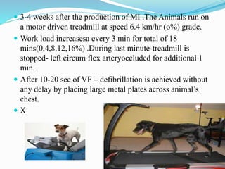
 3-4 weeks after the production of MI .The Animals run on
a motor driven treadmill at speed 6.4 km/hr (o%) grade.
 Work load increasesa every 3 min for total of 18
mins(0,4,8,12,16%) .During last minute-treadmill is
stopped- left circum flex arteryoccluded for additional 1
min.
 After 10-20 sec of VF – defibrillation is achieved without
any delay by placing large metal plates across animal’s
chest.
 X
 Protocol starts with 3 min warm up followed by run @ 6.4
km/hr (0 % grade)
 Grade is increased every 3min run 0% 4% 8% 12% and
16%
 During last min of exercise LCX is occluded, treadmill
stopped & occlusion maintained for additional 1min
 Occlusion is released if VF occurs
 Exercise and ischemia test is repeated after pretreated with
test drug & compared with control reading
EVALUATION:
The exercise plus ischemia test is repeated after
administration of the test drug and compared to control
(saline) group.The effect of drugs intervention on
arrhythmia formation are determined using chi square test
with yates correction.
MECHANICALLYINDUCEDARRHYTHMIA
CORONARY ARTERY OCCLUSION/REPERFUSION
ARRHYTHMIA
PRINCIPLE ; Arrhythmias –induced directly by ischemia
and reperfusion
Animals Used– Dogs (20-25Kg)
Anesthetic – Thiobutobarbital sodium (30mg/kg)
PROCEDURE:
 Animals is selected . Then its is anesthetized
Artificial respiration is given by positive pressu
respirator.
Femoral artery iscannulated and connected to pressure
transducer .
The Chest cavity is opened and LAD is exposed
 Silk suture is placed around LAD.After 45 min(equilibration) –
test/std/control- administered through saphenous vein
 After 20 min- ligature of coronary artery is closed for 90 min
 Occlusion released – reperfusion period maintained for 30 min.
 All the parameters – recorded
 At the end – surviving animals are sacrificed by an overdose
of Pentobarbital sodium.
EVALUATION
• Mortality , Hemodynamics, Arrhythmia
• Ventricular fibrillation
• % animals with VF.
REFERENCES
 Vogel WH, Schölkens BA. Drug Discovery and Evaluation
[Internet]. Springer Berlin Heidelberg; 2002. P.208-29.
 Essentials of medical pharmacology –KD Tripathi
THANKYOU