
Recommended
Recommended
More Related Content
What's hot
What's hot (20)
Similar to SCFE: Slipped Capital Femoral Epiphysis Guide
Similar to SCFE: Slipped Capital Femoral Epiphysis Guide (20)
Recently uploaded
Recently uploaded (20)
SCFE: Slipped Capital Femoral Epiphysis Guide
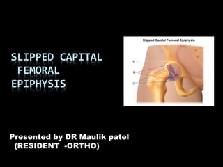
- 1. SLIPPED CAPITAL FEMORAL EPIPHYSIS Presented by DR Maulik patel (RESIDENT -ORTHO)
- 2. Slipped capital femoral epiphysis Slip capital femoral epiphysis is a disorder in which capital femoral epiphysis is displaced from the metaphysis through the physeal plate SCFE is actually a misnomer in that the head is held in the acetabulum by the ligamentum teres,& thus it is actually the neck that comes upward & outwards while the head remains posterior & downward in the acetabulum. A varus relation exists between the head & neck, but occasionally the slip is into valgus, with head displaced superiorly & posteriorly in relation to the neck
- 3. Incidence => 2/100,000 (.002%) Boys(10 – 16 yrs) > girls(10 – 14 yrs) (2.5:1) Blacks > white 5% of parents have had SCFE Left hip is twice as often affected as right hip Bilateral 17-37% in adolescents 50% are simultaneous, 50% sequential Younger child (physically or chronologically) the greater risk of subsequent bilateral involvement a contra-lateral slip will usually present within 2 years 20% are bilateral at time of presentation
- 4. Etiology The causes for the displacement are multifactorial Four important causes are indentified:- 1. Increased height of capital femoral physis. 2. Changes in the geometry of the capital physis and adjacent bone 3. Abnormal loading of growth plate 4. Insufficiency of the tensile (collagen) and (proteoglycans) hydrostatic component of the growth plate. It is not essential that all the factors exists for the slip to occur
- 5. Usually every capital epiphysis does not slip. Growth plate anatomic stability is responsible for holding the physis against the shearing stress these are:- 1. Perichondrium and the perichondrial ring 2. Transphysieal collagen fibres 3. The mamillary processes 4. The central and peripherral countour of the physis 5. Inclination angle of the physis 6. Height of the growth plate.
- 6. Where does it slip? Slip occurs in the zone of hypertrophied cartilage cell layer. Bone marrow epiphysis Bone of epiphysis Zone of resting cartilage Zone of proliferating cartilage Zone of maturing cartilage Zone of calcifying cartilage Developing trabeculae of metaphysis
- 7. Hormonal theory:- slipping of epiphysis occurs only when the growth plate remains open. Growth and maturation of epiphyseal cartilage plate depends on hormonal factors like growth hormone, thyroid hormone and sex hormone. In connection with this Harris observed that the epiphysis tends to slip at time of fast growth.
- 8. Traumatic theory:- The epiphyseal line is the weakest part of the normal adolescent bone. Key pointed out that the slipping of epiphysis is neither in the bone nor in the cartilage, but in the periosteum of the femoral neck. In the childhood the periosteum is thick and is thrown into folds or ridges known as retinacula of Weitbrecht, actually it is the chief factor holding the head in place. In adults the periosteum becomes atrophy to produce a point of weakness in the epiphyseal line. He also pointed out that most of the cases of coxa vara gives a history of very rapid growth and during this the the periosteum crossing the epiphyseal line is stretched and thinned and consequently weaken causing coxa vara.
- 9. Classification Traditional Classification : ( Fahey and O’brain ) Acute < 3 weeks of symptoms Chronic > 3 weeks of symptoms Acute on Chronic> 3 weeks of symptoms + sudden exacerbation Newer Classification ( LODER ) -Unstable – ambulation is impossible, w/ or without crutches -Stable – ambulation is possible w/ or without crutches
- 10. Kallios , ultra sound :- Unstable - effusion is present, physeal instability allows reduction , no metaphyseal resorption or early remodelling Stable - effusion is absent, physeal stability present ,does not allows reduction , metaphyseal resorption or early remodelling present.
- 11. CATEGORIES AS PER SEVERITY OF SLIP Wilson’s Classification:- Pre-slip or grade I: Widening & rarefaction of the physis, but no actual displacement. Minimal slip or grade II: Femoral head displaces up to one third of the superior metaphyseal width of the neck Moderate slip or grade III: Femoral head displaces greater than one third & less than half of the superior metaphyseal width of the neck. Severe slip or grade IV: Femoral head displaces more than 50% of the superior metaphyseal width of the neck.
- 12. GROUP I 1%-32% GROUP II 33-50% GROUP III >50%
- 13. PATHOLOGY The pathologic changes depend on the stage & degree of displacement. Pre-slipping stage: Physis is widened in zone of hypertrophy. Cartilage cells are in disarrayed clusters instead of orderly columns in this layer. Island of unorganized cartilage dispersed irregularly in the proximal metaphysis. Femoral head & acetabulum are normal. Synovial membrane is engorged, edematous & swollen.
- 14. Slipping stage: The slip takes place in layer of hypertrophic cartilage cells adjacent to the zone of provisional calcification. The plane of separation is weaving & irregular due to the irregularity of the contour of the physis. The slipping is usually gradual. Perichondrium remains attached to the femoral neck, stretching & elongating as the physis migrates. In an acute slip the perichondrium is stripped off the neck anteriorly & inferiorly. Capital epiphysis with the acetabulum almost always displace posteriorly & inferiorly. The displaces anteriorly & proximally as a hump. Except in acute traumatic slip there is no hemarthosis.
- 15. Chronic stage:- With healing the inferior angle & anterior portion of the neck adjacent to the physis is filled with callus. When remodeling takes place, the callus becomes incorporated with the neck. The protruding hump becomes round & smooth. These hump impinges against the anterior & superior margins of acetabulum & cause limitation of abduction, medial rotation & full flexion. Swelling & edema of the synovial membrane subsides. Physis ossifies & there is bony union between the head & the neck. Degenerative osteoarthritis sets in the acetabulum.
- 16. Clinical features:- Symptoms:- The onset is gradual and in many cases the earliest symptom is that the patient gets tired after walking or standing. Pain is the main complain, usually confined to hip but may radiates to lower thigh and to knee. Pain is accompanied by limp, the limp may be present even in the absence of pain. The affected leg becomes shorter and smaller and tends to turn laterally, and its movement is restricted. The limb is held in flexion, adduction and external rotation.
- 17. Signs:- Waddling gait is present The patient stands with the leg rotated laterally and slightly adducted, while inspection shows the pelvis to be tilted on the affected side. Scoliosis on the affected side is present Buttock is atrophied and the gluteal fold is lower than on the normal side. On palpation hard mass is felt on which head moves with femur. Trochanter is higher than the sound hip Adduction and lateral rotation are free but abduction and medial rotation and extension are greatly restricted.
- 18. MEASUREMENT OF AMOUNT OF SLIPPING Head shaft angle by Southwick: In lateral view of normal hip the capital femoral physis & femoral neck lie at right angles to each other.The physeal neck angle decreases when slipping takes place. In recent acute slip the distance between the superior edge of the epiphysis & the upper margin of the metaphysis indicated the amount of displacement; which shows the greatest amount of displacement. In chronic slip remodeling of the femoral neck makes it difficult to measure the amount of displacement of the capital femoral epiphysis. Here Southwick’s method is useful,
- 19. Lateral view of hip is taken, First line is drawn between the superior & inferior margins of the metaphyseal surface of the capital femoral physis. Second line is drawn perpendicular to the first line. Third line drawn along the femoral shaft. The head shaft angle formed by the second & third line is measured. The head shaft angle of abnormal side is substracted from the normal side.It is classified as Mild 1-29 degrees Moderate 30-60 degrees Severe > 60 degrees
- 20. Radiological appearance Pre-slipping stage:- In early cases x-ray shows, Minimal slipping indicated by the absence of the normal shoulder on the upper aspect of the neck and head ( i.eTrethowan’s sign) in which a line drawn along the superior surface of the neck will pass above the femoral head rather than through it. The head is more or less sickle shaped instead of hemispherical and its height is diminished. The epiphyseal plate is widened and rarefaction or even streaks of sclerosis may be seen. The lateral view shows the slightest backward displacement better than in AP view.
- 21. Early stage :- The head of the femur is rotated so that it lower and posterior border are displaced downward and laterally. The femoral neck appears in normal relation to the shaft, but is upper border is lengthened and roughly convex upwards, while its lower border is shortened and also appears to be more sharply curved upward than normally. Advanced stage:- The femoral head is atrophied The neck is short and thick and its lower border bowed upward The joint space is clear and there is no evidence of arthritis
- 22. OTHER INVESTIGATIONS Bone scan Ultrasound C-T scan MRI
- 23. Differential diagnosis:- Tuberculosis of hip Perthes hip Congenital dislocation of hip
- 24. Surgical Treatment Goals: – Prevent further slipping close the growth plate – Safely restore normal anatomy reduction or osteotomy
- 25. Treatment Options Pin in situ Reduction and pinning Bone peg epiphyseodesis Osteotomy Reconstruction by arthroplasty, arthrodesis - Each technique has proponents & opponents, & the choice of t/t must be individualized for each child, depending on age, type of slip & severity of displacement
- 26. Pinning In Situ Internal Fixation [single cannulated screws or pins ( Moore or Knowles) ] Screws are extremely effective for stable SCFEs Decreased complications compared to multiple pins, (pin protrusion & chondrolysis) Controversial in the unstable SCFEs Some advocate 2 screws Others have excellent results with 1 screw No biomechanical benefit found with 2 screws The fixation device must enter the epiphysis perpendicular to physeal plate of femoral head & must cross it, but well short of subchondral cortex
- 27. Reduction Very controversial, becoming timely Reduction gives poor results esp. in a chronic SCFE Necessity Mild no Moderate probably no Unstable, severe slips Can make pinning technically easier
- 28. Bone peg epiphysiodesis ADVANTAGES:- Popularity increased after complication followed after pin or screw penetration into joint. Rapid physeal closure & low incidence of complication. Useful in moderate or severe slips. DISADVANTAGES:- Need longer operating time, increased blood loss, longer hospitilization & rehabilitation. Bad results in mild acute & chronic slips.
- 29. Osteotomy Indication Moderately or severely displaced chronic slips Mal union of a chronic slip in poor position
- 31. Femoral neck osteotomies Cuneiform osteotomy of femoral neck (FISH)
- 32. Femoral neck osteotomy Cuneiform osteotomy of femoral neck( DUNN)
- 33. Femoral neck osteotomy Compensatory basilar osteotomy of femoral neck ( KRAMER )
- 34. Femoral neck osteotomy Extra capsular base of neck osteotomy (ABRAHAM )
- 35. Intertrochanteric Osteotomy SCFE, when chronically slipped & united in poor position, a trochanteric osteotomy to produce a opposite deformity may be indicated Biplane wedge osteotomy (Southwick)
- 36. Two boys with SCFE -Taller one post-operative - Shorter one -Shorter one pre-operative
- 37. Complications AVN – Etiology Single most repeatable finding is reduction Stable SCFE - iatrogenic with reduction Unstable SCFE More likely result of slip, not reduction Immediate reduction and fixation vs. traction prior to fixation to reduceAVN One screw vs. 2 Role of pre-treatment bone scan
- 38. Complications Chondrolysis – associations: 5-7% of SCFEs Increased incidence in more severe slips Increased with spica casts Pin penetration
- 39. Complications Chondrolysis - Diagnosis Clinical Pain out of proportion to SCFE severity Radiographic Increased joint space narrowing 50% Persistent juxtaarticular osteoporosis Subchondral errosion of femoral head/acetabulum Bone Scan Marked periarticular uptake
- 40. Complications Chondrolysis - Treatment Remove protruding pins Rest & NWB NSAIDs
- 41. THANKYOU