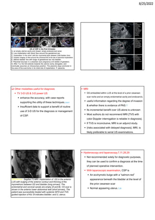
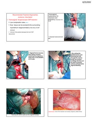
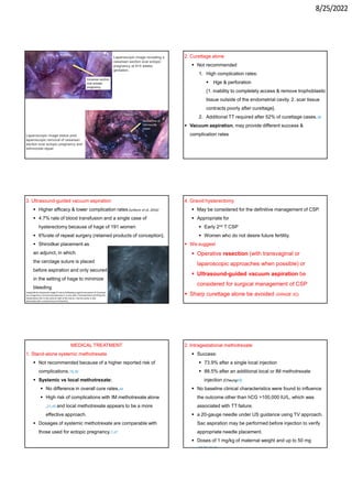
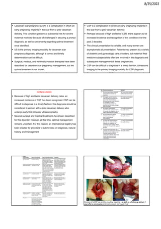
8/25/2022
You can get this lecture and more than 500 lectures from:
1. My scientific page on Face book: Aboubakr Elnashar
Lectures.
https://www.facebook.com/groups/227744884091351/
2. Slide share web site
3. elnashar53@hotmail.com
4. My clinic: Althwara st, Mansura, Egypt
5. WhatsApp: 01555536303
ABOUBAKRELNASHAR
CESAREAN SCAR PREGNANCY
SMFM, ACOG, 2020
Prof Aboubakr Elnashar
Benha university, Egypt
CONTENTS
1. INTRODUCTION
 Definition
 Incidence
 Risk factors
2. PATHOGENESIS & NATURAL HISTORY
3. DIAGNOSIS
 Clinical Picture
 Investigations
4. TREATMENT
5. IMPACT ON FUTURE PREGNANCIES
CONCLUSIONS
1. INTRODUCTION
 DEFINITION
 CSP occurs when an embryo implants in the
fibrous scar tissue of a prior cesarean
hysterotomy.1
 Although at times referred to as a cesarean scar ectopic pregnancy,
these gestations are, in fact, within the uterine cavity and, unlike true
ectopic pregnancies, may result in a liveborn infant.
 INCIDENCE
 The true incidence of CSP is unknown
{underdiagnosed & underreported}.
 1:1800 to 1:2656 of overall pregnancies.3,4
 Increased over time
 Improved imaging with US & MRI
 Increased use of TVS
 Increased physician awareness
RISK FACTORS
 Number of prior CS
 52% of cases occur in a single prior CS. 1,3,14
 Previous delivery for breech presentation 6,11,15,16
{LUS is often less well developed & that a thicker
hysterotomy presents a greater risk for poor healing
: microscopic dehiscence}.
 Closure technique No published data exist
regarding an association between hysterotomy&
CSP.
8/25/2022
2. PATHOGENESIS
 Blastocyst implantation within a microscopic
dehiscence tract in the scar from a prior CS.5e8
 {the fibrous nature of scar tissue: deficient implantation:
 Dehiscence
 Placenta Accreta Spectrum Disorders (PAS)
 Hage as the CSP enlarges.
 CSP& placenta accreta
 have similar disease pathways
 may exist along a common disease continuum.9
The implantation patterns: Types
 Endogenic (“on the scar”) type 1
 GS Growing toward the uterine cavity
 had variable obstetric outcomes
 Exogenic (“in-the-niche”).type 2 11,12
 deeply implanted into the scar
 Growing toward the bladder or abdominal cavity
 All underwent hysterectomy with PAS at
delivery.1
Implantation patterns
A, “On-the-scar,” or
endogenic, form
has a considerable
myometrial layer (clear
space) between the
placenta & anterior
uterine surface (solid
arrow).
B, “In-the-niche,” or
exogenic, form
has a thin myometrial
interphase below the
placenta (between the 2
arrows).
NATURAL HISTORY
 Limited information
 Few CSPs continue to a viable gestational age.
 Small case numbers
 CSPs have expectantly managed resulted
 PAS
 Cesarean hysterectomy: 50-100%
 Massive hemorrhage at delivery 11,15,30 10,31-34
3. DIAGNOSIS
Clinical Picture
 Timing:
 usually presents in the 1st T. the average gestational age at
diagnosis: 7.5 ±2.5 w.11
 2nd T diagnoses have been reported
 The clinical presentation is variable
 One-third: asymptomatic
 One-third: painless vaginal bleeding.11
 One quarter: pain, with or without bleeding.
 Women with ruptured CSP may also present with
hemodynamic collapse.
8/25/2022
Investigations
 US
 The primary imaging modality
 Correct & timely determination can be difficult.
 The initial finding of a low, anteriorly located GS should
 raise concern for a possible CSP
 warrant further investigation.17
 TVS:
 The optimal modality for evaluation of suspected CSP
{provides the highest image resolution}18
 Grayscale combined with color Doppler are
recommended.
 84.6% were detected by TVS
 15.4% incorrectly diagnosed as
 incomplete abortions or
 cervical pregnancies.11
 Combining TVS with TAS with a full bladder to provide
 “panoramic view” of the uterus
 The relationship between the GS & bladder.6
TVS: CSP: A gestational sac can be seen clearly embedded
within a hysterotomy scar.
 Ultrasonographic criteria:
1. Uterus & endocervix: empty
2. Placenta, GS, or both embedded in the hysterotomy scar
3. GS:
 triangular (at 8 w & earlier) or
 rounded or oval (after 8 w) that fills the scar “niche” (the
shallow area representing a healed hysterotomy site)
4. Myometrium between GS & bladder: thin (1-3 mm) or absent
5. Color Doppler: A prominent or rich vascular pattern at or in
the area of a cesarean scar
6. An embryonic or fetal pole, yolk sac, or both with or without
fetal cardiac activity
 All of these criteria may not be observed.
 Especially with very early diagnosis & before f cardiac
activity
 The woman must have confirmation of pregnancy (for
example, a positive pregnancy test result).18
 Bulging or ballooning of the LUS in the midline sagittal TA
view: supportive of CSP diagnosis.19,20
 If CSP is suspected but the diagnosis is not certain
1. Short-interval follow-up,
2. Second opinion, or
3. Additional imaging with MRI should be considered to make
a timely diagnosis without undue delay.
Doppler image of a cesarean scar pregnancy
The image shows a prominent vascular pattern in the area of a
hysterostomy scar
8/25/2022
DD:
 Other clinical entities with a similar US appearance.
 13.6% were originally misdiagnosed as
 Cervical ectopic pregnancies,
 Spontaneous abortions in transit, or
 Low implantation of an intrauterine pregnancy.21
 Given the importance of prompt diagnosis, referral to
an experienced center for a second opinion may be
preferable to ongoing follow-up examinations that are
likely to lead to a delay in diagnosis.
Failed pregnancy
Cx ectopic
CSP
within the cervical canal
anterior LUS
1. GS
normal
thin
2. Overlying anterior
myometrium
positive
negative
3. Sliding organ sign*
lack color flow
vascular flow
around and within
the GS
marked
peritrophoblastic
color Doppler flow
around GS
4. Doppler
Not fixed in
location, not
growing
±growing
5. Short follow up
US
*Gentle pressure with the TV probe: displace GS from its
position within the endocervical canal
 The location of the center of GS relative to the midpoint axis of
the uterus differentiated between IUP and CSP, indicating that
most CSPs are located proximally to the midpoint axis of the
uterus whereas most normal IUPs are located distally from the
midpoint of the uterus.
IUP & SCP
5 -10 W
(Timor-Tritsch et
al, 2016)
Cervical ectopic pregnancy:
Sagittal TAS of the midline
uterus (A): GS centered in the
endocervical canal, normal
myometrial thickness between
GS and bladder (arrow). Sagittal
and TVS of the endocervical
canal (B and C) with vascular
flow around and within the GS
on color Doppler ( C).
Low intrauterine
pregnancies
Miscarriage in
progress
Cervical
pregnancy
 DD:
 Miscarriage
 gestational sac within either the cervix or LUS
 no blood flow on Doppler examination, indicating a
detached gestational sac.
 Cervical pregnancy
 a bulbous region within the cervix
 blood flow surrounding the gestational sac
 layer of myometrium between pregnancy& the bladder.
 CSEP
 limited or no myometrium between pregnancy& bladder
 cervical canal is empty
8/25/2022
US of CSP in the first trimester
A, an empty uterine cavity and closed, empty endocervical canal.
B, Low implantation with blood flow around the gestational sac.
C, Implantation “in the niche” with thin myometrial layer between GS& bladder (line).
D, Doppler imaging: bl flow around the chorionic/GS at the site of placental implantation.
E, Altered bladder line with bulge of gestational sac into bladder.
F, Placental lacunae in a cesarean scar pregnancy at 8 weeks of gestation.
G, After 7 w, the GS extends towards the uterine cavity, elongates, and
eventually assumes an intracavitary position. The placenta stays anchored in
the area of the scar/niche in its initial site of implantation. P, placenta.
 Other modalities useful for diagnosis
 TV 3-D US & 3-D power US
 enhance the accuracy, with case reports
supporting the utility of these techniques.22e24
 Insufficient data to support a benefit of routine
use of 3-D US for the diagnosis or management
of CSP.
 MRI
 GS embedded within LUS at the level of a prior cesarean
scar niche and an empty endometrial cavity and endocervix.
 useful information regarding the degree of invasion
& whether there is evidence of PAS.1
 Its incremental benefit over US alone is unknown
 Most authors do not recommend MRI {TVS with
color Doppler interrogation is reliable in diagnosis}.
 If TVS is inconclusive, MRI is an adjunct study.
 {risks associated with delayed diagnosis}, MRI, is
likely preferable to serial US examinations.
CSP at 9 w: Sagittal T2 MRI: implantation of GS in the anterior
LUS with bulging of the anterior contour and thinning of the
myometrium between GS and bladder (long arrows). The
endometrial and cervical canals are empty (A and B). CS scar is
shown in the anterior lower abdominal wall (short arrows). The
patient was successfully treated with systemic MTX and TVS
guided injection of Kcl. B indicates bladder; and U, uterus.
 Hysteroscopy and laparoscopy.7,11,28,29
 Not recommended solely for diagnostic purposes,
they can be used to confirm a diagnosis at the time
of planned operative intervention.
 With laparoscopic examination, CSP is
 An ecchymotic bulge with a “salmon-red”
appearance beneath the bladder at the level of
the prior cesarean scar
 Normal appearing uterus.7,29
8/25/2022
4. MANAGEMENT
 Expectant management is not recommended
{high risk of severe maternal morbidity}
Pregnancy termination is advised as soon as the
diagnosis is confirmed.1,11,13
 Women who were treated expectantly:
 Most required additional treatment
 More than 50% had severe complications.30
 10 women with 1st T US diagnosis of a pregnancy implanted in or on a prior cesarean scar,
 all the women had PAS diagnosed at the time of repeat cesarean delivery.32
 We recommend against expectant management
(GRADE 1B).
 An exception to the recommendation against
expectant management involves
 Early CSP that is characterized by fetal death or
other evidence of early pregnancy failure:
expectant management may be pursued with
1. Serial US surveillance
2. Quantitative beta-hCG measurements
3. Monitoring for maternal symptoms: bleeding or
pelvic pain.
 Expectant management of nonviable CSP
 Several months to resolve spontaneously
 Development of a uterine arteriovenous
malformation (AVM): 20
 20% (Timor- Tritsch et al,20)
 Persistent, severe vag bleeding and may require
 Uterine artery embolization or even
 Hysterectomy
Treatment Modalities
 The optimal TT is not known, although many different
options: Surgical, medical& minimally invasive
therapies and various combinations bec studies
1. Most are case series, with limited number of RCT
2. Are influenced by variable levels of clinical
experience, institutional capability, provider skill,
and case complexity
3. Lack of head-to-head comparisons between
medical and surgical approaches.
 Surgical: Hysteroscopy, laparoscopy, laparotomy, TV
surgery, curettage
 Medical: methotrexate (both local guided injection and
systemic administration), direct KCl injection
 Others:
 Needle-guided sac decompression
 Uterine artery embolization (UAE)
 High-intensity focused ultrasound imaging
 Use of balloon catheters
 Combinations of these methods.35
 Goal of treatment: Preserving of
 Maternal health
 Fertility when possible.
 Decisions should be determined after considering
 Patient
 Hemodynamic status
 Desire for future fertility
 Compliance
 CSP
 viability
 location
 gestational age
 HCG level
 Surgeon expertise.
 Institutional resources.
8/25/2022
 Treatment selection was influenced by
 Physician specialty, with
 Gynecologic surgeons favoring
 curettage,
 laparoscopy, and
 hysteroscopy
 Obstetricians more readily pursuing
 needle-based injections and
 interventional radiology involvement.21
 Systematic review (Petersen et al, 2016)
 2037 CSP cases, 4 RCT & 48 case series. most
were detected in 1st T
 TT modalities were condensed into 14 main
approaches.
 Success was defined as the efficacy of 1st line
modality to resolve a CSP.
 Major complications were defined as
 hysterectomy
 estimated blood loss >1000 mL, or
 need for blood transfusion.
Complication (%)
Success (%)
54
41
Expectant
13
75
MTX IM
2
77
MTX Local+IM
3
69
UAE+MTX
21
48
D&C
1
95
UAE+D&C+Hysteroscopy
1
99
TV excision
3
94
UAE
0
97
Laparoscopy
 Interventional approaches appeared superior to medical
approaches (Petersen et al, 2016)
 CSEP: Evolution from medical to surgical management (Roche et
al, 2020)
 Although TV resection, UAE, and laparoscopy alone or in
combination appeared to be superior to medical & minimally
invasive treatments
1. These interventions require resources: operating rooms,
advanced equipment, anesthesia, & trained staff.
2. As a result, some of these interventions are not widely
available, & they may be costly.
3. Little high-quality evidence exists that compares these
methods head-to-head with less resource-intensive
modalities, such as
1. Local intragestational injections of methotrexate or KCl.
2. Cervical double-balloon catheter
TVS -guided intragestational-sac injection of MTX in a
live CSP at 6 w, 4 days. The arrow points to the needle
in place. (F = fetus.)
 Cervical double-balloon catheter
 can terminate the pregnancy while compressing the blood
supply to the gestational sac.
 Low rate of complications (4.2%)
 high success rate (97.7%) with this technique.37,38
8/25/2022
Recommended Treatment Approaches
SURGICAL TREATMENT
1. Transvaginal & laparoscopic CSP resection
 Low complication rates.39e41
 Scar tissue can be excised & the surrounding
myometrium reapproximated at the time of CSP
removal.
 Unknown if this practice decreases the risk of CSP
reoccurrence.
Transvaginal
hysterotomy for
cesarean scar
pregnancy (Huanxiao et al,
2015)
1. Exposed vesicocervical
gap.
2. Vaginal fornix mucosa
seen before incision,
exposing the pregnancy
mass scar in the bladder-
cervix gap.
3. By cutting the
gestational sac and
removing the villi
through the uterine
cavity, the suction
curettage decidua can
be used to prevent
profuse postoperative
vaginal bleeding.
Suturing the uterine wall and the vaginal mucosa.
Laparoscopic view of the cesarean scar ectopic pregnancy (black arrow).
8/25/2022
Laparoscopic image revealing a
cesarean section scar ectopic
pregnancy at 9+5 weeks
gestation.
Laparoscopic image status post
laparoscopic removal of cesarean
section scar ectopic pregnancy and
isthmocele repair.
2. Curettage alone
 Not recommended
1. High complication rates:
 Hge & perforation
{1. inability to completely access & remove trophoblastic
tissue outside of the endometrial cavity. 2. scar tissue
contracts poorly after curettage}.
2. Additional TT required after 52% of curettage cases.36
 Vacuum aspiration, may provide different success &
complication rates
3. Ultrasound-guided vacuum aspiration
 Higher efficacy & lower complication rates (Jurkovic et al, 2016)
 4.7% rate of blood transfusion and a single case of
hysterectomy because of hage of 191 women
 6%rate of repeat surgery (retained products of conception}.
 Shirodkar placement as
an adjunct, in which
the cerclage suture is placed
before aspiration and only secured
in the setting of hage to minimize
bleeding
Longitudinal ultrasound image of uterus following surgical evacuation of Cesarean
scar pregnancy. Cervical suture(arrow) is in situ with a hematometra (H) filling the
implantation site in the anterior wall of the uterus. Uterine cavity is also
distended with a small amount of blood (C).
4. Gravid hysterectomy
 May be considered for the definitive management of CSP.
 Appropriate for
 Early 2nd T CSP
 Women who do not desire future fertility.
 We suggest
 Operative resection (with transvaginal or
laparoscopic approaches when possible) or
 Ultrasound-guided vacuum aspiration be
considered for surgical management of CSP
 Sharp curettage alone be avoided (GRADE 2C).
MEDICAL TREATMENT
1. Stand-alone systemic methotrexate
 Not recommended because of a higher reported risk of
complications.16,30
 Systemic vs local methotrexate:
 No difference in overall cure rates,44
 High risk of complications with IM methotrexate alone
,21,45 and local methotrexate appears to be a more
effective approach.
 Dosages of systemic methotrexate are comparable with
those used for ectopic pregnancy.7,47
2. Intragestational methotrexate
 Success:
 73.9% after a single local injection
 88.5% after an additional local or IM methotrexate
injection {Cheung45}
 No baseline clinical characteristics were found to influence
the outcome other than hCG >100,000 IU/L, which was
associated with TT failure.
 a 20-gauge needle under US guidance using TV approach.
Sac aspiration may be performed before injection to verify
appropriate needle placement.
 Doses of 1 mg/kg of maternal weight and up to 50 mg
.18,36,45,46
8/25/2022
TVS -guided intragestational-sac injection of MTX in a
live CSP at 6 w, 4 days. The arrow points to the needle
in place. (F = fetus.)
 We suggest intragestational methotrexate for medical
TT, with or without other TT modalities (GRADE 2C).
 We recommend that systemic methotrexate alone not be
used (GRADE 1C).
 Post medical TT monitoring for
1. Symptoms: hge or uterine AVM development.
2. Interval US:
 Gestational mass take weeks to months to resolve. a
mean time to resolution of 88 days (range, 26-177).18
 Transient increase in beta-hCG levels & CSP mass size
observed after methotrexate therapy.
The patient in the lithotomy position. A
22-gauge, 15 cm Wallace needle was
inserted via vaginal route under
transvaginal ultrasound guidance
Wallace needle was inserted via
vaginal route into the gestational sac.
MTX (1 mg/kg) was injected into the
gestational sac.
ADJUNCT TREATMENT
 UAE
 As a stand-alone procedure & in combination with curettage,
methotrexate, and hysteroscopy
 High success & low complication rates when UAE was
performed without methotrexate or with and without curettage.36
 When methotrexate was added to a UAE strategy: higher
risk (31.4%) that additional TT would be needed.
 Significant reduction in blood loss.51
 Reported outcomes
 vary significantly, and its role as an adjunct to other
management approaches requires further study.
A 30-year-old
womanwithCSPunderwentpreventiveUAE.Transvaginalsagittalsonogramshowsadeepimplantatio
nof
 US-guided placement & inflation of balloon & Foley catheters
 To tamponade a CSP gestational sac that is complicated by
bleeding or
 As a prophylactic measure (Timor-Tritsch et al 37,38,52)
 Well tolerated and effictive, which supports a potential
option that warrants further study.
8/25/2022
TV power Doppler image of the
inflated balloon (B) in a case of a CSP
at 6 w, 4 days, after injection of MXT
The catheter with the balloon inflated with 5 mL
of saline. Inserting a Foley balloon catheter and
inflating it at the site of the CSP. The catheters
come with balloons of different capacity. They
can be used alone (usually in gestations of 5–7
weeks) in the hope of stopping the evolution of
the pregnancy by placing pressure on a small
gestational sac. Even so, this approach is almost
always used in a planned fashion in conjunction
with another treatment or as backup if bleeding
occurs.
6. IMPACT ON FUTURE PREGNANCIES
 Increased risk for recurrence & other severe maternal
morbidities.
 Recurrence rate:
 5% (Ben Nagi et al55)
 15.6% .58
 25% (Sadeghi et al 57)
 40% .59
 Before pregnancy:
1. Waiting 12-24 months , although limited supporting
evidence.4,60
{short interval between CSP TT & subsequent pregnancy
may increase the risk for recurrent CSP or PAS}.14,30
2. Evaluation of the uterus & cesarean scar by SIS. However, it
is not clear whether the detection of a defect is associated with
higher risks
3. Interpregnancy repair or revision of a cesarean scar has
been reported with the use of a variety of surgical modalities.
 However, there are insufficient data to support a benefit
to this practice.21,61e66
 During pregnancy:
1. Close US monitoring to
 confirm the presence of an IU pregnancy
 exclude recurrent CSP or PAS.
 An initial US is recommended at less than 8 w, to confirm a
normal IU location.
2. Repeat CS between 34 0/7 to 35 6/7 w, before the onset of
labor.
3. Betamethasone administration is recommended before
anticipated late preterm delivery.53
4. The delivery team should be prepared for obstetric hge and
the potential need for cesarean hysterectomy.
 Women who consider pregnancy after a CSP should
be informed that there is
 significant risk of recurrence &
 severe maternal morbidity.
 Counseled regarding effective contraceptive
methods, including
 long-acting reversible contraception and
 permanent contraception (GRADE 1C).
 Repeat cesarean delivery between 34 0/7 and 35 6/7
weeks of gestation (GRADE 1C)
You can get this lecture and more than 500 lectures from:
1. My scientific page on Face book: Aboubakr Elnashar
Lectures.
https://www.facebook.com/groups/227744884091351/
2. Slide share web site
3. elnashar53@hotmail.com
4. My clinic: Althwara st, Mansura, Egypt
5. WhatsApp: 01555536303
ABOUBAKRELNASHAR
8/25/2022
1. HYSTEROSCOPY
 To remove CSEP either
 alone or
 with adjuvant medical therapy.
 Methods
 hysteroscopic removal of tissue
 aspiration of GS after medication
 injection of MTX or ethanol into the GS (Gonzalez N, Tulandi et al,
SR, 2017)
 Best indicated in type 1 CSEP.
 Success rate:
 variable rate
 requirement for additional procedures, including
hysterectomy (Maheux-Lacroix et al, SR, 2017)
 Higher in
 lower gravidity/parity
 fewer prior CS
 earlier gestational age at time of procedure.
 ASRM, 2016:
 hysteroscopy could be used to remove CSEP via
direct visualization or US assistance.
 dissection of the CSEP from the uterine wall using
electrosurgery had
 high success rate
 extremely low complication rate
 should be considered safe& effective
 Complications: Rare
 Fluid overload, electrolyte imbalances, perforation, infection, and hospital
admission.
3. LAPAROTOMY
 Very few data on laparotomy as 1st choice
 Performing
 myometrial wedge excision
 Advantages: (Maheux-Lacroix et al, SR, 2017)
 Direct visualization of the lower uterine segment
 Success rate: high
 with a low complication rate
 myometrium between GS& bladder ≥2 mm.
 ACOG 2017
 Laparotomy:
 not considered 1st line treatment
 should be avoided for CSEP management if possible.
1. potential morbidity
 bladder/ureter injuries
 intraoperative blood loss
 wound complications
2. invasive nature
3. longer duration of hospital stay, operating time
4. slower return to normal activity
 MIS should be 1st line if the surgeon is adequately trained
 We recommend against expectant management of cesarean
scar pregnancy (GRADE 1B); we suggest operative resection
(with transvaginal or laparoscopic approaches when possible)
or ultrasound-guided vacuum aspiration be considered for
surgical management of cesarean scar pregnancy and that
sharp curettage alone be avoided (GRADE 2C);
 we suggest intragestational methotrexate for medical treatment
of cesarean scar pregnancy, with or without other treatment
modalities (GRADE 2C);
 Systemic methotrexate alone not be used to treat cesarean
scar pregnancy (GRADE 1C); in women who choose expectant
management and continuation of a cesarean scar pregnancy,
8/25/2022
 Cesarean scar pregnancy {CSP} is a complication in which an
early pregnancy implants in the scar from a prior cesarean
delivery. This condition presents a substantial risk for severe
maternal morbidity because of challenges in securing a prompt
diagnosis, as well as uncertainty regarding optimal treatment
once identified.
 US is the primary imaging modality for cesarean scar
pregnancy diagnosis, although a correct and timely
determination can be difficult.
 Surgical, medical, and minimally invasive therapies have been
described for cesarean scar pregnancy management, but the
optimal treatment is not known.
 Women who decline treatment of a cesarean scar pregnancy
 CSP is a complication in which an early pregnancy implants in
the scar from a prior cesarean delivery.
 Perhaps because of high worldwide CSR, there appears to be
increased incidence and recognition of this condition over the
past 2 decades.
 The clinical presentation is variable, and many women are
asymptomatic at presentation. Patients may present to a variety
of obstetric and gynecologic care providers, but maternal-fetal
medicine subspecialists often are involved in the diagnosis and
subsequent management of these pregnancies.
 CSP can be difficult to diagnose in a timely fashion. Ultrasound
imaging is the primary imaging modality for CSP diagnosis.
 Expectantly managed CSP is associated with high rates of
CONCLUSION
 Because of high worldwide cesarean delivery rates, an
increased incidence of CSP has been recognized. CSP can be
difficult to diagnose in a timely fashion; this diagnosis should be
considered in women with a prior cesarean delivery who
undergo early first-trimester ultrasonography.
 Several surgical and medical treatments have been described
for this disorder; however, at this time, optimal management
remains uncertain. For this reason, an international registry has
been created for providers to submit data on diagnosis, natural
history, and management
Excision of a CSP sac and the resulting repair ( a ) as well as a follow-up picture 1
year after a previously performed excision and repair ( b ).
8/25/2022
Marie-Laure et al, 2020
 Systematic reviews have been inconsistent with regard to the
identification of a single optimal CSP treatment modality that
best balances procedural success and risks.
 In a review by Timor-Tritsch and Monteagudo21 that included
751 reported cases of CSP and
 31 different treatment approaches
 44.1% complication rate was reported overall.
 Complications
 unplanned emergency operations that included
 hysterectomy (4.8%),
 laparotomy (5.3%)
 UAE (2.9%).
 The highest complication rates were observed with
 IM methotrexate alone ( 62.1%),
 Curettage alone or in combination with other modalities
(61.9%)
 UAE alone or in combination with other modalities (46.9%).
 The lowest complication rates
 hysteroscopy alone or in combination (18.4%)
 local intragestational injection of methotrexate or KCl (9.6%).
 Support use of local methotrexate and hysteroscopy-based
approaches to CSP treatment and discouraged the stand-alone
use of systemic methotrexate, curettage, and UAE.
 Of note, most of the available literature does not distinguish
between sharp and suction curettage, although the
 Intragestational KCl in a small number of cases.33
 May be appropriate for the management of CSP heterotopic
pregnancies with a coexisting IU pregnancy {methotrexate
may have embryocidal or teratogenic for the IU cotwin}
 US-guided KCl injection can be accompanied by sac
aspiration.
 5 cases of CSP heterotopic pregnancies treated with local
KCl.48 All resulted in healthy live births of the cotwin,
although 2 cases were complicated by PPH, with 1 case
resulting in hysterectomy because of placenta accreta.
 Hysteroscopic and laparoscopic approaches for treating
CSP heterotopic pregnancies have been described.49,50
 The lowest success rates were observed with
1. Expectant management (41.5% success,53.7%
complications)
2. Curettage (48.1% success, 21% complications)
3. UAE and methotrexate (68.6% success,2.8%
complications)
4. Systemic methotrexate (75.2% success, 13%
complications)
5. Combined local and systemic methotrexate (76.5%
success, 2.3% complications).
8/25/2022
 The highest success rates were observed with
1. Transvaginal CSP resection (99.2% success, 0.9%
complications),
2. laparoscopy (97.1% success, 0% complications)
3. UAE with curettage, hysteroscopy, or both (95.4% success,
1.2% complications)
4. UAE alone (93.6% success, 3.4% complications).
 High rates of complications (Seow et al56)
 7 pregnancies among 14 women with prior CSP
 The mean interval between CSP and subsequent pregnancy
was 13 months (range, 0e34 months).
 4 pregnancies were IU ;CS between 35 and 36 w.
 2 pregnancies complicated by placenta accreta: 1 cesarean
hysterectomy
 1 pregnancy involved a woman who became pregnant 3
months after curettage and cervical balloon TT:
spontaneous uterine rupture and died of hypovolemic shock,
with a stillborn fetus.

CSP ACOG2021.pdf

  • 1.
    8/25/2022 You can getthis lecture and more than 500 lectures from: 1. My scientific page on Face book: Aboubakr Elnashar Lectures. https://www.facebook.com/groups/227744884091351/ 2. Slide share web site 3. elnashar53@hotmail.com 4. My clinic: Althwara st, Mansura, Egypt 5. WhatsApp: 01555536303 ABOUBAKRELNASHAR CESAREAN SCAR PREGNANCY SMFM, ACOG, 2020 Prof Aboubakr Elnashar Benha university, Egypt CONTENTS 1. INTRODUCTION  Definition  Incidence  Risk factors 2. PATHOGENESIS & NATURAL HISTORY 3. DIAGNOSIS  Clinical Picture  Investigations 4. TREATMENT 5. IMPACT ON FUTURE PREGNANCIES CONCLUSIONS 1. INTRODUCTION  DEFINITION  CSP occurs when an embryo implants in the fibrous scar tissue of a prior cesarean hysterotomy.1  Although at times referred to as a cesarean scar ectopic pregnancy, these gestations are, in fact, within the uterine cavity and, unlike true ectopic pregnancies, may result in a liveborn infant.  INCIDENCE  The true incidence of CSP is unknown {underdiagnosed & underreported}.  1:1800 to 1:2656 of overall pregnancies.3,4  Increased over time  Improved imaging with US & MRI  Increased use of TVS  Increased physician awareness RISK FACTORS  Number of prior CS  52% of cases occur in a single prior CS. 1,3,14  Previous delivery for breech presentation 6,11,15,16 {LUS is often less well developed & that a thicker hysterotomy presents a greater risk for poor healing : microscopic dehiscence}.  Closure technique No published data exist regarding an association between hysterotomy& CSP.
  • 2.
    8/25/2022 2. PATHOGENESIS  Blastocystimplantation within a microscopic dehiscence tract in the scar from a prior CS.5e8  {the fibrous nature of scar tissue: deficient implantation:  Dehiscence  Placenta Accreta Spectrum Disorders (PAS)  Hage as the CSP enlarges.  CSP& placenta accreta  have similar disease pathways  may exist along a common disease continuum.9 The implantation patterns: Types  Endogenic (“on the scar”) type 1  GS Growing toward the uterine cavity  had variable obstetric outcomes  Exogenic (“in-the-niche”).type 2 11,12  deeply implanted into the scar  Growing toward the bladder or abdominal cavity  All underwent hysterectomy with PAS at delivery.1 Implantation patterns A, “On-the-scar,” or endogenic, form has a considerable myometrial layer (clear space) between the placenta & anterior uterine surface (solid arrow). B, “In-the-niche,” or exogenic, form has a thin myometrial interphase below the placenta (between the 2 arrows). NATURAL HISTORY  Limited information  Few CSPs continue to a viable gestational age.  Small case numbers  CSPs have expectantly managed resulted  PAS  Cesarean hysterectomy: 50-100%  Massive hemorrhage at delivery 11,15,30 10,31-34 3. DIAGNOSIS Clinical Picture  Timing:  usually presents in the 1st T. the average gestational age at diagnosis: 7.5 ±2.5 w.11  2nd T diagnoses have been reported  The clinical presentation is variable  One-third: asymptomatic  One-third: painless vaginal bleeding.11  One quarter: pain, with or without bleeding.  Women with ruptured CSP may also present with hemodynamic collapse.
  • 3.
    8/25/2022 Investigations  US  Theprimary imaging modality  Correct & timely determination can be difficult.  The initial finding of a low, anteriorly located GS should  raise concern for a possible CSP  warrant further investigation.17  TVS:  The optimal modality for evaluation of suspected CSP {provides the highest image resolution}18  Grayscale combined with color Doppler are recommended.  84.6% were detected by TVS  15.4% incorrectly diagnosed as  incomplete abortions or  cervical pregnancies.11  Combining TVS with TAS with a full bladder to provide  “panoramic view” of the uterus  The relationship between the GS & bladder.6 TVS: CSP: A gestational sac can be seen clearly embedded within a hysterotomy scar.  Ultrasonographic criteria: 1. Uterus & endocervix: empty 2. Placenta, GS, or both embedded in the hysterotomy scar 3. GS:  triangular (at 8 w & earlier) or  rounded or oval (after 8 w) that fills the scar “niche” (the shallow area representing a healed hysterotomy site) 4. Myometrium between GS & bladder: thin (1-3 mm) or absent 5. Color Doppler: A prominent or rich vascular pattern at or in the area of a cesarean scar 6. An embryonic or fetal pole, yolk sac, or both with or without fetal cardiac activity  All of these criteria may not be observed.  Especially with very early diagnosis & before f cardiac activity  The woman must have confirmation of pregnancy (for example, a positive pregnancy test result).18  Bulging or ballooning of the LUS in the midline sagittal TA view: supportive of CSP diagnosis.19,20  If CSP is suspected but the diagnosis is not certain 1. Short-interval follow-up, 2. Second opinion, or 3. Additional imaging with MRI should be considered to make a timely diagnosis without undue delay. Doppler image of a cesarean scar pregnancy The image shows a prominent vascular pattern in the area of a hysterostomy scar
  • 4.
    8/25/2022 DD:  Other clinicalentities with a similar US appearance.  13.6% were originally misdiagnosed as  Cervical ectopic pregnancies,  Spontaneous abortions in transit, or  Low implantation of an intrauterine pregnancy.21  Given the importance of prompt diagnosis, referral to an experienced center for a second opinion may be preferable to ongoing follow-up examinations that are likely to lead to a delay in diagnosis. Failed pregnancy Cx ectopic CSP within the cervical canal anterior LUS 1. GS normal thin 2. Overlying anterior myometrium positive negative 3. Sliding organ sign* lack color flow vascular flow around and within the GS marked peritrophoblastic color Doppler flow around GS 4. Doppler Not fixed in location, not growing ±growing 5. Short follow up US *Gentle pressure with the TV probe: displace GS from its position within the endocervical canal  The location of the center of GS relative to the midpoint axis of the uterus differentiated between IUP and CSP, indicating that most CSPs are located proximally to the midpoint axis of the uterus whereas most normal IUPs are located distally from the midpoint of the uterus. IUP & SCP 5 -10 W (Timor-Tritsch et al, 2016) Cervical ectopic pregnancy: Sagittal TAS of the midline uterus (A): GS centered in the endocervical canal, normal myometrial thickness between GS and bladder (arrow). Sagittal and TVS of the endocervical canal (B and C) with vascular flow around and within the GS on color Doppler ( C). Low intrauterine pregnancies Miscarriage in progress Cervical pregnancy  DD:  Miscarriage  gestational sac within either the cervix or LUS  no blood flow on Doppler examination, indicating a detached gestational sac.  Cervical pregnancy  a bulbous region within the cervix  blood flow surrounding the gestational sac  layer of myometrium between pregnancy& the bladder.  CSEP  limited or no myometrium between pregnancy& bladder  cervical canal is empty
  • 5.
    8/25/2022 US of CSPin the first trimester A, an empty uterine cavity and closed, empty endocervical canal. B, Low implantation with blood flow around the gestational sac. C, Implantation “in the niche” with thin myometrial layer between GS& bladder (line). D, Doppler imaging: bl flow around the chorionic/GS at the site of placental implantation. E, Altered bladder line with bulge of gestational sac into bladder. F, Placental lacunae in a cesarean scar pregnancy at 8 weeks of gestation. G, After 7 w, the GS extends towards the uterine cavity, elongates, and eventually assumes an intracavitary position. The placenta stays anchored in the area of the scar/niche in its initial site of implantation. P, placenta.  Other modalities useful for diagnosis  TV 3-D US & 3-D power US  enhance the accuracy, with case reports supporting the utility of these techniques.22e24  Insufficient data to support a benefit of routine use of 3-D US for the diagnosis or management of CSP.  MRI  GS embedded within LUS at the level of a prior cesarean scar niche and an empty endometrial cavity and endocervix.  useful information regarding the degree of invasion & whether there is evidence of PAS.1  Its incremental benefit over US alone is unknown  Most authors do not recommend MRI {TVS with color Doppler interrogation is reliable in diagnosis}.  If TVS is inconclusive, MRI is an adjunct study.  {risks associated with delayed diagnosis}, MRI, is likely preferable to serial US examinations. CSP at 9 w: Sagittal T2 MRI: implantation of GS in the anterior LUS with bulging of the anterior contour and thinning of the myometrium between GS and bladder (long arrows). The endometrial and cervical canals are empty (A and B). CS scar is shown in the anterior lower abdominal wall (short arrows). The patient was successfully treated with systemic MTX and TVS guided injection of Kcl. B indicates bladder; and U, uterus.  Hysteroscopy and laparoscopy.7,11,28,29  Not recommended solely for diagnostic purposes, they can be used to confirm a diagnosis at the time of planned operative intervention.  With laparoscopic examination, CSP is  An ecchymotic bulge with a “salmon-red” appearance beneath the bladder at the level of the prior cesarean scar  Normal appearing uterus.7,29
  • 6.
    8/25/2022 4. MANAGEMENT  Expectantmanagement is not recommended {high risk of severe maternal morbidity} Pregnancy termination is advised as soon as the diagnosis is confirmed.1,11,13  Women who were treated expectantly:  Most required additional treatment  More than 50% had severe complications.30  10 women with 1st T US diagnosis of a pregnancy implanted in or on a prior cesarean scar,  all the women had PAS diagnosed at the time of repeat cesarean delivery.32  We recommend against expectant management (GRADE 1B).  An exception to the recommendation against expectant management involves  Early CSP that is characterized by fetal death or other evidence of early pregnancy failure: expectant management may be pursued with 1. Serial US surveillance 2. Quantitative beta-hCG measurements 3. Monitoring for maternal symptoms: bleeding or pelvic pain.  Expectant management of nonviable CSP  Several months to resolve spontaneously  Development of a uterine arteriovenous malformation (AVM): 20  20% (Timor- Tritsch et al,20)  Persistent, severe vag bleeding and may require  Uterine artery embolization or even  Hysterectomy Treatment Modalities  The optimal TT is not known, although many different options: Surgical, medical& minimally invasive therapies and various combinations bec studies 1. Most are case series, with limited number of RCT 2. Are influenced by variable levels of clinical experience, institutional capability, provider skill, and case complexity 3. Lack of head-to-head comparisons between medical and surgical approaches.  Surgical: Hysteroscopy, laparoscopy, laparotomy, TV surgery, curettage  Medical: methotrexate (both local guided injection and systemic administration), direct KCl injection  Others:  Needle-guided sac decompression  Uterine artery embolization (UAE)  High-intensity focused ultrasound imaging  Use of balloon catheters  Combinations of these methods.35  Goal of treatment: Preserving of  Maternal health  Fertility when possible.  Decisions should be determined after considering  Patient  Hemodynamic status  Desire for future fertility  Compliance  CSP  viability  location  gestational age  HCG level  Surgeon expertise.  Institutional resources.
  • 7.
    8/25/2022  Treatment selectionwas influenced by  Physician specialty, with  Gynecologic surgeons favoring  curettage,  laparoscopy, and  hysteroscopy  Obstetricians more readily pursuing  needle-based injections and  interventional radiology involvement.21  Systematic review (Petersen et al, 2016)  2037 CSP cases, 4 RCT & 48 case series. most were detected in 1st T  TT modalities were condensed into 14 main approaches.  Success was defined as the efficacy of 1st line modality to resolve a CSP.  Major complications were defined as  hysterectomy  estimated blood loss >1000 mL, or  need for blood transfusion. Complication (%) Success (%) 54 41 Expectant 13 75 MTX IM 2 77 MTX Local+IM 3 69 UAE+MTX 21 48 D&C 1 95 UAE+D&C+Hysteroscopy 1 99 TV excision 3 94 UAE 0 97 Laparoscopy  Interventional approaches appeared superior to medical approaches (Petersen et al, 2016)  CSEP: Evolution from medical to surgical management (Roche et al, 2020)  Although TV resection, UAE, and laparoscopy alone or in combination appeared to be superior to medical & minimally invasive treatments 1. These interventions require resources: operating rooms, advanced equipment, anesthesia, & trained staff. 2. As a result, some of these interventions are not widely available, & they may be costly. 3. Little high-quality evidence exists that compares these methods head-to-head with less resource-intensive modalities, such as 1. Local intragestational injections of methotrexate or KCl. 2. Cervical double-balloon catheter TVS -guided intragestational-sac injection of MTX in a live CSP at 6 w, 4 days. The arrow points to the needle in place. (F = fetus.)  Cervical double-balloon catheter  can terminate the pregnancy while compressing the blood supply to the gestational sac.  Low rate of complications (4.2%)  high success rate (97.7%) with this technique.37,38
  • 8.
    8/25/2022 Recommended Treatment Approaches SURGICALTREATMENT 1. Transvaginal & laparoscopic CSP resection  Low complication rates.39e41  Scar tissue can be excised & the surrounding myometrium reapproximated at the time of CSP removal.  Unknown if this practice decreases the risk of CSP reoccurrence. Transvaginal hysterotomy for cesarean scar pregnancy (Huanxiao et al, 2015) 1. Exposed vesicocervical gap. 2. Vaginal fornix mucosa seen before incision, exposing the pregnancy mass scar in the bladder- cervix gap. 3. By cutting the gestational sac and removing the villi through the uterine cavity, the suction curettage decidua can be used to prevent profuse postoperative vaginal bleeding. Suturing the uterine wall and the vaginal mucosa. Laparoscopic view of the cesarean scar ectopic pregnancy (black arrow).
  • 9.
    8/25/2022 Laparoscopic image revealinga cesarean section scar ectopic pregnancy at 9+5 weeks gestation. Laparoscopic image status post laparoscopic removal of cesarean section scar ectopic pregnancy and isthmocele repair. 2. Curettage alone  Not recommended 1. High complication rates:  Hge & perforation {1. inability to completely access & remove trophoblastic tissue outside of the endometrial cavity. 2. scar tissue contracts poorly after curettage}. 2. Additional TT required after 52% of curettage cases.36  Vacuum aspiration, may provide different success & complication rates 3. Ultrasound-guided vacuum aspiration  Higher efficacy & lower complication rates (Jurkovic et al, 2016)  4.7% rate of blood transfusion and a single case of hysterectomy because of hage of 191 women  6%rate of repeat surgery (retained products of conception}.  Shirodkar placement as an adjunct, in which the cerclage suture is placed before aspiration and only secured in the setting of hage to minimize bleeding Longitudinal ultrasound image of uterus following surgical evacuation of Cesarean scar pregnancy. Cervical suture(arrow) is in situ with a hematometra (H) filling the implantation site in the anterior wall of the uterus. Uterine cavity is also distended with a small amount of blood (C). 4. Gravid hysterectomy  May be considered for the definitive management of CSP.  Appropriate for  Early 2nd T CSP  Women who do not desire future fertility.  We suggest  Operative resection (with transvaginal or laparoscopic approaches when possible) or  Ultrasound-guided vacuum aspiration be considered for surgical management of CSP  Sharp curettage alone be avoided (GRADE 2C). MEDICAL TREATMENT 1. Stand-alone systemic methotrexate  Not recommended because of a higher reported risk of complications.16,30  Systemic vs local methotrexate:  No difference in overall cure rates,44  High risk of complications with IM methotrexate alone ,21,45 and local methotrexate appears to be a more effective approach.  Dosages of systemic methotrexate are comparable with those used for ectopic pregnancy.7,47 2. Intragestational methotrexate  Success:  73.9% after a single local injection  88.5% after an additional local or IM methotrexate injection {Cheung45}  No baseline clinical characteristics were found to influence the outcome other than hCG >100,000 IU/L, which was associated with TT failure.  a 20-gauge needle under US guidance using TV approach. Sac aspiration may be performed before injection to verify appropriate needle placement.  Doses of 1 mg/kg of maternal weight and up to 50 mg .18,36,45,46
  • 10.
    8/25/2022 TVS -guided intragestational-sacinjection of MTX in a live CSP at 6 w, 4 days. The arrow points to the needle in place. (F = fetus.)  We suggest intragestational methotrexate for medical TT, with or without other TT modalities (GRADE 2C).  We recommend that systemic methotrexate alone not be used (GRADE 1C).  Post medical TT monitoring for 1. Symptoms: hge or uterine AVM development. 2. Interval US:  Gestational mass take weeks to months to resolve. a mean time to resolution of 88 days (range, 26-177).18  Transient increase in beta-hCG levels & CSP mass size observed after methotrexate therapy. The patient in the lithotomy position. A 22-gauge, 15 cm Wallace needle was inserted via vaginal route under transvaginal ultrasound guidance Wallace needle was inserted via vaginal route into the gestational sac. MTX (1 mg/kg) was injected into the gestational sac. ADJUNCT TREATMENT  UAE  As a stand-alone procedure & in combination with curettage, methotrexate, and hysteroscopy  High success & low complication rates when UAE was performed without methotrexate or with and without curettage.36  When methotrexate was added to a UAE strategy: higher risk (31.4%) that additional TT would be needed.  Significant reduction in blood loss.51  Reported outcomes  vary significantly, and its role as an adjunct to other management approaches requires further study. A 30-year-old womanwithCSPunderwentpreventiveUAE.Transvaginalsagittalsonogramshowsadeepimplantatio nof  US-guided placement & inflation of balloon & Foley catheters  To tamponade a CSP gestational sac that is complicated by bleeding or  As a prophylactic measure (Timor-Tritsch et al 37,38,52)  Well tolerated and effictive, which supports a potential option that warrants further study.
  • 11.
    8/25/2022 TV power Dopplerimage of the inflated balloon (B) in a case of a CSP at 6 w, 4 days, after injection of MXT The catheter with the balloon inflated with 5 mL of saline. Inserting a Foley balloon catheter and inflating it at the site of the CSP. The catheters come with balloons of different capacity. They can be used alone (usually in gestations of 5–7 weeks) in the hope of stopping the evolution of the pregnancy by placing pressure on a small gestational sac. Even so, this approach is almost always used in a planned fashion in conjunction with another treatment or as backup if bleeding occurs. 6. IMPACT ON FUTURE PREGNANCIES  Increased risk for recurrence & other severe maternal morbidities.  Recurrence rate:  5% (Ben Nagi et al55)  15.6% .58  25% (Sadeghi et al 57)  40% .59  Before pregnancy: 1. Waiting 12-24 months , although limited supporting evidence.4,60 {short interval between CSP TT & subsequent pregnancy may increase the risk for recurrent CSP or PAS}.14,30 2. Evaluation of the uterus & cesarean scar by SIS. However, it is not clear whether the detection of a defect is associated with higher risks 3. Interpregnancy repair or revision of a cesarean scar has been reported with the use of a variety of surgical modalities.  However, there are insufficient data to support a benefit to this practice.21,61e66  During pregnancy: 1. Close US monitoring to  confirm the presence of an IU pregnancy  exclude recurrent CSP or PAS.  An initial US is recommended at less than 8 w, to confirm a normal IU location. 2. Repeat CS between 34 0/7 to 35 6/7 w, before the onset of labor. 3. Betamethasone administration is recommended before anticipated late preterm delivery.53 4. The delivery team should be prepared for obstetric hge and the potential need for cesarean hysterectomy.  Women who consider pregnancy after a CSP should be informed that there is  significant risk of recurrence &  severe maternal morbidity.  Counseled regarding effective contraceptive methods, including  long-acting reversible contraception and  permanent contraception (GRADE 1C).  Repeat cesarean delivery between 34 0/7 and 35 6/7 weeks of gestation (GRADE 1C) You can get this lecture and more than 500 lectures from: 1. My scientific page on Face book: Aboubakr Elnashar Lectures. https://www.facebook.com/groups/227744884091351/ 2. Slide share web site 3. elnashar53@hotmail.com 4. My clinic: Althwara st, Mansura, Egypt 5. WhatsApp: 01555536303 ABOUBAKRELNASHAR
  • 12.
    8/25/2022 1. HYSTEROSCOPY  Toremove CSEP either  alone or  with adjuvant medical therapy.  Methods  hysteroscopic removal of tissue  aspiration of GS after medication  injection of MTX or ethanol into the GS (Gonzalez N, Tulandi et al, SR, 2017)  Best indicated in type 1 CSEP.  Success rate:  variable rate  requirement for additional procedures, including hysterectomy (Maheux-Lacroix et al, SR, 2017)  Higher in  lower gravidity/parity  fewer prior CS  earlier gestational age at time of procedure.  ASRM, 2016:  hysteroscopy could be used to remove CSEP via direct visualization or US assistance.  dissection of the CSEP from the uterine wall using electrosurgery had  high success rate  extremely low complication rate  should be considered safe& effective  Complications: Rare  Fluid overload, electrolyte imbalances, perforation, infection, and hospital admission. 3. LAPAROTOMY  Very few data on laparotomy as 1st choice  Performing  myometrial wedge excision  Advantages: (Maheux-Lacroix et al, SR, 2017)  Direct visualization of the lower uterine segment  Success rate: high  with a low complication rate  myometrium between GS& bladder ≥2 mm.  ACOG 2017  Laparotomy:  not considered 1st line treatment  should be avoided for CSEP management if possible. 1. potential morbidity  bladder/ureter injuries  intraoperative blood loss  wound complications 2. invasive nature 3. longer duration of hospital stay, operating time 4. slower return to normal activity  MIS should be 1st line if the surgeon is adequately trained  We recommend against expectant management of cesarean scar pregnancy (GRADE 1B); we suggest operative resection (with transvaginal or laparoscopic approaches when possible) or ultrasound-guided vacuum aspiration be considered for surgical management of cesarean scar pregnancy and that sharp curettage alone be avoided (GRADE 2C);  we suggest intragestational methotrexate for medical treatment of cesarean scar pregnancy, with or without other treatment modalities (GRADE 2C);  Systemic methotrexate alone not be used to treat cesarean scar pregnancy (GRADE 1C); in women who choose expectant management and continuation of a cesarean scar pregnancy,
  • 13.
    8/25/2022  Cesarean scarpregnancy {CSP} is a complication in which an early pregnancy implants in the scar from a prior cesarean delivery. This condition presents a substantial risk for severe maternal morbidity because of challenges in securing a prompt diagnosis, as well as uncertainty regarding optimal treatment once identified.  US is the primary imaging modality for cesarean scar pregnancy diagnosis, although a correct and timely determination can be difficult.  Surgical, medical, and minimally invasive therapies have been described for cesarean scar pregnancy management, but the optimal treatment is not known.  Women who decline treatment of a cesarean scar pregnancy  CSP is a complication in which an early pregnancy implants in the scar from a prior cesarean delivery.  Perhaps because of high worldwide CSR, there appears to be increased incidence and recognition of this condition over the past 2 decades.  The clinical presentation is variable, and many women are asymptomatic at presentation. Patients may present to a variety of obstetric and gynecologic care providers, but maternal-fetal medicine subspecialists often are involved in the diagnosis and subsequent management of these pregnancies.  CSP can be difficult to diagnose in a timely fashion. Ultrasound imaging is the primary imaging modality for CSP diagnosis.  Expectantly managed CSP is associated with high rates of CONCLUSION  Because of high worldwide cesarean delivery rates, an increased incidence of CSP has been recognized. CSP can be difficult to diagnose in a timely fashion; this diagnosis should be considered in women with a prior cesarean delivery who undergo early first-trimester ultrasonography.  Several surgical and medical treatments have been described for this disorder; however, at this time, optimal management remains uncertain. For this reason, an international registry has been created for providers to submit data on diagnosis, natural history, and management Excision of a CSP sac and the resulting repair ( a ) as well as a follow-up picture 1 year after a previously performed excision and repair ( b ).
  • 14.
    8/25/2022 Marie-Laure et al,2020  Systematic reviews have been inconsistent with regard to the identification of a single optimal CSP treatment modality that best balances procedural success and risks.  In a review by Timor-Tritsch and Monteagudo21 that included 751 reported cases of CSP and  31 different treatment approaches  44.1% complication rate was reported overall.  Complications  unplanned emergency operations that included  hysterectomy (4.8%),  laparotomy (5.3%)  UAE (2.9%).  The highest complication rates were observed with  IM methotrexate alone ( 62.1%),  Curettage alone or in combination with other modalities (61.9%)  UAE alone or in combination with other modalities (46.9%).  The lowest complication rates  hysteroscopy alone or in combination (18.4%)  local intragestational injection of methotrexate or KCl (9.6%).  Support use of local methotrexate and hysteroscopy-based approaches to CSP treatment and discouraged the stand-alone use of systemic methotrexate, curettage, and UAE.  Of note, most of the available literature does not distinguish between sharp and suction curettage, although the  Intragestational KCl in a small number of cases.33  May be appropriate for the management of CSP heterotopic pregnancies with a coexisting IU pregnancy {methotrexate may have embryocidal or teratogenic for the IU cotwin}  US-guided KCl injection can be accompanied by sac aspiration.  5 cases of CSP heterotopic pregnancies treated with local KCl.48 All resulted in healthy live births of the cotwin, although 2 cases were complicated by PPH, with 1 case resulting in hysterectomy because of placenta accreta.  Hysteroscopic and laparoscopic approaches for treating CSP heterotopic pregnancies have been described.49,50  The lowest success rates were observed with 1. Expectant management (41.5% success,53.7% complications) 2. Curettage (48.1% success, 21% complications) 3. UAE and methotrexate (68.6% success,2.8% complications) 4. Systemic methotrexate (75.2% success, 13% complications) 5. Combined local and systemic methotrexate (76.5% success, 2.3% complications).
  • 15.
    8/25/2022  The highestsuccess rates were observed with 1. Transvaginal CSP resection (99.2% success, 0.9% complications), 2. laparoscopy (97.1% success, 0% complications) 3. UAE with curettage, hysteroscopy, or both (95.4% success, 1.2% complications) 4. UAE alone (93.6% success, 3.4% complications).  High rates of complications (Seow et al56)  7 pregnancies among 14 women with prior CSP  The mean interval between CSP and subsequent pregnancy was 13 months (range, 0e34 months).  4 pregnancies were IU ;CS between 35 and 36 w.  2 pregnancies complicated by placenta accreta: 1 cesarean hysterectomy  1 pregnancy involved a woman who became pregnant 3 months after curettage and cervical balloon TT: spontaneous uterine rupture and died of hypovolemic shock, with a stillborn fetus.