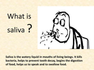
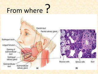
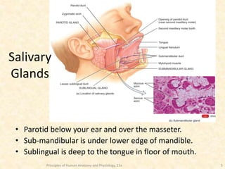
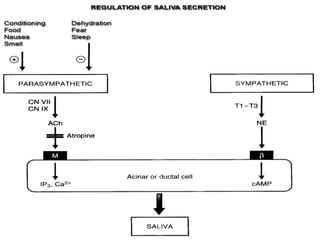
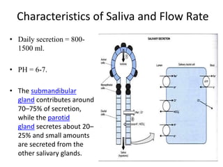
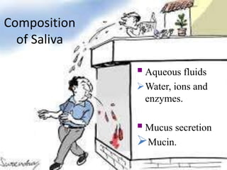
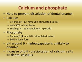
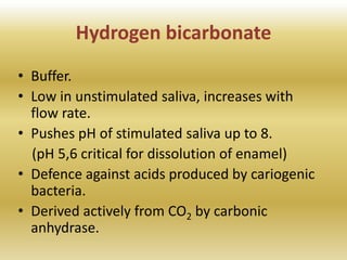
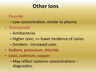
SALIVA
• COMPOSITION AND FUNCTION
What is
saliva ?
Saliva is the watery liquid in mouths of living beings. It kills
bacteria, helps to prevent tooth decay, begins the digestion
of food, helps us to speak and to swallow food.
From where ?
Principles of Human Anatomy and Physiology, 11e 5
• Parotid below your ear and over the masseter.
• Sub-mandibular is under lower edge of mandible.
• Sublingual is deep to the tongue in floor of mouth.
Salivary
Glands
Principles of Human Anatomy and Physiology, 11e 6
Salivary Glands’ Cellular Structure
• Cells in acini (clusters).
• Serous cells secrete a watery fluid.
• Mucous cells (pale staining) secrete a slimy,
mucus secretion.
Secretory Unit (salivon)
The basic unit “salivon” consists of:
 Acinus -initial secretory process
 Intercalated duct
 Striated duct -modification of secretory
product
 Myoepithelial cells
surround acinus and intercalated duct
contraction moves saliva, prevents
development of back pressure
TWO STAGE HYPOTHESIS
OF SALIVA FORMATION
Water &
electrolytes
Isotonic
primary saliva
Most proteins
Some proteins electrolytes
Na+ Cl- resorbed
K+ secreted
Hypotonic
final saliva
into mouth
Control of Secretion
Unique aspects of control of salivary
secretion
Secretion rate depends entirely on neural control
–ANS.
Both Parasympathetic and Sympathetic systems
lead to increased secretion.
Composition modified by Aldosterone.
increases Na, Cl reabsoption
increases K secretion
Parasympathetic
Origin:
salivary nucleus in medulla.
Outflow:
CN VII & IX.
Transmitter:
Ach.
Increased stimulation in response to-
 conditioned reflexes (taste, smell).
Decreased stimulation due to-
 sleep, fear, dehydration.
Sectioning of parasympathetic markedly decreases flow &
leads to atrophy.
Clinical significance- Parasympathetic
Sympathetic
Origin:
Intermedio-lateral gray area of T1-T3.
Transmitter:
Norepinephrine.
Stimulates:
- Secretion (enzymatic)
- Contraction of myoepithelial cells.
- Metabolic rate.
Sympathetic
Stimulates :
- Growth.
Sectioning of sympathetic nerves has minimal impact
on secretion of saliva.
Characteristics of Saliva and Flow Rate
• Daily secretion = 800-
1500 ml.
• PH = 6-7.
• The submandibular
gland contributes around
70–75% of secretion,
while the parotid
gland secretes about 20–
25% and small amounts
are secreted from the
other salivary glands.
A CONSIDERABLE VOLUME OF SALIVA IS
PRODUCED…
 0.5 to 1.5 liter of fluid is secreted in a
day.
 This represents about 1/5 of the total
plasma volume.
 This fluid is not lost as most of it is
swallowed and reabsorbed by the gut.
Variations in salivary composition
• Unstimulated flow
– Submandibular g. 70%
– Parotid g. 20%
– Accesory g. 7%
– Sublingual 2%
• Acid stimulation
– Submandibular g. 45%
– Parotid g. 45%
• Chewing
– Submandibular g. 30%
– Parotid g. 60%
0
1
2
3
4
5
6
7
sub
mandibular
parotid
sub lingual
20
CIRCAIDIAN RHYTHM OF SALIVA FLOW
No sleep
sleep
12 am 6 am 12 pm 6 pm 12 am 6 am 12 pm 6 pm 12 am
30
20
10
 Time of day
21
Effect of feeding on salivary secretion
0
0.005
0.01
0.015
0.02
0.025
0.03
0.035
Volumeofsalivacollectecdeach10min
10 min collection periods
Meal
during
this
period
Chemical composition and functions
of saliva
Composition
of Saliva
Aqueous fluids
Water, ions and
enzymes.
Mucus secretion
Mucin.
Chronology of defining salivary
components and functions
• Beginning in 1950’s whole saliva was evaluated
(antimicrobial properties, role in microbial
attachment, mineralization, taste and
lubrication)
• In 1970’s individual components isolated and
biochemically characterized.
• In mid-1980’s beginning to map functional
domains (peptide synthesis and recombinant
approaches).
Major salivary components
Mucin 1 (MG1)
sIGA
Mucin 2 (MG2)
Lactoferrin
Peroxidases
Amylases
Carbonic anhydrases
Proline-rich proteins
Lysozyme
Statherins
Histatins
1 10 100 1000 10000
Size (kDa)
Inorganic components
Saliva compositon
Calcium and phosphate
• Help to prevent dissolution of dental enamel.
• Calcium
– 1.4 mmol/l (1.7 mmol/l in stimulated saliva)
– only 50% in ionic form
– sublingual > submandibular > parotid
• Phosphate
– 6 mmol/l (4 mmol/l in stimulated saliva)
– 90% in ionic form
• pH around 6 - hydroxyapatite is unlikely to
dissolve
• Increase of pH - precipitation of calcium salts
=> dental calculus
Hydrogen bicarbonate
• Buffer.
• Low in unstimulated saliva, increases with
flow rate.
• Pushes pH of stimulated saliva up to 8.
(pH 5,6 critical for dissolution of enamel)
• Defence against acids produced by cariogenic
bacteria.
• Derived actively from CO2 by carbonic
anhydrase.
Other Ions
• Fluoride
–Low concentration, similar to plasma.
• Thiocyanate
–Antibacterial.
–Higher conc. => lower incidence of caries.
–Smokers - increased conc.
• Sodium, potassium, chloride
• Lead, cadmium, copper
–May reflect systemic concentrations –
diagnostics.
Organic components
Saliva composition
Organic components of saliva
• Mucins.
• Proline-rich proteins.
• Amylase.
• Lipase.
• Peroxidase.
• Lysozyme.
• Lactoferrin.
• sIgA.
• Histatins.
• Statherin.
• Blood group substances, sugars, steroid hormones, amino
acids, ammonia, urea.
MAJOR FUNCTIONS OF SALIVA
• Solvent.
• Buffering.
• Lubrication.
• Remineralization.
• Digestion.
• Anti-bacterial.
• Anti-fungal.
• Temperature regulation.
• Production of growth factors and other regulatory peptides.
Multifunctionality
Salivary
Functions
Anti-
Bacterial
Buffering
Digestion
Mineral-
ization
Lubricat-
ion &Visco-
elasticity
Tissue
Coating
Anti-
Fungal
Anti-
Viral
Carbonic anhydrases,
Histatins
Amylases,
Mucins, Lipase
Cystatins,
Histatins, Proline-
rich proteins,
Statherins
Mucins, Statherins
Amylases,
Cystatins, Mucins,
Proline-rich proteins, Statherins
Histatins
Cystatins,
Mucins
Amylases, Cystatins,
Histatins, Mucins,
Peroxidases
adapted from M.J. Levine, 1993
Mucins
• Lubrication.
• Hydrophilic, entrapping water (resists
dehydration).
• Unique rheological properties (e.g., high
elasticity, adhesiveness, and low solubility).
• Two major mucins (MG1 and MG2).
• Tissue Coating
– Protective coating about hard and soft tissues.
– Primary role in formation of acquired pellicle.
– Concentrates anti-microbial molecules at mucosal
interface.
Amylases
• Calcium metalloenzymes.
• Hydrolyzes (1-4) bonds of starches such as amylose and
amylopectin.
• Maltose is the major end-product (20% is glucose).
• 30% of total protein is from parotid gland.
• “Appears” to have digestive function - inactivated in stomach,
provides disaccharides for acid-producing bacteria.
• A role in modulating bacterial adherence.
Lingual Lipase
• Secreted by lingual glands and parotid
glands.
• Involved in the first phase of fat digestion.
• Hydrolyzes medium- to long-chain
triglycerides.
• Important in digestion of milk fat in new-
borns.
• Unlike other mammalian lipases, it is highly
hydrophobic and readily enters fat globules
Statherins
• Supersaturation of calcium phosphates
maintain enamel integrity.
• Statherins prevent precipitation or
crystallization of supersaturated calcium
phosphate in ductal saliva and oral fluid.
• Produced by acinar cells in salivary glands.
• Also an effective lubricant.
Proline-rich Proteins (PRPs)
• 40% of AAs is proline.
• Inhibitors of calcium phosphate crystal growth.
• Subdivided into three groups.
– Acidic 45%
– Basic 30%
– Glycosylated 25%
Lactoferrin
• Iron-binding protein.
• Batericidal, fungicidal, anti- protozoal, anti-
viral, catalytic, anti allergic, anti cancer and
radio protective function.
• It belongs to innate immune system.
Lysozyme
• Present in numerous organs and most body
fluids.
• Also called muramidase.
• hydrolysis of (1-4) bond between N-
acetylmuramic acid and N-acetylglucosamine
in the peptidoglycan layer of bacteria.
– Gram negative bacteria generally more resistant
than gram positive because of outer LPS layer
Histatins
• A group of small histidine-rich proteins.
• Potent inhibitors of Candida albicans
growth.
Cystatins
• Are inhibitors of cysteine-proteases.
• Are ubiquitous in many body fluids.
• Considered to be protective against unwanted
proteolysis
– bacterial proteases
– lysed leukocytes
• May inhibit proteases in periodontal tissues.
• Also have an effect on calcium phosphate
precipitation.
Salivary peroxidase systems
• Sialoperoxidase (SP, salivary peroxidase)
– Produced in acinar cells of parotid glands.
– Also present in submandibular saliva.
– Readily adsorbed to various surfaces of mouth
• enamel, salivary sediment, bacteria, dental plaque
• Myeloperoxidase (MP)
– From leukocytes entering via gingival crevice
– 15-20% of total peroxidase in whole saliva
Interesting facts about saliva.
A mosquito injects a saliva containing anticoagulant when it bites.
Some birds make a sticky saliva which helps them build their
nests. The saliva is used to stick materials together.
Some swiftlets make their nests entirely out of their saliva, which
is gummy and hardens when exposed to air.
Summary - Clinical Highlights
• Understanding of salivary mechanisms at
fundamental level a prerequisite for
–effective treatment of salivary gland
dysfunctions.
–modulation of bacterial colonization.
–development of artificial saliva other
“cutting edge” approaches to salivary
dysfunctions and diseases.
CLASSIFICATION OF SALIVARYGLAND
DISORDERS:
Developmental disorders
 Aberrancy
 Aplasia
 Hypoplasia
Hyperplasia
 Artesia
 Accessory ducts
 Diverticuli
Congenital fistula
Functional disorders
 Sialorrhoea
 Xerostomia
Obstructive disorders
Sialolithiasis
Mucus plug Stricture
Stenosis
Foreign bodies
Extra ductal causes
Cyst
Mucocele
Ranula
Asymptomatic enlargement
 Sialosis
 Allergic Associated with malnutrition and alcoholism
Infection
Viral
Bacterial
Mycotic
Autoimmune disorders
 Sjogren’s syndrome
 Mikulicz’s disease
Uveoparotid fever
Recurrent non specific
parotitis
Analysis of Saliva is done for the diagnosis of the
following
1.Hereditary diseases
2.Auto immune disease
3.Infection
4.Malignancy
5.Monitoring hormone levels
6.Monitoring drug levels
7.Bone turnover markers
8.Forensic evidence
9.Dental caries and periodontal diseases
10.Diagnosis of oral diseases with relevance for
systemic diseases
Spit screening
Granger et al have proposed a saliva test to replace the standard
blood test. According to the researchers, spit contains the same
protein, CRP, that indicates a risk of heart disease when found in
blood at elevated levels.
Genetic spitprint
Your spit contains your entire genetic blueprint, and in a form that
may be easier to work with than DNA extracted by other methods
Spit exposure to infants
It may be coming back in vogue in the West, too, thanks to
research indicating a mother's saliva helps boost her infant's
immune system.
TAKE HOME SOCIAL MESSGAE
NO MATTER WHO YOU ARE,
SPITTING IN PUBLIC IS NOT
OKAY
REFERENCES
1. Guyton’s Physiology. chapter 2. Table 64-2
2. Orban’s book of oral Histology. 4th ed.
3. Peter A. Reichart, HansPeter Philipsen 2000.
4. A.J.M. Ligtenberg, E.C.I. Veerman – 2014.
5. Neville’s Oral Pathology. 6th Ed.
6. Shafer’s Oral Pathology 6th Ed.
S
L
I
M
E P
L
E
A S
E

Saliva.1

  • 1.
  • 2.
    What is saliva ? Salivais the watery liquid in mouths of living beings. It kills bacteria, helps to prevent tooth decay, begins the digestion of food, helps us to speak and to swallow food.
  • 3.
  • 4.
    Principles of HumanAnatomy and Physiology, 11e 5 • Parotid below your ear and over the masseter. • Sub-mandibular is under lower edge of mandible. • Sublingual is deep to the tongue in floor of mouth. Salivary Glands
  • 5.
    Principles of HumanAnatomy and Physiology, 11e 6 Salivary Glands’ Cellular Structure • Cells in acini (clusters). • Serous cells secrete a watery fluid. • Mucous cells (pale staining) secrete a slimy, mucus secretion.
  • 6.
    Secretory Unit (salivon) Thebasic unit “salivon” consists of:  Acinus -initial secretory process  Intercalated duct  Striated duct -modification of secretory product  Myoepithelial cells surround acinus and intercalated duct contraction moves saliva, prevents development of back pressure
  • 7.
    TWO STAGE HYPOTHESIS OFSALIVA FORMATION Water & electrolytes Isotonic primary saliva Most proteins Some proteins electrolytes Na+ Cl- resorbed K+ secreted Hypotonic final saliva into mouth
  • 8.
  • 9.
    Unique aspects ofcontrol of salivary secretion Secretion rate depends entirely on neural control –ANS. Both Parasympathetic and Sympathetic systems lead to increased secretion. Composition modified by Aldosterone. increases Na, Cl reabsoption increases K secretion
  • 10.
    Parasympathetic Origin: salivary nucleus inmedulla. Outflow: CN VII & IX. Transmitter: Ach. Increased stimulation in response to-  conditioned reflexes (taste, smell). Decreased stimulation due to-  sleep, fear, dehydration.
  • 11.
    Sectioning of parasympatheticmarkedly decreases flow & leads to atrophy. Clinical significance- Parasympathetic
  • 12.
    Sympathetic Origin: Intermedio-lateral gray areaof T1-T3. Transmitter: Norepinephrine. Stimulates: - Secretion (enzymatic) - Contraction of myoepithelial cells. - Metabolic rate.
  • 13.
    Sympathetic Stimulates : - Growth. Sectioningof sympathetic nerves has minimal impact on secretion of saliva.
  • 15.
    Characteristics of Salivaand Flow Rate • Daily secretion = 800- 1500 ml. • PH = 6-7. • The submandibular gland contributes around 70–75% of secretion, while the parotid gland secretes about 20– 25% and small amounts are secreted from the other salivary glands.
  • 16.
    A CONSIDERABLE VOLUMEOF SALIVA IS PRODUCED…  0.5 to 1.5 liter of fluid is secreted in a day.  This represents about 1/5 of the total plasma volume.  This fluid is not lost as most of it is swallowed and reabsorbed by the gut.
  • 17.
    Variations in salivarycomposition • Unstimulated flow – Submandibular g. 70% – Parotid g. 20% – Accesory g. 7% – Sublingual 2% • Acid stimulation – Submandibular g. 45% – Parotid g. 45% • Chewing – Submandibular g. 30% – Parotid g. 60% 0 1 2 3 4 5 6 7 sub mandibular parotid sub lingual
  • 19.
    20 CIRCAIDIAN RHYTHM OFSALIVA FLOW No sleep sleep 12 am 6 am 12 pm 6 pm 12 am 6 am 12 pm 6 pm 12 am 30 20 10  Time of day
  • 20.
    21 Effect of feedingon salivary secretion 0 0.005 0.01 0.015 0.02 0.025 0.03 0.035 Volumeofsalivacollectecdeach10min 10 min collection periods Meal during this period
  • 21.
    Chemical composition andfunctions of saliva
  • 22.
    Composition of Saliva Aqueous fluids Water,ions and enzymes. Mucus secretion Mucin.
  • 23.
    Chronology of definingsalivary components and functions • Beginning in 1950’s whole saliva was evaluated (antimicrobial properties, role in microbial attachment, mineralization, taste and lubrication) • In 1970’s individual components isolated and biochemically characterized. • In mid-1980’s beginning to map functional domains (peptide synthesis and recombinant approaches).
  • 24.
    Major salivary components Mucin1 (MG1) sIGA Mucin 2 (MG2) Lactoferrin Peroxidases Amylases Carbonic anhydrases Proline-rich proteins Lysozyme Statherins Histatins 1 10 100 1000 10000 Size (kDa)
  • 25.
  • 26.
    Calcium and phosphate •Help to prevent dissolution of dental enamel. • Calcium – 1.4 mmol/l (1.7 mmol/l in stimulated saliva) – only 50% in ionic form – sublingual > submandibular > parotid • Phosphate – 6 mmol/l (4 mmol/l in stimulated saliva) – 90% in ionic form • pH around 6 - hydroxyapatite is unlikely to dissolve • Increase of pH - precipitation of calcium salts => dental calculus
  • 27.
    Hydrogen bicarbonate • Buffer. •Low in unstimulated saliva, increases with flow rate. • Pushes pH of stimulated saliva up to 8. (pH 5,6 critical for dissolution of enamel) • Defence against acids produced by cariogenic bacteria. • Derived actively from CO2 by carbonic anhydrase.
  • 28.
    Other Ions • Fluoride –Lowconcentration, similar to plasma. • Thiocyanate –Antibacterial. –Higher conc. => lower incidence of caries. –Smokers - increased conc. • Sodium, potassium, chloride • Lead, cadmium, copper –May reflect systemic concentrations – diagnostics.
  • 29.
  • 30.
    Organic components ofsaliva • Mucins. • Proline-rich proteins. • Amylase. • Lipase. • Peroxidase. • Lysozyme. • Lactoferrin. • sIgA. • Histatins. • Statherin. • Blood group substances, sugars, steroid hormones, amino acids, ammonia, urea.
  • 31.
    MAJOR FUNCTIONS OFSALIVA • Solvent. • Buffering. • Lubrication. • Remineralization. • Digestion. • Anti-bacterial. • Anti-fungal. • Temperature regulation. • Production of growth factors and other regulatory peptides.
  • 32.
    Multifunctionality Salivary Functions Anti- Bacterial Buffering Digestion Mineral- ization Lubricat- ion &Visco- elasticity Tissue Coating Anti- Fungal Anti- Viral Carbonic anhydrases, Histatins Amylases, Mucins,Lipase Cystatins, Histatins, Proline- rich proteins, Statherins Mucins, Statherins Amylases, Cystatins, Mucins, Proline-rich proteins, Statherins Histatins Cystatins, Mucins Amylases, Cystatins, Histatins, Mucins, Peroxidases adapted from M.J. Levine, 1993
  • 33.
    Mucins • Lubrication. • Hydrophilic,entrapping water (resists dehydration). • Unique rheological properties (e.g., high elasticity, adhesiveness, and low solubility). • Two major mucins (MG1 and MG2).
  • 34.
    • Tissue Coating –Protective coating about hard and soft tissues. – Primary role in formation of acquired pellicle. – Concentrates anti-microbial molecules at mucosal interface.
  • 35.
    Amylases • Calcium metalloenzymes. •Hydrolyzes (1-4) bonds of starches such as amylose and amylopectin. • Maltose is the major end-product (20% is glucose). • 30% of total protein is from parotid gland. • “Appears” to have digestive function - inactivated in stomach, provides disaccharides for acid-producing bacteria. • A role in modulating bacterial adherence.
  • 36.
    Lingual Lipase • Secretedby lingual glands and parotid glands. • Involved in the first phase of fat digestion. • Hydrolyzes medium- to long-chain triglycerides. • Important in digestion of milk fat in new- borns. • Unlike other mammalian lipases, it is highly hydrophobic and readily enters fat globules
  • 37.
    Statherins • Supersaturation ofcalcium phosphates maintain enamel integrity. • Statherins prevent precipitation or crystallization of supersaturated calcium phosphate in ductal saliva and oral fluid. • Produced by acinar cells in salivary glands. • Also an effective lubricant.
  • 38.
    Proline-rich Proteins (PRPs) •40% of AAs is proline. • Inhibitors of calcium phosphate crystal growth. • Subdivided into three groups. – Acidic 45% – Basic 30% – Glycosylated 25%
  • 39.
    Lactoferrin • Iron-binding protein. •Batericidal, fungicidal, anti- protozoal, anti- viral, catalytic, anti allergic, anti cancer and radio protective function. • It belongs to innate immune system.
  • 40.
    Lysozyme • Present innumerous organs and most body fluids. • Also called muramidase. • hydrolysis of (1-4) bond between N- acetylmuramic acid and N-acetylglucosamine in the peptidoglycan layer of bacteria. – Gram negative bacteria generally more resistant than gram positive because of outer LPS layer
  • 41.
    Histatins • A groupof small histidine-rich proteins. • Potent inhibitors of Candida albicans growth.
  • 42.
    Cystatins • Are inhibitorsof cysteine-proteases. • Are ubiquitous in many body fluids. • Considered to be protective against unwanted proteolysis – bacterial proteases – lysed leukocytes • May inhibit proteases in periodontal tissues. • Also have an effect on calcium phosphate precipitation.
  • 43.
    Salivary peroxidase systems •Sialoperoxidase (SP, salivary peroxidase) – Produced in acinar cells of parotid glands. – Also present in submandibular saliva. – Readily adsorbed to various surfaces of mouth • enamel, salivary sediment, bacteria, dental plaque • Myeloperoxidase (MP) – From leukocytes entering via gingival crevice – 15-20% of total peroxidase in whole saliva
  • 44.
    Interesting facts aboutsaliva. A mosquito injects a saliva containing anticoagulant when it bites. Some birds make a sticky saliva which helps them build their nests. The saliva is used to stick materials together. Some swiftlets make their nests entirely out of their saliva, which is gummy and hardens when exposed to air.
  • 45.
    Summary - ClinicalHighlights • Understanding of salivary mechanisms at fundamental level a prerequisite for –effective treatment of salivary gland dysfunctions. –modulation of bacterial colonization. –development of artificial saliva other “cutting edge” approaches to salivary dysfunctions and diseases.
  • 46.
    CLASSIFICATION OF SALIVARYGLAND DISORDERS: Developmentaldisorders  Aberrancy  Aplasia  Hypoplasia Hyperplasia  Artesia  Accessory ducts  Diverticuli Congenital fistula
  • 47.
    Functional disorders  Sialorrhoea Xerostomia Obstructive disorders Sialolithiasis Mucus plug Stricture Stenosis Foreign bodies Extra ductal causes
  • 48.
    Cyst Mucocele Ranula Asymptomatic enlargement  Sialosis Allergic Associated with malnutrition and alcoholism Infection Viral Bacterial Mycotic
  • 49.
    Autoimmune disorders  Sjogren’ssyndrome  Mikulicz’s disease Uveoparotid fever Recurrent non specific parotitis
  • 50.
    Analysis of Salivais done for the diagnosis of the following 1.Hereditary diseases 2.Auto immune disease 3.Infection 4.Malignancy 5.Monitoring hormone levels 6.Monitoring drug levels 7.Bone turnover markers 8.Forensic evidence 9.Dental caries and periodontal diseases 10.Diagnosis of oral diseases with relevance for systemic diseases
  • 51.
    Spit screening Granger etal have proposed a saliva test to replace the standard blood test. According to the researchers, spit contains the same protein, CRP, that indicates a risk of heart disease when found in blood at elevated levels. Genetic spitprint Your spit contains your entire genetic blueprint, and in a form that may be easier to work with than DNA extracted by other methods Spit exposure to infants It may be coming back in vogue in the West, too, thanks to research indicating a mother's saliva helps boost her infant's immune system.
  • 52.
    TAKE HOME SOCIALMESSGAE NO MATTER WHO YOU ARE, SPITTING IN PUBLIC IS NOT OKAY
  • 53.
    REFERENCES 1. Guyton’s Physiology.chapter 2. Table 64-2 2. Orban’s book of oral Histology. 4th ed. 3. Peter A. Reichart, HansPeter Philipsen 2000. 4. A.J.M. Ligtenberg, E.C.I. Veerman – 2014. 5. Neville’s Oral Pathology. 6th Ed. 6. Shafer’s Oral Pathology 6th Ed.
  • 54.