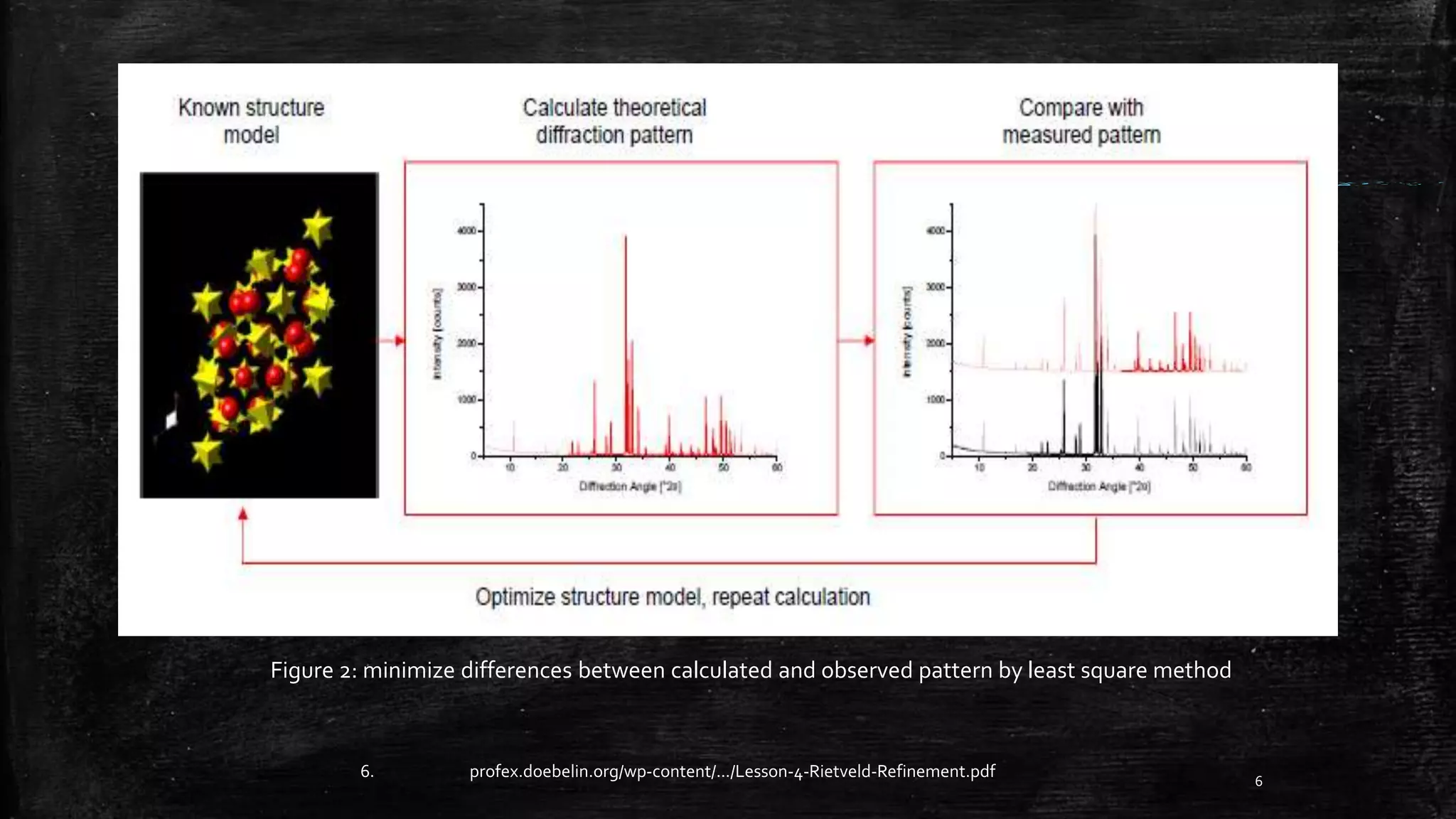
Rietveld refinement is a widely used technique for determining crystal structures and quantifying crystalline materials from powder diffraction data. It works by minimizing the difference between observed and calculated diffraction patterns using least squares refinement. Key aspects include modeling the background, peak shape, unit cell parameters, atomic positions, and other structural details. Common software packages are used to perform the iterative refinement calculations.
![What comes before Rietveld method:
• Debye & Paul Scherer
Large and randomly oriented grains
Difficulty in measuring intensities
Increasing complexities
4
2. Fabio pulizzi, powder struggle, nature, (2014),
7.http://pd.chem.ucl.ac.uk/pdnn/mod1/ip.htm
Figure 1: Debye-Scherrer rings[7]](https://image.slidesharecdn.com/6ff210d3-e276-4311-b0a7-4e730b933ba3-160827002536/75/Rietveld-Refinements-ppt-4-2048.jpg)
![What is Rietveld Refinement?
5
• Structure refinement technique
• use of least square method:
𝑀𝑖𝑛 = 𝑖=0
𝑛=1
[𝑊𝑖(𝑌𝑜𝑏𝑠 𝑖 − 𝑌𝑐𝑎𝑙𝑐 𝑖)2
]
𝑌𝑐𝑎𝑙𝑐 𝑖 can be expressed by following formation.
𝑌 𝑐𝑎𝑙𝑐 𝑖= 𝑝ℎ=1
𝑝ℎ𝑎𝑠𝑒𝑠
𝑆 𝑝ℎ ℎ𝑘𝑙(𝑝ℎ)(𝐾ℎ𝑘𝑙|𝐹ℎ𝑘𝑙
2|⏀ℎ𝑘𝑙(2θ𝑖−2θℎ𝑘𝑙)
3. Eric J. Mittemeijer ,U welzel, Modern Diffraction Methods,Chapter-2,WILEYVCH.](https://image.slidesharecdn.com/6ff210d3-e276-4311-b0a7-4e730b933ba3-160827002536/75/Rietveld-Refinements-ppt-5-2048.jpg)

![Le-bail Method:
• Use for Profile refinement
• Crystal structure determination
How to use?
Modify Rietveld code to set all 𝐹ℎ𝑘𝑙 𝑐𝑎𝑙𝑐 = 1
Extract 𝐹ℎ𝑘𝑙 𝑜𝑏𝑠 using Rietveld algorithm
Set 𝐹ℎ𝑘𝑙 𝑐𝑎𝑙𝑐 from extracted 𝐹ℎ𝑘𝑙 𝑜𝑏𝑠
Repeat 𝐹ℎ𝑘𝑙 𝑜𝑏𝑠 extraction now with 𝐹ℎ𝑘𝑙 𝑐𝑎𝑙𝑐
9
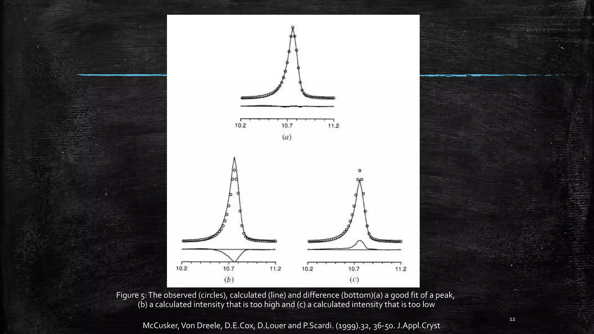
Figure 3: Peak fits of three selected reflections of LaB6.[3]
3. Eric J. Mittemeijer ,U welzel, Modern Diffraction Methods, Chapter-2,WILEYVCH.](https://image.slidesharecdn.com/6ff210d3-e276-4311-b0a7-4e730b933ba3-160827002536/75/Rietveld-Refinements-ppt-9-2048.jpg)