
Recommended
More Related Content
Similar to Anatomy of the Ribs.pptx
Similar to Anatomy of the Ribs.pptx (20)
Recently uploaded
Recently uploaded (12)
Anatomy of the Ribs.pptx
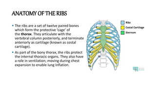
- 1. ANATOMY OF THE RIBS The ribs are a set of twelve paired bones which form the protective ‘cage’ of the thorax. They articulate with the vertebral column posteriorly, and terminate anteriorly as cartilage (known as costal cartilage). As part of the bony thorax, the ribs protect the internal thoracic organs. They also have a role in ventilation; moving during chest expansion to enable lung inflation.
- 2. RIB STRUCTURE There are two classifications of ribs – atypical and typical. The typical ribs have a generalized structure, while the atypical ribs have variations on this structure. 1-TYPICAL RIBS: The typical rib consists of a head, neck and body: The head is wedge shaped, and has two articular facets separated by a wedge of bone. One facet articulates with the numerically corresponding vertebra, and the other articulates with the vertebra above. The neck contains no bony prominences, but simply connects the head with the body. Where the neck meets the body there is a roughed tubercle, with a facet for articulation with the transverse process of the corresponding vertebra. The body, or shaft of the rib is flat and curved. The internal surface of the shaft has a groove for the neurovascular supply of the thorax, protecting the vessels and nerves from damage.
- 4. 2-Atypical Ribs: Ribs 1, 2, 10 11 and 12 can be described as ‘atypical’ – they have features that are not common to all the ribs. Rib 1 is shorter and wider than the other ribs. It only has one facet on its head for articulation with its corresponding vertebra. The superior surface is marked by two grooves, which make way for the subclavian vessels. Rib 2 is thinner and longer than rib 1, and has two articular facets on the head as normal. It has a roughened area on its upper surface, from which the serratus anterior muscle originates. Rib 10 only has one facet – for articulation with its numerically corresponding vertebra. Ribs 11 and 12 have no neck, and only contain one facet, which is for articulation with their corresponding vertebra.
- 5. ARTICULATIONS The majority of the ribs have an anterior and posterior articulation. Posterior: All the twelve ribs articulate posteriorly with the vertebra of the spine. Each rib forms two joints: Costotransverse joint – Between the tubercle of the rib, and the transverse costal facet of the corresponding vertebra. Costovertebral joint – Between the head of the rib, superior costal facet of the corresponding vertebra, and the inferior costal facet of the vertebra above.
- 6. Anterior: The anterior attachment of the ribs vary Ribs 1-7 attach independently to the sternum. Ribs 8 – 10 attach to the costal cartilages superior to them. Ribs 11 and 12 do not have an anterior attachment and end in the abdominal musculature. Because of this, they are sometimes called ‘floating ribs’.
- 7. THE MUSCLES OF THE THORACIC CAGE There are five muscles that make up the thoracic cage; • INTERCOSTALS (external, internal and innermost), • SUBCOSTALS • TRANSVERSUS THORACIS These muscles act to change the volume of the thoracic cavity during respiration.
- 8. INTERCOSTALS The intercostal muscles lie in the intercostal spaces between ribs. They are organised into three layers. 1. External Intercostals There are 11 pairs of external intercostal muscles. They run inferoanteriorly from the rib above to the rib below, and are continuous with the external oblique of the abdomen. Actions: Elevates the ribs, increasing the thoracic volume.
- 9. 2. Internal Intercostals These flat muscles lie deep to the external intercostals. They are continuous with the internal oblique muscle of the abdominal wall. Actions: The interosseous part reduces the thoracic volume by depressing the ribcage, and the interchondral part elevates the ribs. 3. Innermost Intercostals These muscles are the deepest of the intercostal muscles, and are similar in structure to the internal intercostals. Actions: The interosseous part reduces the thoracic volume by depressing the ribcage, and the interchondral part elevates the ribs.
- 10. TRANSVERSUS THORACIS These muscles of the thoracic cage are continuous with transversus abdominis inferiorly. Actions: Weakly depress the ribs. SUBCOSTALS The subcostal muscles are found in the inferior portion of the thoracic wall. Actions: Share the action of the internal intercostals
- 11. LIGAMENTS Costotransverse ligaments: The main ligamentous attachment to the ribs is the costotransverse ligament. This is a fairly complex ligament which is made up of three parts. The first part is known as ‘The costotransverse ligament’ and it fills the gap between the rib and the corresponding transverse process. Lateral Costotransverse ligaments: The next group of fibres are known as the lateral costotransverse ligament, which lies posteriorly and attaches the transverse process of the vertebra to the rib, just distal to the tubercle. Superior costotransverse ligament: The final part is the superior costotransverse ligament which is a two-layered ligament with the fibres orientated at right angles. This passes from the superior margin of the neck of the rib to the transverse process of the above vertebra.