
Recommended
More Related Content
Similar to Thorax.pptx
Similar to Thorax.pptx (20)
Recently uploaded
Recently uploaded (20)
Thorax.pptx
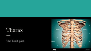
- 2. BONES AND JOINTS OF THE THORACIC WALL
- 3. STERNUM the anterior median part of the thoracic cage.
- 4. PARTS OF A STERNUM It consist of three parts 1.Manubrium sterni 2.Body 3.Xiphoid process
- 5. Anatomical position ● Quadrilateral manubrium is directed upwards & triangular xiphoid process is directed downwards. ● Anterior surface of the manubrium is convex from side to side and concave from above downwards. ● Anterior surface of the body is flat and marked by three transverse ridge and the posterior surface of the body is smooth& slightly concave.
- 6. Features ● It is morphologically a flat bone. It's ossification is intra cartilaginous ossification. ● Manubrium has two surface & Four borders. ● The superior border ia thik, rounded and marked by jugular notch or suprasternal notch on middle & clavicular notch on each side. ● Manubrium makes a slight angle with the body of sternum called sternal angle of Luis. ● The lateral border of the sternum contain 7 pair of costal facet.
- 7. Attachment Manubrium Anterior surface- sternocleidomastoid muscle & pectoralis major muscle . Posterior surface - sternohyoid (upper) & Sternothyroid (lower) Body of the sternum Anterior surface- pectoralis major muscle (on each side) Posterior surface - sternocostalis muscle (Lower lateral part) Lateral border - internal intercostal muscle & external intercostal membrane Xiphoid process Anterior surface - rectus abdominalis muscle Posterior surface - The diaphragm
- 8. Relations Manubrium Posterior surface: Arch of aorta (Lower half), branches of arch of aorta (upper half)& branches are brachiocephalic trunk, left common carotid and left subclavian artery Body of the sternum Right side: Right lung & right pleura Left side: left lung & left pleura (upper half)
- 9. Joints 1.Sternoclavicular joint is a saddle synovial joint. 2.Manubrio-sternal joint is a secondary cartilaginous joint. 3. Xiphisternal joint is primary cartilaginous joint 4.1st chondrosternal joint is a primary cartilaginous joint. 5.2nd-7th chondrosternal joint is synovial joint.
- 10. Clinical importances of sternum ● Bone marrow collection: from Sternum cause it contains red bone marrow throughout the life.It is done in upper part of manubrium to prevent injury to arch of aorta which lies behind its lower half.
- 11. Ribs Lateral boundary of thoracic cage
- 12. Classifications According to their features 1.Typical ribs-3rd to 9th 2.Atypical ribs
- 13. Typical ribs
- 14. Ribs of similar features.
- 15. Identifications 1.Costal groove at the lower border of internal surface. 2.Twisted,angulated and curved shaft
- 16. Anatomical position ❄️Posterior end is higher and medial than the anterior end. ❄️The anterior end bears a concave depression to receive its own costal cartilage. ❄️The shaft is thin,flat and curved with convexity directed outward. ❄️The inner surface of the shaft is marked by costal groove near the lower border
- 17. Parts 1.Two ends- Anterior end bears a concave depression that forms the costo- chondral joint.Posterior end bears - a)Head bears two facets separated by crest b)Neck possesses two surfaces-anterior and posterior.Two borders-upper and lower.c)Tubercle bears two parts-Medial articular part that forms costo- transverse joint and Lateral non-articular part. 2.Shaft- It is curved with convexity directed outward and twisted.It possesses two surfaces-external surface is convex and smooth and internal surface is smooth .Two borders- rounded upper and sharp lower borders.
- 18. Attachments 1.Radiate ligament,intra-articular ligament at the head. 2. Inferior costotransverse ligament at the rough posterior part, Superior costotransverse ligament at the crest of the neck. The anterior surface is covered by costal pleura. 3.The Lateral costotransverse ligament is attached to the lateral non-articular part of Tubercle. 4.The thoracolumbar fascia, lateral fibres of Sacrospinalis muscle,lere attached to the posterior angle. The anterior angle separates the origin of External oblique and serratus anterior in 5th-8th ribs and External oblique from Latissimus dorsi in 9th- 10th rib.
- 19. Attachment 5. The Internal intercostal muscle arises from the floor of costal groove and the Innermost intercostal muscle arises from the middle two-fourths of the ridge above the groove. 6. The External intercostal muscle is atteched on the outer lip and Internal & Innermost intercostal muscle are attached on the inner lip of the upper border of the shaft.
- 20. Relations Internal surface lodges intercostal vein,artery and nerve from above downwards.
- 22. Ossifications 1.One primary centre-for shaft. 2.Three secondary centres-for head and two tubercle.
- 24. The first rib
- 25. Identifications 1.Single circular facet 2.No costal grooves 3.Scalene tubercle in inner border 4.Shortest,broadest,flat,curved 5.No twist,no angle
- 26. Anatomical position 1.Anterior end is larger,thicker and pitted 2.Posterior end is small and rounded. 3.Both ends touch the surface. 4.Posterior end is higher than the anterior end.
- 27. Parts 1.Two ends - Anterior end is largest and thickest which articulates with 1st costal cartilage. Posterior end bears a)Head bears single circular facete. b)Neck c)Tubercle is prominent and thik.It faces upwards and backwards. 2.Shaft It possesse two surfaces -Upper surface is marked by two shallow grooves that ends in scalene tubercle.Lower surface is smooth and no costal grooves.Two borders-convex outer and concave inner border.
- 28. Attachments 1.Capsular ligament and radiate ligament on head 2.Inferior costo-transverse ligament and superior costo-transverse ligament on neck. 3.Lateral costo-transverse ligament on tubercles. 4.Scalenus anterior,subclavius,scalenus medius, costo-clavicular ligament on upper surface.External and Internal intercostal muscle on lower surface.1st digitation of serratus anterior on outer border. Supra-pleural membrane and scalenus anterior on inner border.
- 29. Relations 1.Sympathetic trunk,1st posterior intercoastal vein,superior intercoastal artery,1st thoracic nerve,lower trunk of branchial plexus lies anteriorly on neck from medial to lateral. 2.Subclavian vein,artery and lower trunk of branchial plexus on upper surface. 3.1st intercoastal vein and artery on outer border.
- 30. The second rib
- 31. Identifications 1.More curved 2.Twice longer than 1st rib 3.Head is small and bears two facets . 4.Non-articular part of tubercle is small 5.No twist,slight angle,slight costal groove on posterior internal surface. 6.Angle is close to the tubercle.
- 32. Anatomical position 1.Both ends touch surface 2.Posterior end is higher and medial than the anterior end. 3.Tubercle is in middle of the external surface.
- 33. Parts 1.Two ends- Anterior end lies forward and downward.Posterior End lies backward,upward and medially. 2.Shaft Convex external surface faces upwards and outwards.Concave Internal surface faces downwards and inwards.
- 34. Attachments Scalenus posterior and serratus posterior superior on angle. Half of the 1st and whole of the 2nd digitation of serratus anterior. Others are as typical ribs.
- 35. ◾10th Rib
- 36. Anatomical Points ❄️The posterior end is higher and nearer to the medial plane. ❄️ Posterior end comprises head,neck,tubercle and the anterior end bears a concave depression to receive its own cartilage. ❄️The inner surface is smooth and marked by a costal groove close to its lower border.
- 37. Joints ❄️Anterior end articulate with costal cartilage → costochondral junction. ❄️Head articulates with body of vertebrae → costovertebral joint.
- 38. Attachment and Relation Attachment ❄️ Upper lip→innermost intercostal ❄️Lower lip→external intercostal Relation of costal groove ❄️ Intercostal vein ❄️Intercostal artery ❄️Intercostal nerve
- 39. 11th and 12th Rib Termed as Floating Rib
- 40. Floating Rib They are floating ribs becuase anterior ends are free. They have no communication with the sternum
- 41. Anatomical Position ❄️The posterior end is higher and nearer to the medial plane. ❄️Posterior end comprises only head. There is no neck and tubercle. ❄️The inner surface is marked by shallow costal groove only on its posterior aspect. ❄️There is an angle on the shaft. ❄️Anterior end is pointed.
- 42. Attachment of 12th Rib
- 43. Attachment 1. External surface → From medial to lateral a. Lavaetor Costae b. Longissimus Thoracis c. Illio-costalis →Laterally a.Serratus posterior anterior b.Lattimus dorsi c.External oblique
- 44. Attachment 2. Internal surface a.Upper part→ Diaphragm b. medial part→ Quadratus lumborum 3. Upper border→ External and internal intercostal muscles 4. Lower border→ Lumbocostal ligament
- 45. Thoracic vertebrae Posterior boundary of thoracic cage
- 47. Classifications According to features 1.Typical vertebra 2.Atypical vertebra
- 48. Typical thoracic vertebra Vertebra are the interlocking bones that form the spinal column. Among the 33 individual, 12 are thoracic vertebra. Out of 12 thoracic vertebta, the second to eighth are marked as the typical thoracic vertebra because they carry some common carecteristic features Vertebra are the interlocking bones that form the spinal column. Among the 33 individual, 12 are thoracic vertebra. Out of 12 thoracic vertebta, the second to eighth are marked as the typical thoracic vertebra because they carry some common carecteristic features Vertebra are the interlocking bones that form the spinal column. Among the 33 individual, 12 are thoracic vertebra. Out of 12 thoracic vertebta, the second to eighth are marked as the typical thoracic vertebra because they carry some common carecteristic features
- 49. Identification ● The thoracic vertebra are identified by two demifacets on the both sides of the body. ● the typical thoracic vertebra are identified by costal facets on the anterior surface of transverse process
- 50. Anatomical position ● The body lies anteriorly ● The arch lies posteriorly ● The large transverse process is directed backward and laterally from the junction of pedicle and lamina. ● The long spinous process is directed backwards and downwards .
- 51. Features ● The body is heart shapef.That is the anteroposterior diameter and the transverse diameter are almost equal. On each sidel,it bears two demi facets. ● The vertebral foramen is circular and small. The pedicles are short.The superior articular process is not well marked but the inferior articular process is conspicuous. The laminae are short, thick and board and overlap from above downwards. ● And the large transverse process arises from the junction of pedical and lamina and directed backwards and somewhat laterally. In the anterior surface, it has a concave costal facet ● The spinous process is long and directed backwards and downwards
- 52. Attachment ● The anterior part of the body is attached to the anterior longitudinal ligament and the posterior part of the body is attached to the posterior longitudinal ligament. ● The laminae of adjacent vertebra are connected with a series of fibro-elastic membrane which is named ligamenta flava. ● The transverse process gives attachment to ● . The lateral costotransverse ligament at the tip. ● . The intertransverse muscles and ligaments along the upper and lower border. ● . The superior costotransverse ligament along the lower border ● . The inferior costotransverse ligament along the anterior surface ● . The levator costae and deep muscle of the back to the posterior surface
- 53. The spinous process gives attachment to the supraspinous and interspinous ligaments They also give attachment to several muscles including : Trapezius Rhomboids major and minor Latissimus dorsi serratus posterior superior serratus posterior inferior multifidus sacrospinalis semispinalis capitis the content of vertebral foramen in spinal cord with its meninges
- 54. Joints ● Facet joint or zygacophyseal joint : the joint between superior articular process and inferior articular process of adjoining vertebrae.It is a plane type of Synovial Joint. ● Intervertebral joint :the joint between the body of two corresponding vertebra. it is a secondary cartilaginous joint. ● Costovertebral joint: the joint between superior costal demifacet of a vertebra and head of corresponding rib. It’s a plane type of synovial joint. ● Costotransverse joint: The joint between costal facet of a vertebra and tubercle of corresponding rib.It is also a plane type of synovial joint
- 57. First Thoracic vertebra 1.Body is cervical in form as transverse diameter is twice than anterior-posterior diameter. 2.Upper costal facetes are circular and lower costal facetes are semicircular 3.Spine is thik amd lies horizontally 4.No foramen transversarium
- 58. 9th Thoracic vertebra 1.Large semicircular facate above 2.Small semilunar facate below or may ne absent 10th Thoracic vertebra 1.Large facetes above and lower facetes are absent.Facetes of transverse processmay be absent 2.Upper facetes enroach upon the pedicle
- 59. 11th Thoracoc vertebra 1.Complete circular facet which extends back on pedicle 2.No facet on transverse process 12t Thoracic vertebra 1. circular facet which enroaches on pedicle 2.Body is large and resembles tjat of lumbar vertebra 3.No facet on transverse process 4.Inferior articular facet is convex and twisted laterally. 5..Transverse process has three tubercles-(a).Superior, which corresponds to mamillary process (b) Lateral, which corresponds to transverse process and (c)Inferior, which corresponds to accessory process.
- 60. Thank you slide Student’s most favourite slide