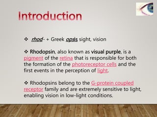
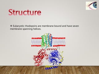
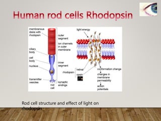
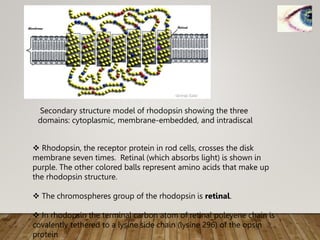
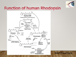
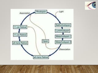
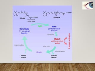
 rhod- + Greek opsis sight, vision
 Rhodopsin, also known as visual purple, is a
pigment of the retina that is responsible for both
the formation of the photoreceptor cells and the
first events in the perception of light.
 Rhodopsins belong to the G-protein coupled
receptor family and are extremely sensitive to light,
enabling vision in low-light conditions.
When we see the word rhodopsin, our thoughts first go to the light
sensitive pigments of rods in our own eyes.
Very similar proteins, however, are present also in our cones and
eyes of various insects, molluscs and other animals
 Eukaryotic rhodopsins are membrane bound and have seven
membrane spanning helices.
Rod cell structure and effect of light on
rhodopsin
 Rhodopsin, the receptor protein in rod cells, crosses the disk
membrane seven times. Retinal (which absorbs light) is shown in
purple. The other colored balls represent amino acids that make up
the rhodopsin structure.
 The chromospheres group of the rhodopsin is retinal.
 In rhodopsin the terminal carbon atom of retinal poleyene chain is
covalently tethered to a lysine side chain (lysine 296) of the opsin
protein
Secondary structure model of rhodopsin showing the three
domains: cytoplasmic, membrane-embedded, and intradiscal
Wald’s Visual Cycle
rhodopsin.pptx
rhodopsin.pptx

rhodopsin.pptx

  • 2.
     rhod- +Greek opsis sight, vision  Rhodopsin, also known as visual purple, is a pigment of the retina that is responsible for both the formation of the photoreceptor cells and the first events in the perception of light.  Rhodopsins belong to the G-protein coupled receptor family and are extremely sensitive to light, enabling vision in low-light conditions.
  • 3.
    When we seethe word rhodopsin, our thoughts first go to the light sensitive pigments of rods in our own eyes. Very similar proteins, however, are present also in our cones and eyes of various insects, molluscs and other animals
  • 4.
     Eukaryotic rhodopsinsare membrane bound and have seven membrane spanning helices.
  • 5.
    Rod cell structureand effect of light on rhodopsin
  • 6.
     Rhodopsin, thereceptor protein in rod cells, crosses the disk membrane seven times. Retinal (which absorbs light) is shown in purple. The other colored balls represent amino acids that make up the rhodopsin structure.  The chromospheres group of the rhodopsin is retinal.  In rhodopsin the terminal carbon atom of retinal poleyene chain is covalently tethered to a lysine side chain (lysine 296) of the opsin protein Secondary structure model of rhodopsin showing the three domains: cytoplasmic, membrane-embedded, and intradiscal
  • 7.